

miR-1202 regulates BPH-1 cell proliferation, apoptosis, and epithelial-to-mesenchymal transition through targeting HMGCL
- PMID: 38551020
- PMCID: PMC11177111
- DOI: 10.3724/abbs.2024001
miR-1202 regulates BPH-1 cell proliferation, apoptosis, and epithelial-to-mesenchymal transition through targeting HMGCL
Abstract
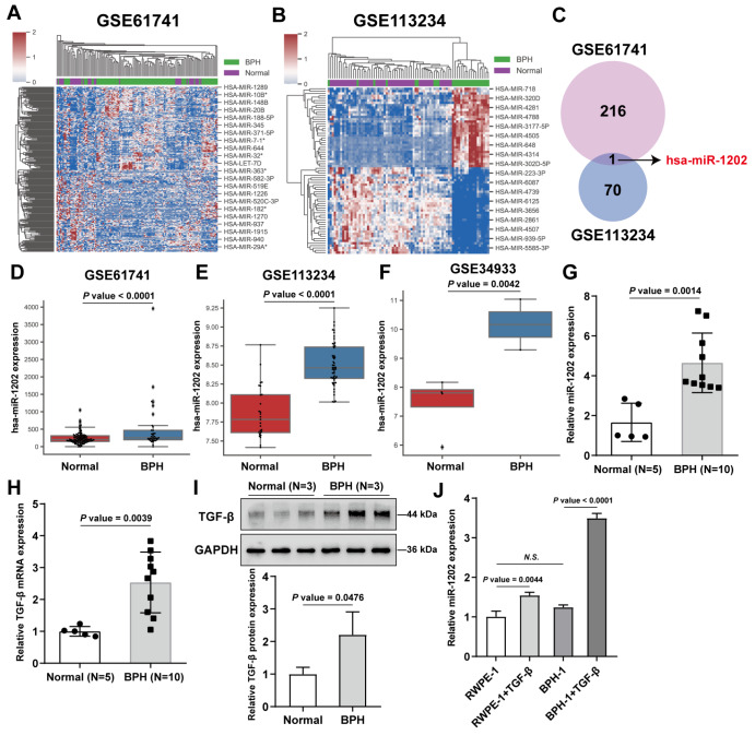
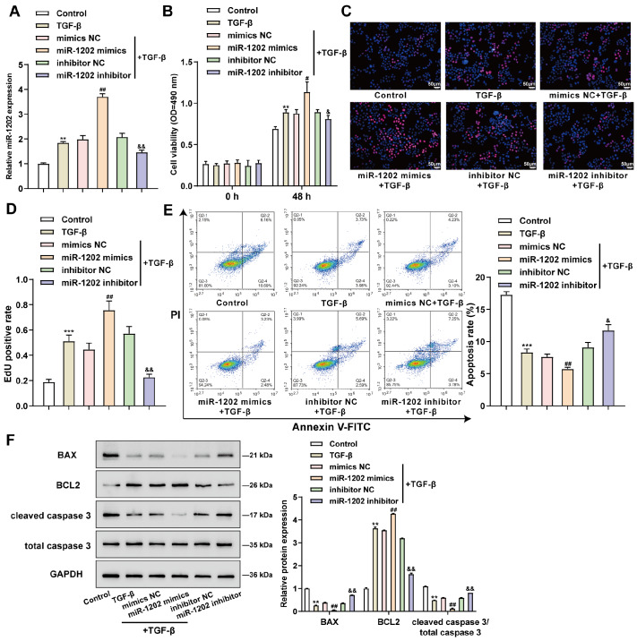
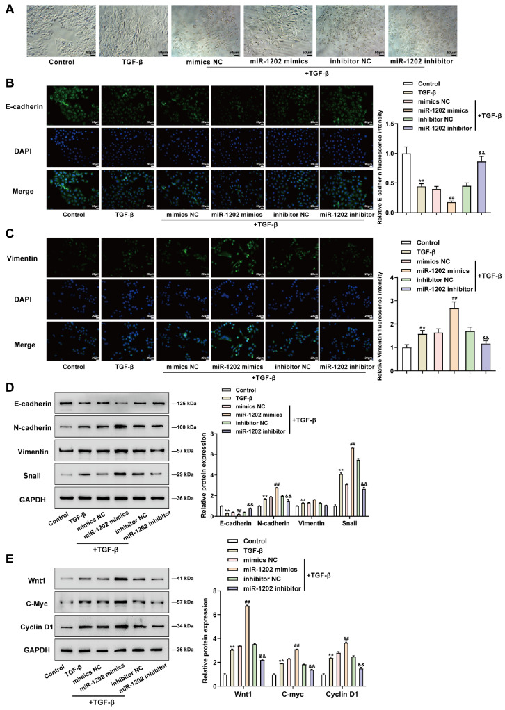
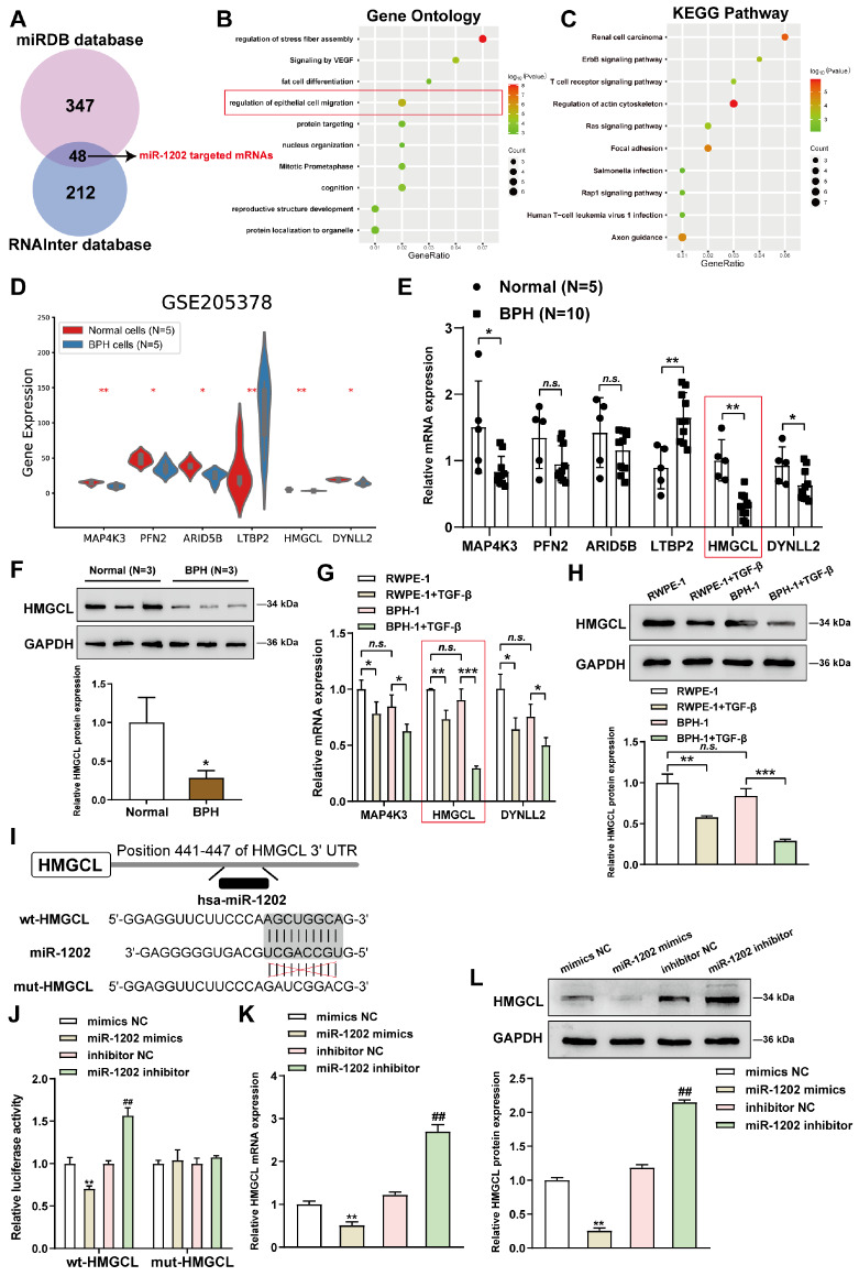
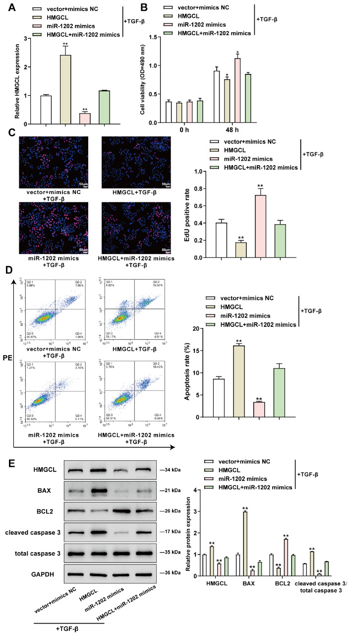
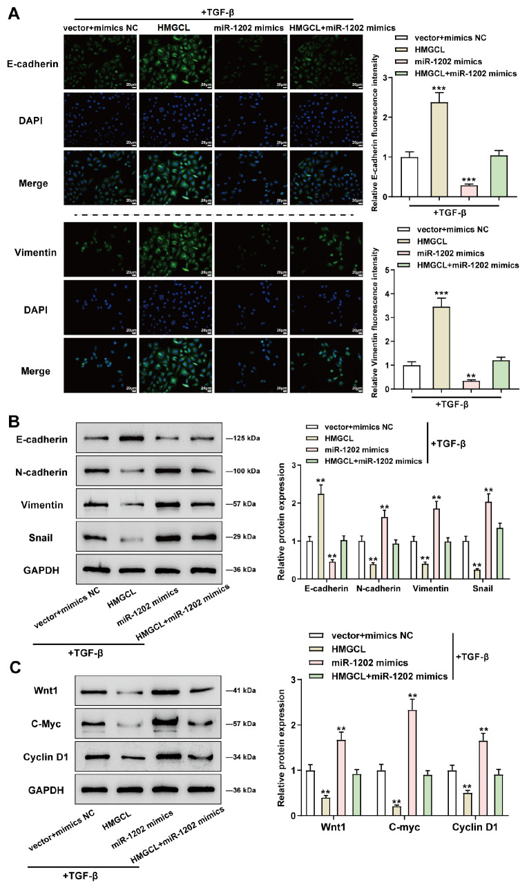
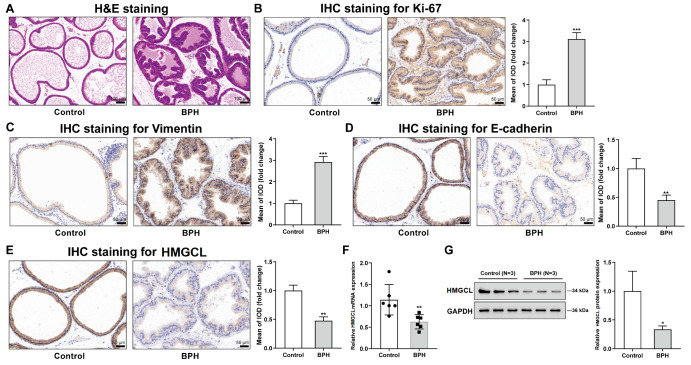
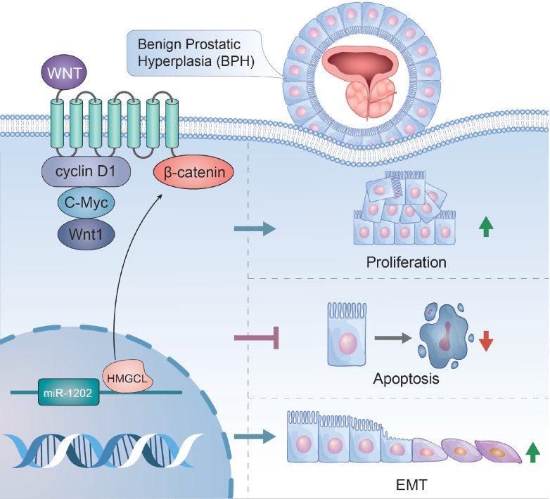
Benign prostatic hyperplasia (BPH) is the expansion of the prostate gland that results in urinary symptoms. Both the epithelial-to-mesenchymal transition (EMT) and the Wnt signaling pathway are associated with BPH pathology. In this study, we find that miR-1202 is increased in BPH samples. Overexpression of miR-1202 in TGF-β-treated BPH-1 cells enhances cell survival and DNA synthesis and inhibits cell apoptosis, whereas miR-1202 inhibition partially abolishes the effects of TGF-β on BPH-1 cells. miR-1202 overexpression reduces E-cadherin level but elevates vimentin, N-cadherin, and snail levels, whereas miR-1202 inhibition partially attenuates the effects of TGF-β on EMT markers. Regarding the Wnt/β-catenin pathway, miR-1202 overexpression significantly enhances, whereas miR-1202 inhibition partially decreases, the promotive effects of TGF-β on Wnt1, c-Myc, and cyclin D1 proteins. 3-Hydroxy-3-methylglutaryl-CoA lyase (HMGCL) is a direct downstream target of miR-1202, and miR-1202 inhibits HMGCL expression through binding to its 3'UTR. Overexpression of HMGCL significantly reduces the effect of miR-1202 overexpression on the phenotypes of BPH-1 cells by inhibiting cell survival and promoting apoptosis. Similarly, HMGCL overexpression has the opposite effects on EMT markers and the Wnt/β-catenin signaling, and markedly alleviates the effects of miR-1202 overexpression. Finally, in the BPH rat model, Ki67 and vimentin levels are elevated, but E-cadherin and HMGCL levels are reduced. In conclusion, miR-1202 is upregulated in benign prostatic hyperplasia; miR-1202 enhances epithelial cell proliferation, suppresses cell apoptosis, and promotes EMT by targeting HMGCL. The Wnt/β-catenin pathway may participate in the miR-1202/HMGCL axis-mediated regulation of BPH-1 cell phenotypes.
Keywords: 3-hydroxy-3-methylglutaryl-CoA lyase (HMGCL); Wnt/β-catenin pathway; benign prostate hyperplasia (BPH); epithelial-to-mesenchymal transition (EMT); miR-1202.
Conflict of interest statement
The authors declare that they have no conflict of interest.
Figures
Similar articles
-
MIR663AHG as a competitive endogenous RNA regulating TGF-β-induced epithelial proliferation and epithelial-mesenchymal transition in benign prostate hyperplasia.J Biochem Mol Toxicol. 2023 Sep;37(9):e23391. doi: 10.1002/jbt.23391. Epub 2023 Jul 30. J Biochem Mol Toxicol. 2023. PMID: 37518988
-
The miR-223-3p/MAP1B axis aggravates TGF-β-induced proliferation and migration of BPH-1 cells.Cell Signal. 2021 Aug;84:110004. doi: 10.1016/j.cellsig.2021.110004. Epub 2021 Apr 8. Cell Signal. 2021. PMID: 33839256
-
Y-27632 targeting ROCK1&2 modulates cell growth, fibrosis and epithelial-mesenchymal transition in hyperplastic prostate by inhibiting β-catenin pathway.Mol Biomed. 2024 Oct 26;5(1):52. doi: 10.1186/s43556-024-00216-9. Mol Biomed. 2024. PMID: 39455522 Free PMC article.
-
Exploring TGF-β signaling in benign prostatic hyperplasia: from cellular senescence to fibrosis and therapeutic implications.Biogerontology. 2025 Mar 30;26(2):79. doi: 10.1007/s10522-025-10226-x. Biogerontology. 2025. PMID: 40159577 Review.
-
Epithelial to Mesenchymal Transition and Cell Biology of Molecular Regulation in Endometrial Carcinogenesis.J Clin Med. 2019 Mar 30;8(4):439. doi: 10.3390/jcm8040439. J Clin Med. 2019. PMID: 30935077 Free PMC article. Review.
Cited by
-
Identification and Characterization of Differentially Expressed MicroRNAs in Benign Prostatic Hyperplasia.Cancer Rep (Hoboken). 2025 Apr;8(4):e70178. doi: 10.1002/cnr2.70178. Cancer Rep (Hoboken). 2025. PMID: 40223182 Free PMC article.
References
-
- Kim EH, Larson JA, Andriole GL. Management of benign prostatic hyperplasia. Annu Rev Med. . 2016;67:137–151. doi: 10.1146/annurev-med-063014-123902. - DOI - PubMed
-
- McNeal JE. Regional morphology and pathology of the prostate. Am J Clin Pathol. . 1968;49:347–357. doi: 10.1093/ajcp/49.3.347. - DOI - PubMed
-
- Alonso-Magdalena P, Brössner C, Reiner A, Cheng G, Sugiyama N, Warner M, Gustafsson JÅ. A role for epithelial-mesenchymal transition in the etiology of benign prostatic hyperplasia. Proc Natl Acad Sci USA. . 2009;106:2859–2863. doi: 10.1073/pnas.0812666106. - DOI - PMC - PubMed
MeSH terms
Substances
LinkOut - more resources
Full Text Sources
Medical
Molecular Biology Databases
Research Materials
