. 2024 Dec;13(1):2337671.
doi: 10.1080/22221751.2024.2337671. Epub 2024 Apr 12.

Rat hepatitis E virus (Rocahepevirus ratti) exposure in cats and dogs, Hong Kong

Affiliations

Rat hepatitis E virus (Rocahepevirus ratti) exposure in cats and dogs, Hong Kong

Estie Hon-Kiu Shun et al. Emerg Microbes Infect. 2024 Dec.

Abstract

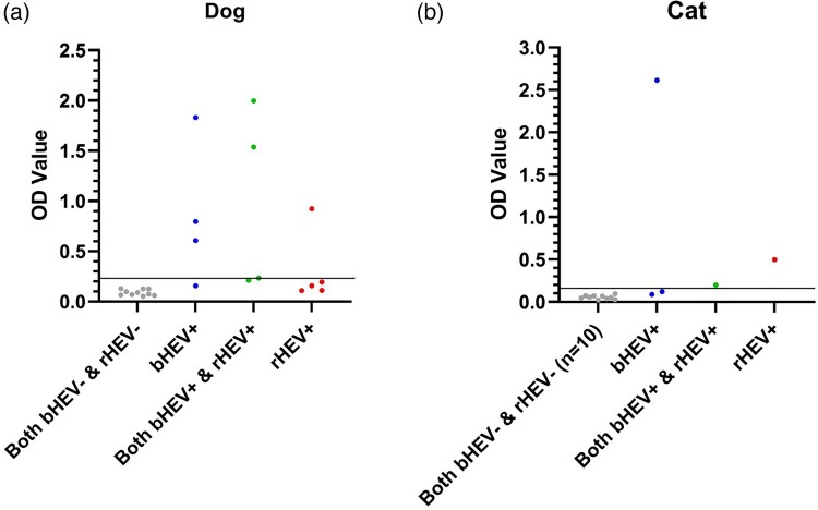
Hepatitis E virus (HEV) variants infecting humans belong to two species: Paslahepevirus balayani (bHEV) and Rocahepevirus ratti (rat hepatitis E virus; rHEV). R. ratti is a ubiquitous rodent pathogen that has recently been recognized to cause hepatitis in humans. Transmission routes of rHEV from rats to humans are currently unknown. In this study, we examined rHEV exposure in cats and dogs to determine if they are potential reservoirs of this emerging human pathogen. Virus-like particle-based IgG enzymatic immunoassays (EIAs) capable of differentiating rHEV & bHEV antibody profiles and rHEV-specific real-time RT-PCR assays were used for this purpose. The EIAs could detect bHEV and rHEV patient-derived IgG spiked in dog and cat sera. Sera from 751 companion dogs and 130 companion cats in Hong Kong were tested with these IgG enzymatic immunoassays (EIAs). Overall, 13/751 (1.7%) dogs and 5/130 (3.8%) cats were sero-reactive to HEV. 9/751 (1.2%) dogs and 2/130 (1.5%) cats tested positive for rHEV IgG, which was further confirmed by rHEV immunoblots. Most rHEV-seropositive animals were from areas in or adjacent to districts reporting human rHEV infection. Neither 881 companion animals nor 652 stray animals carried rHEV RNA in serum or rectal swabs. Therefore, we could not confirm a role for cats and dogs in transmitting rHEV to humans. Further work is required to understand the reasons for low-level seropositivity in these animals.

Keywords: Hepatitis E virus; cat diseases; dogs; hepatitis antibodies; rats.


Conflict of interest statement

No potential conflict of interest was reported by the author(s).

Figures

Figure 1.
Figure 1.
Characteristics of HEV-A4-P239 and HEV-C1-P241 peptides. (A) Genome map of hepatitis E virus (HEV) showing Open Reading Frame 2 (ORF2) region corresponding to HEV-A4-P239 and HEV-C1-P241 peptides. S: shell, M: middle, P: protruding domains of ORF2 are highlighted in pink. (B) Sodium dodecyl sulphate-polyacrylamide gel electrophoresis of HEV-A4-P239 and HEV-C1-P241 showing bands at 40–55 kDa. (C) Immunoblots based on HEV-A4-P239 and HEV-C1-P241 showing that Paslahepevirus balayani (bHEV) infected patient sera (lane 1: bHEV genotype 3, lane 2: bHEV genotype 4) reacted in HEV-A4-P239 immunoblot, but not HEV-C1-P241 immunoblot. Rocahepevirus ratti (rHEV) infected patient sera (lane 3) reacted in HEV-C1-P241 immunoblot, but not HEV-A4-P239 immunoblot. N: blank lane; P: anti-His antibody control. (D) Optical density (OD) values of bHEV-positive and rHEV-positive patient-derived IgG spiked into dog and cat serum pools using rHEV and bHEV enzymatic immunoassays. Purple lines represent rHEV-patient IgG spiked into cat serum pool. Orange lines represent rHEV-patient IgG spiked into dog serum pool. Pink lines represent bHEV-patient IgG spiked into cat serum pool. Brown lines represent rHEV-patient IgG spiked into dog serum pool. Black lines represent HEV-negative patient IgG spiked into HEV-negative dog serum pool. Green lines represent HEV-negative patient IgG spiked into HEV-negative cat serum pool. Red lines represent IgG purified from the HEV-negative dog serum pool. Gray lines represent IgG purified from the HEV-negative cat serum pool.
Figure 2.
Figure 2.
OD values of (A) dog and (B) cat serum samples tested in bHEV and rHEV EIAs. Blue dots and lines: sera testing positive only in bHEV EIA; red dots and lines: sera testing positive only in rHEV EIA; green dots and lines: sera testing positive in both EIAs. Horizontal black lines represent respective assay cutoffs (mean OD + 3 standard deviations of the group).
Figure 3.
Figure 3.
Optical Density (OD) values bHEV and rHEV EIA positive companion (A) dog and (B) cat sera in Wantai assay. Gray dots: bHEV and rHEV EIA negative control samples (n = 10); blue dots: sera testing positive only in bHEV EIA; red dots: sera testing positive only in rHEV EIA; green dots: sera testing positive in both EIAs. Horizontal lines represent cutoffs derived from mean OD + 3 standard deviations of 40 companion animal sera testing negative in both bHEV and rHEV EIAs.
Figure 4.
Figure 4.
HEV-C1-P241 immunoblot of rHEV EIA-positive companion animal sera samples. (A) Immunoblot of rHEV EIA-positive dog sera. (B) Immunoblot of rHEV EIA-positive cat sera. N: blank lane; P: anti-His antibody control. Blot assays were performed in a MiniProtean II Apparatus, which enables lane-by-lane separation of the immunoblot without cutting into strips.

References

    1. Rein DB, Stevens GA, Theaker J, et al. . The global burden of hepatitis E virus genotypes 1 and 2 in 2005. Hepatology. 2012 Apr;55(4):988–997. doi:10.1002/hep.25505 - DOI - PubMed
    1. Sridhar S, Teng JLL, Chiu TH, et al. . Hepatitis E virus genotypes and evolution: emergence of camel hepatitis E variants. Int J Mol Sci. 2017 Apr 20;18(4):869. doi:10.3390/ijms18040869 - DOI - PMC - PubMed
    1. Kamar N, Dalton HR, Abravanel F, et al. . Hepatitis E virus infection. Clin Microbiol Rev. 2014 Jan;27(1):116–138. doi:10.1128/CMR.00057-13 - DOI - PMC - PubMed
    1. Kamar N, Selves J, Mansuy JM, et al. . Hepatitis E virus and chronic hepatitis in organ-transplant recipients. N Engl J Med. 2008 Feb 21;358(8):811–817. doi:10.1056/NEJMoa0706992 - DOI - PubMed
    1. Lee GH, Tan BH, Teo EC, et al. . Chronic infection with camelid hepatitis E virus in a liver transplant recipient who regularly consumes camel meat and milk. Gastroenterology. 2016 Feb;150(2):355–357.e3. doi:10.1053/j.gastro.2015.10.048 - DOI - PubMed

Substances