. 2024 Apr 17;90(4):e0236323.
doi: 10.1128/aem.02363-23. Epub 2024 Mar 29.

Long-duration environmental biosensing by recording analyte detection in DNA using recombinase memory

Affiliations

Long-duration environmental biosensing by recording analyte detection in DNA using recombinase memory

Prashant Bharadwaj Kalvapalle et al. Appl Environ Microbiol. .

Abstract

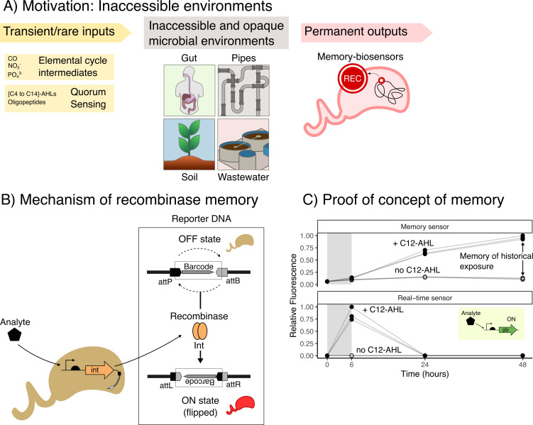
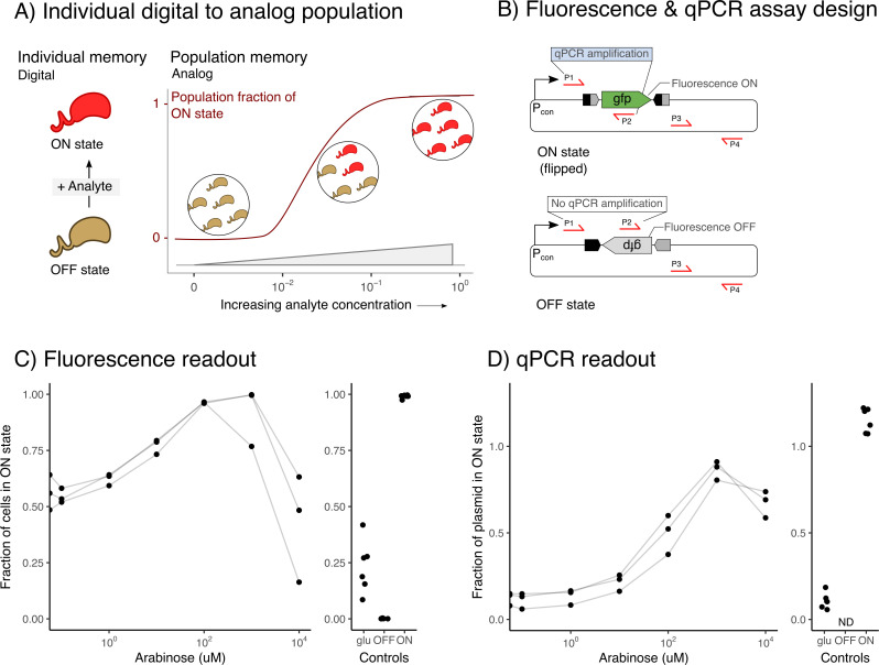
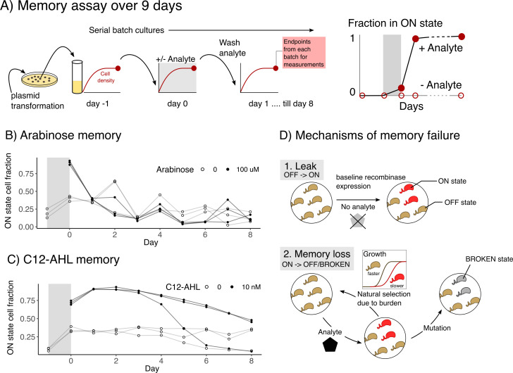
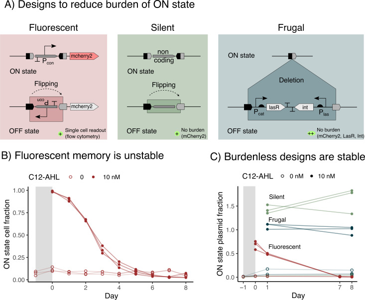
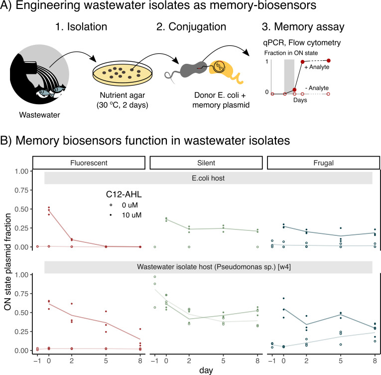
Microbial biosensors that convert environmental information into real-time visual outputs are limited in their sensing abilities in complex environments, such as soil and wastewater, due to optical inaccessibility. Biosensors that could record transient exposure to analytes within a large time window for later retrieval represent a promising approach to solve the accessibility problem. Here, we test the performance of recombinase-memory biosensors that sense a sugar (arabinose) and a microbial communication molecule (3-oxo-C12-L-homoserine lactone) over 8 days (~70 generations) following analyte exposure. These biosensors sense the analyte and trigger the expression of a recombinase enzyme which flips a segment of DNA, creating a genetic memory, and initiates fluorescent protein expression. The initial designs failed over time due to unintended DNA flipping in the absence of the analyte and loss of the flipped state after exposure to the analyte. Biosensor performance was improved by decreasing recombinase expression, removing the fluorescent protein output, and using quantitative PCR to read out stored information. Application of memory biosensors in wastewater isolates achieved memory of analyte exposure in an uncharacterized Pseudomonas isolate. By returning these engineered isolates to their native environments, recombinase-memory systems are expected to enable longer duration and in situ investigation of microbial signaling, cross-feeding, community shifts, and gene transfer beyond the reach of traditional environmental biosensors.IMPORTANCEMicrobes mediate ecological processes over timescales that can far exceed the half-lives of transient metabolites and signals that drive their collective behaviors. We investigated strategies for engineering microbes to stably record their transient exposure to a chemical over many generations through DNA rearrangements. We identify genetic architectures that improve memory biosensor performance and characterize these in wastewater isolates. Memory biosensors are expected to be useful for monitoring cell-cell signals in biofilms, detecting transient exposure to chemical pollutants, and observing microbial cross-feeding through short-lived metabolites within cryptic methane, nitrogen, and sulfur cycling processes. They will also enable in situ studies of microbial responses to ephemeral environmental changes, or other ecological processes that are currently challenging to monitor non-destructively using real-time biosensors and analytical instruments.

Keywords: biosensor; genetic memory; integrase; quorum sensing; recombinase; synthetic biology; wastewater.


Conflict of interest statement

The authors declare no conflict of interest.

Figures

Fig 1
Fig 1
Recombinase memory stores information of historical analyte exposure. (A) Memory biosensors can be deployed into inaccessible, hard-to-image environments for undisturbed monitoring of ecological processes by coupling the detection of transient and rare chemical inputs into permanent DNA modifications. (B) A recombinase-memory biosensor works by conditionally expressing a recombinase enzyme when it senses an analyte. The recombinase binds the DNA attachment sites attP and attB and reverses the DNA segment flanked by the sites, thereby encoding information as a genetic memory in the DNA. (C) A memory sensor provides information on historical exposures to environmental chemicals (top), in contrast to a real-time reporter which only provides information during chemical exposure (bottom). Data are shown for reporters of the quorum-sensing molecule, 3-oxo-C12-L-homoserine lactone (C12-AHL). Sensor outputs were measured indirectly by monitoring cellular fluorescence arising from DNA flipping. Three independent cultures of both sensors were exposed to 1 µM C12-AHL for 6 hours in the exponential phase (gray), prior to washing and subculturing for two passages. While the memory sensor retained the fluorescence after the analyte was removed, the real-time sensor’s output was not significantly different from baseline at 48 hours (paired sample two-tailed t-test, P = 0.84). This loss of signal was expected with the real-time sensor, since this biosensor only synthesizes the fluorescent protein in the presence of the analyte. After analyte removal, the fluorescent protein reporter is subject to degradation and dilution as cells grow.
Fig 2
Fig 2
Recombinase memory stores analog information. (A) A digital memory storage device coded within individual bacteria can code analog information at the population level. Exposure of cells in the OFF state (brown) to low analyte concentrations marks a subset of the cells with the memory, switching them to the ON state (red), while higher concentrations increase the fraction of cells in the population to the ON state. (B) Primer sets were designed to read out memory using qPCR. The first primer pair anneals outside the region that is flipped to code a memory (P1) and inside the flipped DNA (P2), such that a product is only formed with the ON state. The second primer set (P3 and P4) was designed to quantify the total reporter plasmid for normalization. (C) The ON state was read out using fluorescence after 24 hours of exposure to a range of concentrations of the input analyte, arabinose. Flow cytometry data from three independent replicate cultures are shown, where the ON state is defined as fluorescence values >99 percentile of the OFF state control. The line connects data from related cultures. (D) The ON state fraction was quantified using qPCR as the copies of flipped state DNA normalized to total copies of the plasmid. Controls are shown in the adjacent sub-panel. The positive control (ON) represents cells harboring the reporter plasmid already in the ON state. The negative controls (OFF and glu) represent cells harboring the OFF state reporter plasmid without the integrase plasmid and the memory biosensor grown with glucose which represses the pBAD promoter. ND, non-detectable signal.
Fig 3
Fig 3
Week-long serial culturing led to memory loss. (A) Experimental design for testing memory stability. Biosensors were grown for ~72 generations using serial batch cultures, with the analyte present only on the first day (shaded region). (B) The arabinose-memory sensor was tested using three independent replicate cultures following 1 day of exposure to 100 µM arabinose (filled circles) or no arabinose (open circles). Samples from the same biological replicates are connected by a line. The fraction of ON state cells expressing GFP were read out by flow cytometry at the end of each day (Fig. S4). The ON state fraction following analyte exposure was unstable and decayed with a half-life of 0.98 ± 0.09 days. Additionally, the ON state fraction remained significantly higher than no arabinose levels only on days 0 and 1 (paired sample one-tailed t-test, P < 0.03). The OFF state was also unstable, as the uninduced cells presented fluorescence. (C) The C12-AHL memory sensor was characterized using 10 nM analyte. Both the ON and OFF states were unstable. However, the memory loss was slower and decayed starting at day 2 with a half-life of 2.8 ± 0.78 days. There was a significant difference between AHL exposed and unexposed samples until day 6 (paired sample one-tailed t-test, P < 0.03). (D) Mechanisms predicted to underlie memory instability. (1) Leaky recombinase expression in the absence of the analyte is predicted to yield a signal in the OFF state (brown). With time, the recombinase accumulates to sufficient levels to flip the memory to ON state (red). (2) The metabolic burden of fluorescent protein expression is predicted to make the ON state unstable. Cells in the ON state are either outcompeted by cells in the OFF state, or they accumulate mutations that abolish fluorescent protein expression resulting in a BROKEN state (gray). Half-lives were obtained by fitting a single exponential, c·e-kt, to the data.
Fig 4
Fig 4
Memory designs can extend the duration of memory utility. (A) The Fluorescent (red), Silent (green), and Frugal (blue) biosensor designs all use a broad host range origin to enable studies in diverse bacterial species, and they lack repeat sequences that can lead to deleterious homologous recombination. Upon sensing the analyte, the Fluorescent design produces fluorescence similar to the parent design, the Silent design flips a non-functional DNA region, and the Frugal design deletes the sensor components (lasR and int recombinase) to yield a smaller plasmid that is expected to minimize cellular burden. (B) The Fluorescent memory was tested for stability using three independent replicate cultures following a 1 day of exposure to 10 nM C12-AHL (filled circles) or no C12-AHL (open circles). The y-axis shows the fraction of cells in the ON state, gated by fluorescence (Fig. S8), over the duration of the experiment. (C) For each design, the memory output is shown for the same experiment, measured by qPCR. The fraction of the plasmids in the ON state is defined as the copies of flipped state DNA normalized to the total copies of the plasmid; the latter was measured at the start and end of the experiment. To evaluate stability, we assessed whether the signal was significantly different when comparing days 1 and 8 of the experiment (paired sample one-sided t-test on C12-AHL exposed samples, for d1 > d8: Fluorescent: P = 1e − 4, Silent: P = 0.8, Frugal: P = 0.16). The ON state plasmid fraction values that exceed the theoretical maximum of 1 are thought to arise because they were calculated using the absolute copy numbers of two different amplicons, which were quantified using two different standard curves (Fig. S11).
Fig 5
Fig 5
Memory designs function in wastewater microbes. (A) To test memory in wastewater isolates: (1) wastewater was streaked on nutrient agar plates to isolate colonies and characterized using 16S rRNA sequencing (2), the memory-biosensor plasmids were conjugated from E. coli donors into the wastewater isolates, and (3) memory performance was characterized using qPCR. (B) A comparison of the memory designs in E. coli (top) and one wastewater Pseudomonas isolate (bottom). The performance of the Fluorescent (red), Silent (green), and Frugal (blue) designs are compared. For each experiment, we measured the fraction of plasmid in the ON state as in Fig. 4, with data from three independent cultures shown as points, and a line connecting the averages. With the Fluorescent design, analyte exposure yielded a signal that decayed with a half-life of 1.0 ± 0.1 days with E. coli and 3.6 ± 0.8 days with the isolate (fit to a single exponential model). With the Silent design, analyte exposure yielded a significantly higher signal than without exposure when pooled across days in E. coli (P = 2e − 8) but not in the isolate (P = 0.74). With the Frugal design, analyte exposure yielded a significantly higher signal than the control on all days with E. coli (P < 0.04) and with the isolate (P < 0.033). Isolate without exposure had a significantly higher signal on days 5 and 8, than day −1 (P < 0.048). All the P-values were obtained using a paired sample one-tailed t-test. Missing points for Silent in E. coli indicate that there was no qPCR detection for flipped.

Similar articles

Cited by

References

    1. Libis V, Delépine B, Faulon JL. 2016. Sensing new chemicals with bacterial transcription factors. Curr Opin Microbiol 33:105–112. doi:10.1016/j.mib.2016.07.006 - DOI - PubMed
    1. Chang C, Stewart RC. 1998. The two-component system: regulation of diverse signaling pathways in prokaryotes and eukaryotes. Plant Physiol 117:723–731. doi:10.1104/pp.117.3.723 - DOI - PMC - PubMed
    1. Koretke KK, Lupas AN, Warren PV, Rosenberg M, Brown JR. 2000. Evolution of two-component signal transduction. Mol Biol Evol 17:1956–1970. doi:10.1093/oxfordjournals.molbev.a026297 - DOI - PubMed
    1. Del Valle I, Fulk EM, Kalvapalle P, Silberg JJ, Masiello CA, Stadler LB. 2021. Translating new synthetic biology advances for biosensing into the earth and environmental sciences. Front Microbiol 11:618373. doi:10.3389/fmicb.2020.618373 - DOI - PMC - PubMed
    1. Bereza-Malcolm LT, Mann G, Franks AE. 2015. Environmental sensing of heavy metals through whole cell microbial biosensors: a synthetic biology approach. ACS Synth Biol 4:535–546. doi:10.1021/sb500286r - DOI - PubMed

Publication types

LinkOut - more resources