

Femtosecond pulsed laser photodynamic therapy activates melanin and eradicates malignant melanoma
- PMID: 38551838
- PMCID: PMC10998568
- DOI: 10.1073/pnas.2316303121
Femtosecond pulsed laser photodynamic therapy activates melanin and eradicates malignant melanoma
Abstract
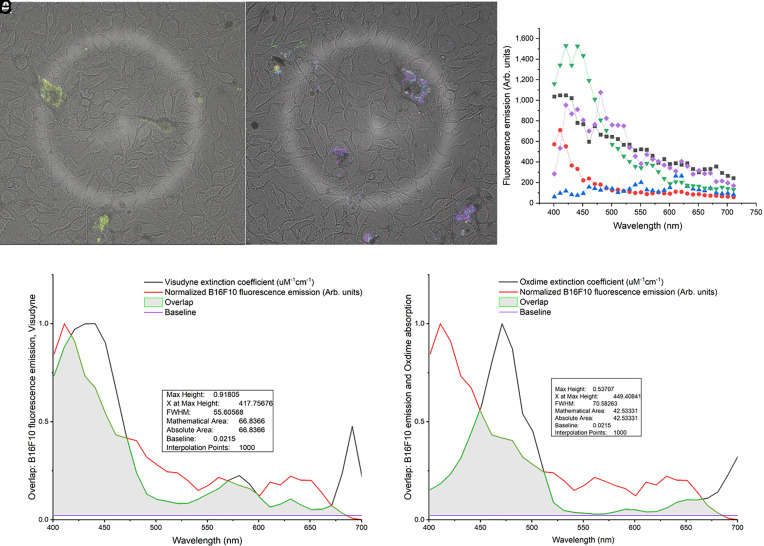
Photodynamic therapy (PDT) relies on a series of photophysical and photochemical reactions leading to cell death. While effective for various cancers, PDT has been less successful in treating pigmented melanoma due to high light absorption by melanin. Here, this limitation is addressed by 2-photon excitation of the photosensitizer (2p-PDT) using ~100 fs pulses of near-infrared laser light. A critical role of melanin in enabling rather than hindering 2p-PDT is elucidated using pigmented and non-pigmented murine melanoma clonal cell lines in vitro. The photocytotoxicities were compared between a clinical photosensitizer (Visudyne) and a porphyrin dimer (Oxdime) with ~600-fold higher σ2p value. Unexpectedly, while the 1p-PDT responses are similar in both cell lines, 2p activation is much more effective in killing pigmented than non-pigmented cells, suggesting a dominant role of melanin 2p-PDT. The potential for clinical translational is demonstrated in a conjunctival melanoma model in vivo, where complete eradication of small tumors was achieved. This work elucidates the melanin contribution in multi-photon PDT enabling significant advancement of light-based treatments that have previously been considered unsuitable in pigmented tumors.
Keywords: melanin; melanoma; multiphoton; ocular melanoma; photodynamic therapy.
Conflict of interest statement
Competing interests statement:The authors declare no competing interest.
Figures


Similar articles
-
Photodynamic treatment induces cell death by apoptosis or autophagy depending on the melanin content in two B16 melanoma cell lines.Oncol Rep. 2013 Mar;29(3):1196-200. doi: 10.3892/or.2012.2190. Epub 2012 Dec 14. Oncol Rep. 2013. PMID: 23242333
-
Melanogenesis and DNA damage following photodynamic therapy in melanoma with two meso-substituted porphyrins.J Photochem Photobiol B. 2016 Aug;161:402-10. doi: 10.1016/j.jphotobiol.2016.06.012. Epub 2016 Jun 8. J Photochem Photobiol B. 2016. PMID: 27314538
-
St John's Wort (Hypericum perforatum L.) photomedicine: hypericin-photodynamic therapy induces metastatic melanoma cell death.PLoS One. 2014 Jul 30;9(7):e103762. doi: 10.1371/journal.pone.0103762. eCollection 2014. PLoS One. 2014. PMID: 25076130 Free PMC article.
-
Melanoma resistance to photodynamic therapy: new insights.Biol Chem. 2013 Feb;394(2):239-50. doi: 10.1515/hsz-2012-0228. Biol Chem. 2013. PMID: 23152406 Free PMC article. Review.
-
Photodynamic therapy in melanoma--an update.J Physiol Pharmacol. 2012 Apr;63(2):109-18. J Physiol Pharmacol. 2012. PMID: 22653896 Review.
Cited by
-
Emerging ultrafast technologies in biotechnology.3 Biotech. 2025 May;15(5):142. doi: 10.1007/s13205-025-04309-2. Epub 2025 Apr 24. 3 Biotech. 2025. PMID: 40292246 Review.
-
Nanomedicine-Based Treatments for Rare and Aggressive Ocular Cancers: Advances in Drug Delivery.Curr Treat Options Oncol. 2025 Jul;26(7):569-586. doi: 10.1007/s11864-025-01330-8. Epub 2025 May 22. Curr Treat Options Oncol. 2025. PMID: 40399581 Review.
-
Comparative analysis of ALA mediated sonodynamic therapy considering tumor size, light combination and ultrasound delivery in murine cutaneous melanoma.Sci Rep. 2025 Aug 22;15(1):30859. doi: 10.1038/s41598-025-16366-x. Sci Rep. 2025. PMID: 40846765 Free PMC article.
-
Targeting ocular malignancies using a novel light-activated virus-like drug conjugate.Adv Ophthalmol Pract Res. 2024 Dec 3;5(1):49-57. doi: 10.1016/j.aopr.2024.12.001. eCollection 2025 Feb-Mar. Adv Ophthalmol Pract Res. 2024. PMID: 39911685 Free PMC article. Review.
References
-
- Allison R. R., et al. , Photosensitizers in clinical PDT. Photodiagn. Photodyn. Ther. 1, 27–42 (2004). - PubMed
-
- NCI, Photodynamic therapy to treat cancer (NCI, 2011) (27 January 2023).
-
- Andersen R., Loebel N., Hammond D., Wilson M., Treatment of periodontal disease by photodisinfection compared to scaling and root planing. J. Clin. Dent. 18, 34–38 (2007). - PubMed
MeSH terms
Substances
Grants and funding
- 2013-07276-1/Fundação de Amparo à Pesquisa do Estado de São Paulo (FAPESP)
- 305795/2016-3/Conselho Nacional de Desenvolvimento Científico e Tecnológico (CNPq)
- N/A/CNPq | Ciência sem Fronteiras (CsF)
- N/A/Coordenação de Aperfeiçoamento de Pessoal de Nível Superior (CAPES)
- N/A/Universidade de Sao Paulo - University of Toronto Joint Program
- N/A/Banting Fellowship from the Canadian Institutes of Health Research
- N/A/Princess Margaret Cancer Centre Foundation Invest-in-Research Fund
- N/A/Princess Margaret Cancer Centre Foundation Excellence Fellowship Award
- N/A/Henry Farrugia Research Fund
- N/A/Vision Science Research Program Award
- M20301556/Cancer Prevention and Research Institute of Texas (CPRIT)
- M230930/Governor's University Research Initiative
LinkOut - more resources
Full Text Sources
Medical
