. 2024 Jun:162:9-27.
doi: 10.1016/j.clinph.2024.03.008. Epub 2024 Mar 18.

Cortical and white matter substrates supporting visuospatial working memory

Affiliations

Cortical and white matter substrates supporting visuospatial working memory

Riyo Ueda et al. Clin Neurophysiol. 2024 Jun.

Abstract

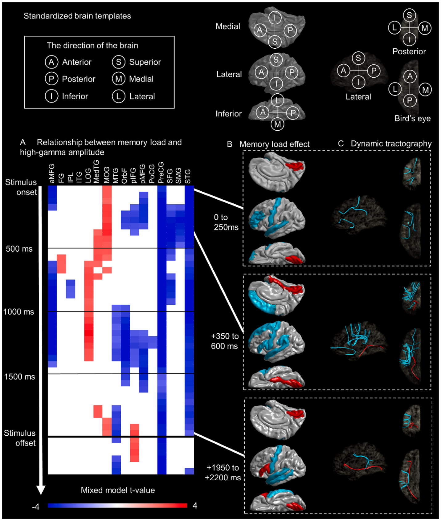
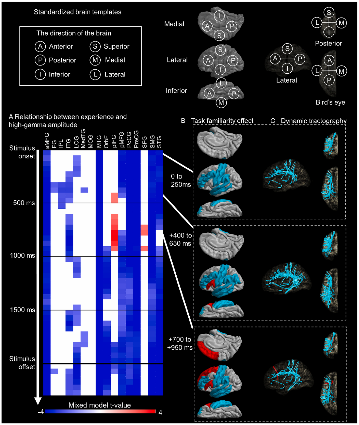
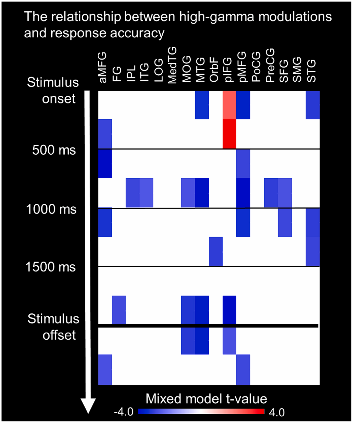
Objective: In tasks involving new visuospatial information, we rely on working memory, supported by a distributed brain network. We investigated the dynamic interplay between brain regions, including cortical and white matter structures, to understand how neural interactions change with different memory loads and trials, and their subsequent impact on working memory performance.

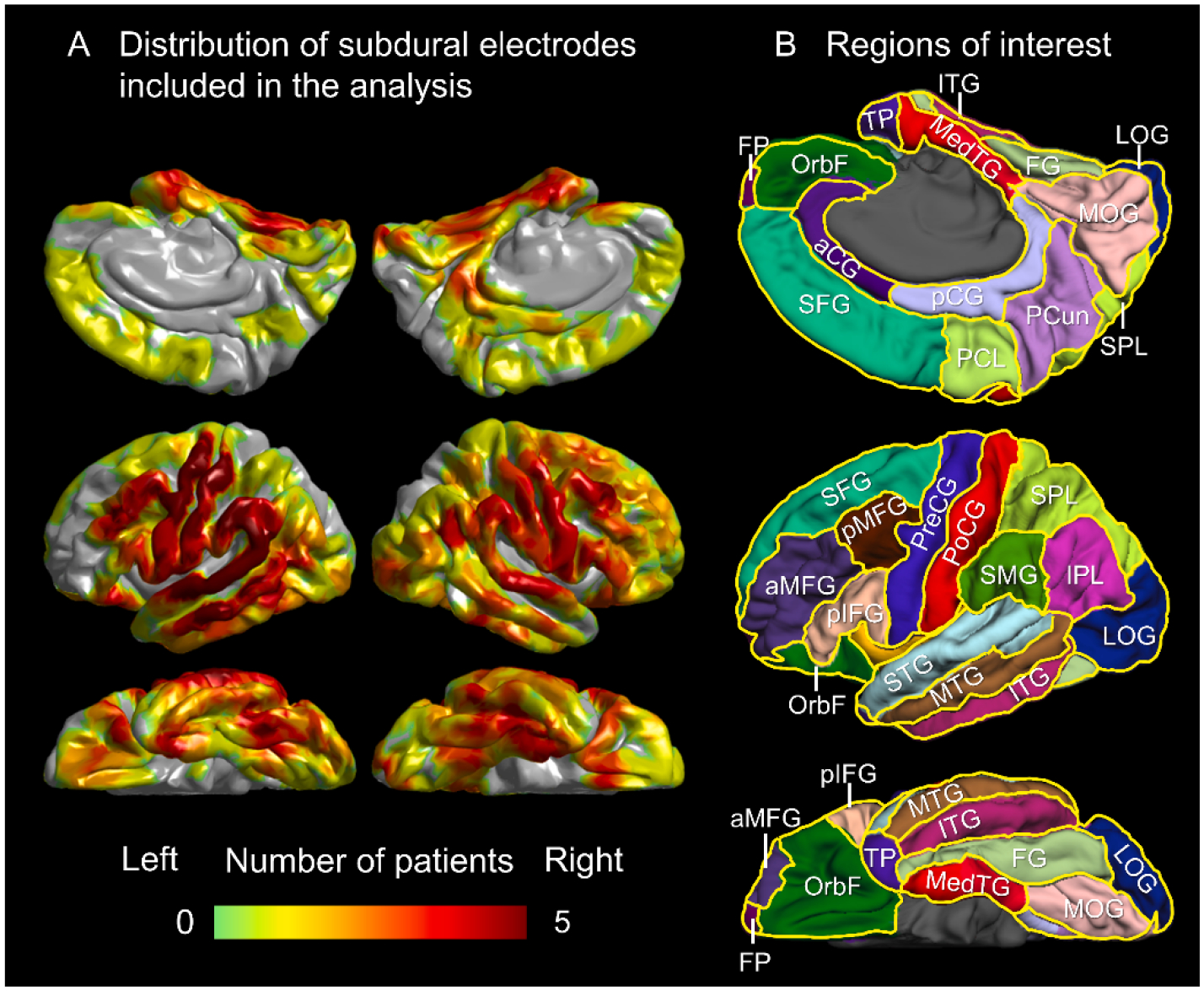
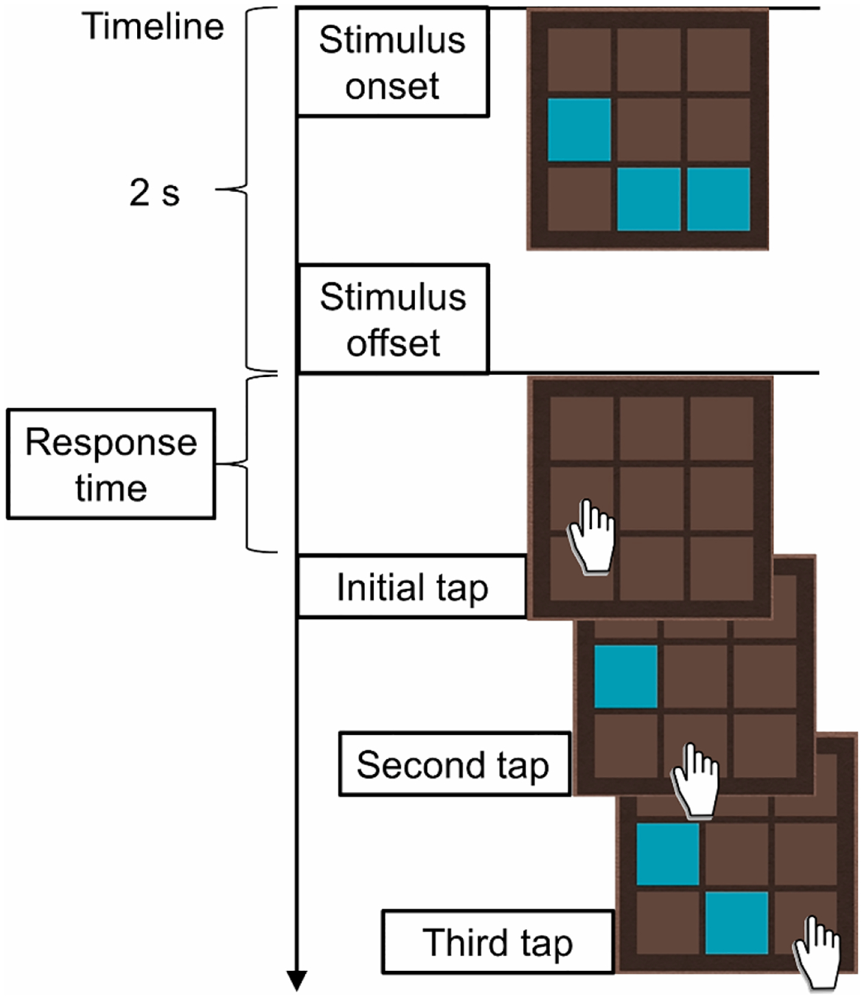
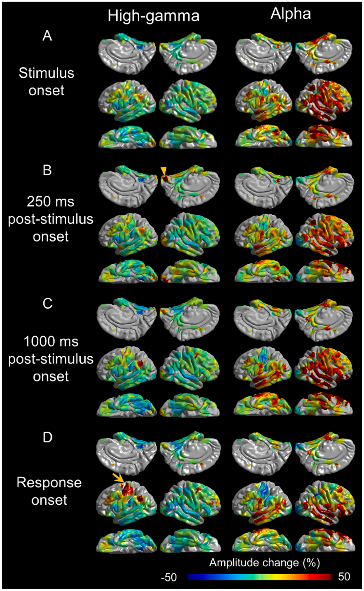
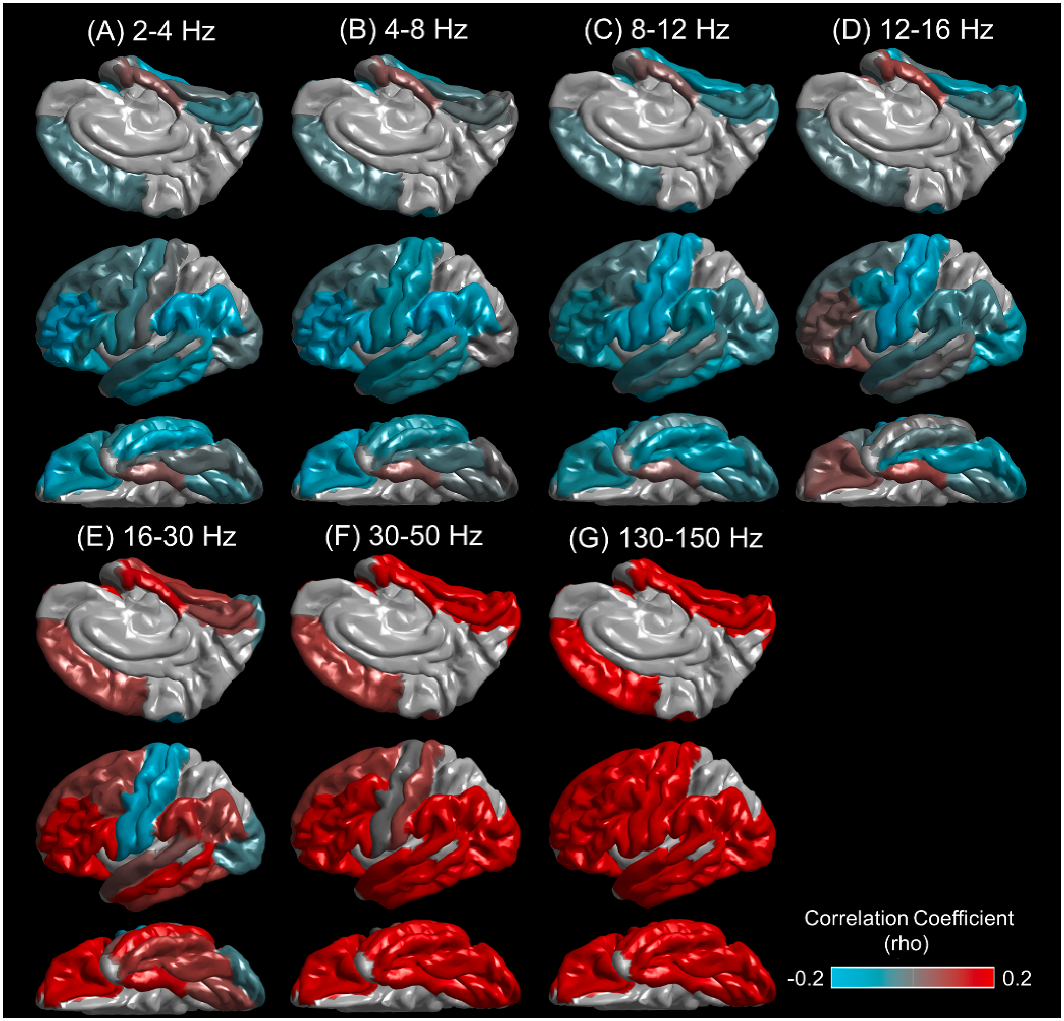
Methods: Patients undertook a task of immediate spatial recall during intracranial EEG monitoring. We charted the dynamics of cortical high-gamma activity and associated functional connectivity modulations in white matter tracts.

Results: Elevated memory loads were linked to enhanced functional connectivity via occipital longitudinal tracts, yet decreased through arcuate, uncinate, and superior-longitudinal fasciculi. As task familiarity grew, there was increased high-gamma activity in the posterior inferior-frontal gyrus (pIFG) and diminished functional connectivity across a network encompassing frontal, parietal, and temporal lobes. Early pIFG high-gamma activity was predictive of successful recall. Including this metric in a logistic regression model yielded an accuracy of 0.76.

Conclusions: Optimizing visuospatial working memory through practice is tied to early pIFG activation and decreased dependence on irrelevant neural pathways.

Significance: This study expands our knowledge of human adaptation for visuospatial working memory, showing the spatiotemporal dynamics of cortical network modulations through white matter tracts.

Keywords: Broadband high-frequency activity; Functional brain mapping; Intracranial EEG recording; Pediatric epilepsy surgery; Physiological high-frequency oscillations (HFOs).


Conflict of interest statement

Declarations of Interest

None of the authors have potential conflicts of interest to be disclosed.

Figures

Fig. 1.
Fig. 1.. Distribution of subdural electrode sites across regions of interest (ROIs).
(A) The pooled distribution of electrode sites from 10 patients. (B) ROI locations. PreCG: precentral gyrus. PoCG: postcentral gyrus. STG: superior-temporal gyrus. aMFG and pMFG: anterior and posterior middle-frontal gyri. SMG: supramarginal gyrus. SFG: superior-frontal gyrus. FG: fusiform gyrus. pIFG: posterior inferior-frontal gyrus (summation of pars opercularis and pars triangularis). MedTG: medial temporal gyrus (summation of entorhinal and parahippocampal gyri). MTG: middle-temporal gyrus. LOG: lateral occipital gyrus. IPL: inferior parietal lobule. ITG: inferior-temporal gyrus. OrbF: orbitofrontal gyrus (summation of pars orbitalis and medial and lateral orbitofrontal gyri). MOG: medial occipital gyrus (summation of cuneus and lingual gyri). SPL: superior parietal lobule. A total of 16 regions of interest mentioned above were included in the group-level ROI analysis as they contained at least 20 electrode sites. In turn, the ROI analysis excluded the following regions that were sampled by fewer than 20 electrode sites. aCG: anterior cingulate gyrus. PCun: precuneus gyrus. pCG: posterior cingulate gyrus. PCL: paracentral lobule. FP: frontal pole. Supplementary Table 1 presents the precise count of electrode sites in the respective ROIs.
Fig. 2.
Fig. 2.. Memory Matrix: a visuospatial working memory game.
Each trial involved remembering the locations of blue-painted tiles visible for two seconds. After the blue tiles disappeared, patients tapped on the remembered locations. A feedback sign and sound were given immediately following each tap to indicate if the response was correct. After the n-th tap (in this case, n = 3), the screen was refreshed and the next trial began. The stimulus period was defined as the two-second period between stimulus onset and offset, while the response period was defined as the time between stimulus offset and the n-th tap. Supplementary Video 1 demonstrates how to play this iPad-based memory game. As the number of blue-painted tiles increased, the matrix size (row × column) increased as follows: 3 tiles: 3×3; 4 tiles: 3×4; 5 tiles: 4×4; 6 tiles: 4×5; 7 tiles: 5×5; 8–9 tiles: 5×6; 10–13 tiles: 6×6.
Fig. 3.
Fig. 3.. Memory load-dependent high-gamma amplitude modulations and white matter substrates.
(A) Spatiotemporal characteristics of the relationship between memory load and high-gamma amplitude. The red cells indicate mixed model t-values at regions of interest (ROIs) and corresponding 250-ms time windows (sliding every 50 ms) where increased memory loads were significantly associated with higher high-gamma amplitudes (i.e., FDR-corrected p-value < 0.05). The blue cells indicate spatiotemporal locations where increased memory loads were significantly associated with decreased high-gamma amplitudes. (B) Snapshots of memory load-dependent high-gamma modulations on the cortical surface. Color-coded ROIs indicate that increased memory loads were significantly associated with increased (red) or decreased (blue) high-gamma amplitudes. (C) Snapshots of white matter tracts underlying memory load-dependent functional connectivity modulations. Color-coded streamlines indicate functional connectivity enhancement (red) and diminution (blue) between ROIs. 0 ms: a 250-ms time window immediately after stimulus onset. Please refer to Fig. 1 for the meaning of each abbreviation.
Fig. 4.
Fig. 4.. Task familiarity-dependent high-gamma amplitude modulations and white matter substrates.
(A) Spatiotemporal characteristics of the relationship between task familiarity and high-gamma amplitude. The red cells indicate mixed model t-values at regions of interest (ROIs) and corresponding 250-ms time windows where increased trial numbers were significantly associated with increased high-gamma amplitudes. The blue cells indicate time windows where increased trial numbers were significantly associated with decreased high-gamma amplitudes. (B) Snapshots of task familiarity-dependent high-gamma modulations. Color-coded ROIs indicate that increased trial numbers were significantly associated with increased (red) or decreased (blue) high-gamma amplitudes. (C) Snapshots of white matter tracts underlying task familiarity-dependent functional connectivity modulations. Color-coded streamlines indicate functional connectivity enhancement (red) and diminution (blue) between ROIs. 0 ms: a 250-ms time window immediately after stimulus onset. Please refer to Fig. 1 for the meaning of each abbreviation.
Fig. 5.
Fig. 5.. Relationship between high-gamma modulations and response accuracy.
(A) In this matrix of 16 regions of interest (ROIs) and ten 250-ms time windows, the mixed model t-values are shown in the cells, representing the strength of the association between high-gamma amplitude and the likelihood of successful trials. Red cells indicate a significant positive association between high-gamma amplitude and achieving a successful trial. Blue cells indicate a significant negative association, where reduced high-gamma amplitude was associated with a higher chance of successful trials. Please refer to Fig. 1 for the meaning of each abbreviation.
Fig. 6.
Fig. 6.. Task-related high-gamma and alpha amplitude modulations.
The snapshots showcase the percent change of high-gamma70–110 Hz and alpha8–12 Hz amplitudes in comparison to the baseline mean. (A) Stimulus onset. (B) 250 ms after stimulus onset. The arrowhead indicates increased high-gamma amplitude in the right medial occipital region. (C) 1000 ms after stimulus onset. (D) Response onset (i.e., the onset of the first tap response). The arrow indicates increased high-gamma amplitude in the left precentral and postcentral gyri. For a comprehensive overview of the iEEG high-gamma and alpha amplitude changes, please refer to Supplementary Video 2.
Fig. 7.
Fig. 7.. Correlation between task-related high-gamma and other frequency band amplitudes.
The mean Spearman’s rho is presented, which reflects the degree of correlation between high-gamma amplitude at 70–110 Hz and one of the following frequency band amplitudes within a given region of interest (ROI). (A) Delta at 2–4 Hz. (B) Theta at 4–8 Hz. (C) Alpha at 8–12 Hz. (D) Sigma at 12–16 Hz. (E) Beta at 16–30 Hz. (F) Low-gamma at 30–50 Hz. (G) Very high-gamma at 130–150 Hz.

Similar articles

Cited by

References

    1. Agam Y, Sekuler R. Interactions between working memory and visual perception: An ERP/EEG study. Neuroimage 2007;36:933–942. 10.1016/j.neuroimage.2007.04.014. - DOI - PMC - PubMed
    1. Akanuma N, Alarcón G, Lum F, Kissani N, Koutroumanidis M, Adachi N, et al. Lateralising value of neuropsychological protocols for presurgical assessment of temporal lobe epilepsy. Epilepsia 2003;44:408–418. 10.1046/j.1528-1157.2003.24502.x. - DOI - PubMed
    1. AlSkaif T, Dev S, Visser L, Hossari M, van Sark W. A systematic analysis of meteorological variables for PV output power estimation. Renew Energy 2020;153:12–22. 10.1016/j.renene.2020.01.150. - DOI
    1. Arya R, Horn PS, Crone NE. ECoG high-gamma modulation versus electrical stimulation for presurgical language mapping. Epilepsy Behav 2018;79:26–33. 10.1016/j.yebeh.2017.10.044. - DOI - PMC - PubMed
    1. Arya R, Roth C, Leach JL, Middeler D, Wilson JA, Vannest J et al. Neuropsychological outcomes after resection of cortical sites with visual naming associated electrocorticographic high-gamma modulation. Epilepsy Res 2019;151:17–23. 10.1016/j.eplepsyres.2019.01.011. - DOI - PubMed