. 2024 Apr 11;187(8):1907-1921.e16.
doi: 10.1016/j.cell.2024.03.005. Epub 2024 Mar 28.

Mastigoneme structure reveals insights into the O-linked glycosylation code of native hydroxyproline-rich helices

Affiliations

Mastigoneme structure reveals insights into the O-linked glycosylation code of native hydroxyproline-rich helices

Jin Dai et al. Cell. .

Abstract

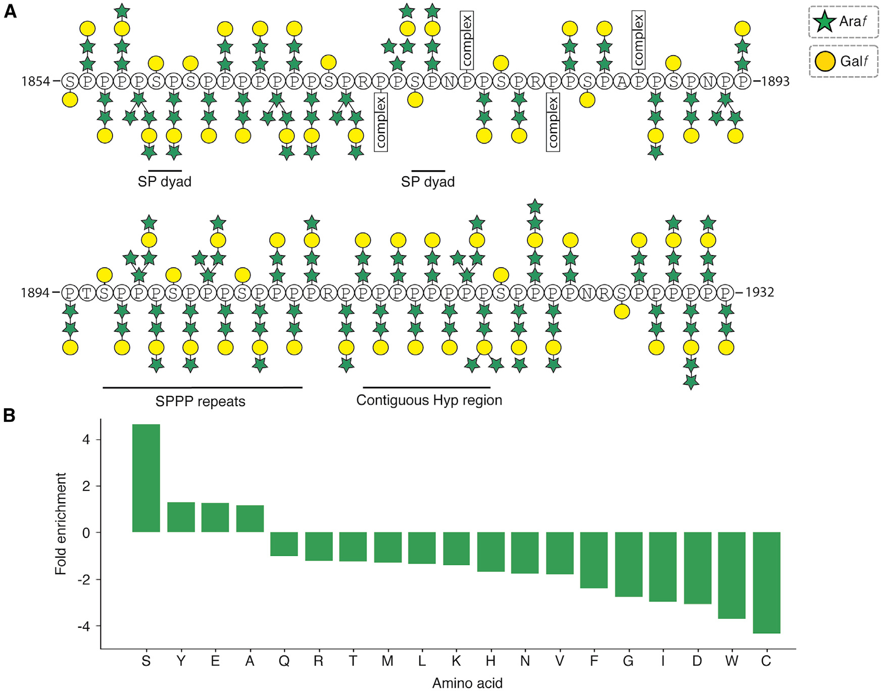
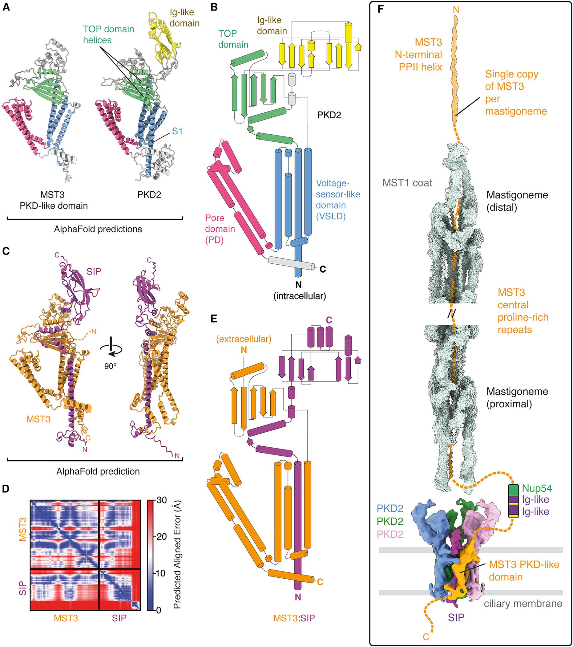
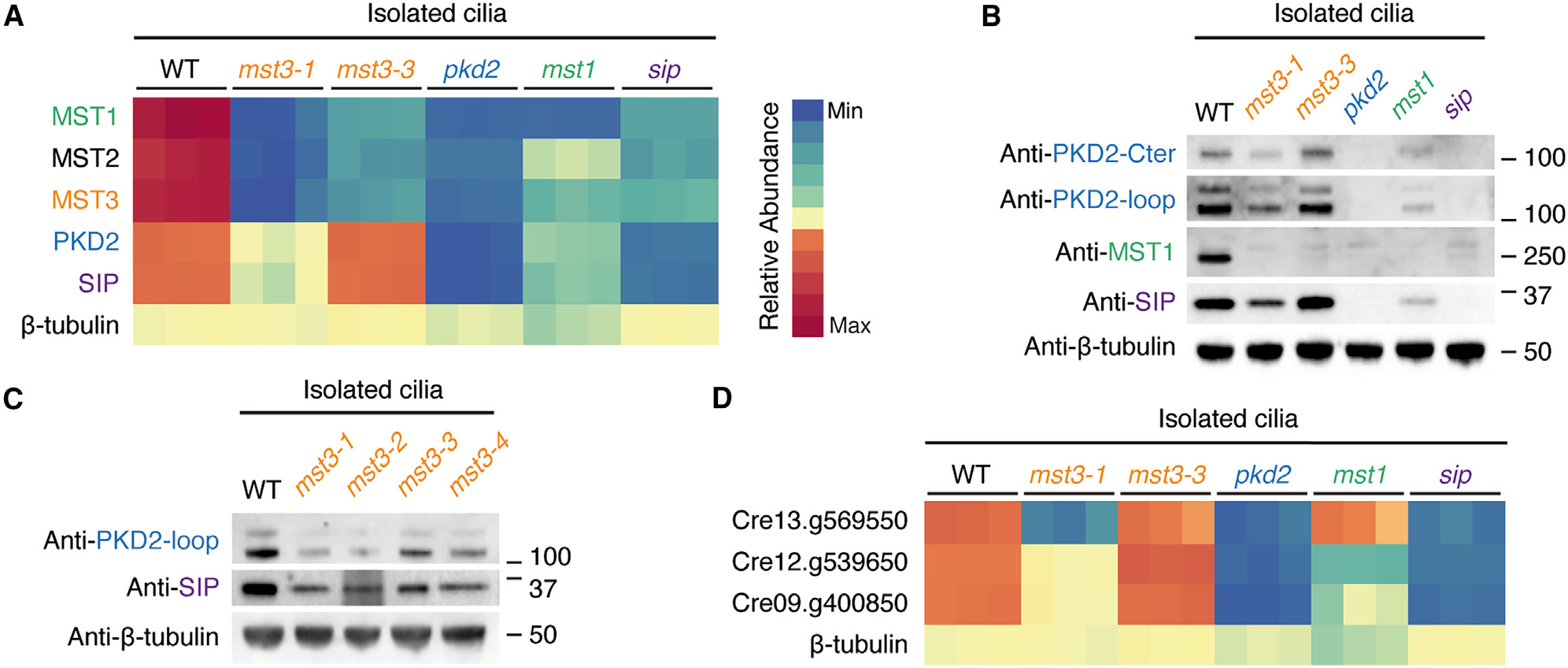
Hydroxyproline-rich glycoproteins (HRGPs) are a ubiquitous class of protein in the extracellular matrices and cell walls of plants and algae, yet little is known of their native structures or interactions. Here, we used electron cryomicroscopy (cryo-EM) to determine the structure of the hydroxyproline-rich mastigoneme, an extracellular filament isolated from the cilia of the alga Chlamydomonas reinhardtii. The structure demonstrates that mastigonemes are formed from two HRGPs (a filament of MST1 wrapped around a single copy of MST3) that both have hyperglycosylated poly(hydroxyproline) helices. Within the helices, O-linked glycosylation of the hydroxyproline residues and O-galactosylation of interspersed serine residues create a carbohydrate casing. Analysis of the associated glycans reveals how the pattern of hydroxyproline repetition determines the type and extent of glycosylation. MST3 possesses a PKD2-like transmembrane domain that forms a heteromeric polycystin-like cation channel with PKD2 and SIP, explaining how mastigonemes are tethered to ciliary membranes.

Keywords: O-linked glycosylation; cilia; cryo-EM; hydroxyproline; ion channels; mass spectrometry; mastigoneme; polycystic kidney disease; post-translational modifications.


Conflict of interest statement

Declaration of interests The authors declare no competing interests.

Figures

Figure 1.
Figure 1.. Cryo-EM structure of the C. reinhardtii mastigoneme
(A) Schematic of C. reinhardtii showing mastigonemes decorating the distal two-thirds of both cilia. Mastigonemes are approximately 800 nm long. (B) Left, electron micrograph showing a mastigoneme filament and doublet microtubules (DMTs) isolated from purified C. reinhardtii cilia. Right, two-dimensional class averages of mastigoneme filaments. (C) Helical reconstruction of the mastigoneme filament. MST1 monomers are colored by opposing polarity and in different shades of blue and green. (D) Two MST1 monomers related by apparent D1 symmetry. (E) Domain organization of C. reinhardtii MST1. The first 42 residues are predicted to be a cleaved signal peptide (SP). Residues 43–1,176 form nine tandem immunoglobulin-like domains (D1–D9). Residues 1,176–1,854 form a cysteine-rich domain that contains 26 disulfide bonds (shown as connected yellow circles). Residues 1,854–1,933 form a poly(proline)-rich [p(P)] region. N-glycosylation sites are marked with a teal circle. (F) Atomic model of MST1 colored by domain. The C-terminal proline-rich region forms a glycosylated poly(hydroxyproline) type II helix. See Figures S1 and S2.
Figure 2.
Figure 2.. Interactions involved in the filamentation of MST1
(A) MST1 forms two non-polar strands arranged as a double helix. Repeating units of MST1 are visualized here with the immunoglobulin-like domains and the poly(proline) helix hidden to better display the strand-like architecture of the cysteine-rich domains. The boxes indicate the regions shown in (B)–(D). (B) The fundamental repeat unit of the mastigoneme is an antiparallel MST1 homodimer. Only the cysteine-rich domains are shown for clarity. (C) Intrastrand interactions between the C-terminal ends of cysteine-rich domains from adjacent homodimers. Interacting residues, which are predominantly hydrophobic, are labeled. (D) The C terminus of the poly(proline) type II (PPII) helix binds near the interstrand interface between neighboring cysteine-rich domains. The box indicates the region shown in detail in (E). (E) Details of the interactions between the C terminus of MST1 and the interstrand interface. See also Figure S1.
Figure 3.
Figure 3.. Details of the glycosylated PPII helix of MST1
(A) Cryo-EM map and atomic model of the MST1 PPII helix. The polypeptide backbone is shown as a transparent blue isosurface to reveal the atomic model. The glycans are displayed as opaque gray isosurfaces at the same contour level. Black arrowheads indicate the positions of some of the non-proline/serine residues. (B) Transparent cryo-EM map showing the density and atomic models for a tri- and tetrasaccharide bound to hydroxyproline residues within the PPII helix of MST1. Individual saccharide units are labeled according to the Symbol Nomenclature for Glycans (SNFG). An unbuilt saccharide unit in the tetrasaccharide is marked with an asterisk. (C) Proposed chemical structure of the trisaccharide glycan linked to hydroxyproline. The glycan consists of two L-arabinofuranoside moieties attached through a β1→2 glycosidic linkage to α-D-galactofuranoside (α-D-Galf). (D) An example of a branched oligosaccharide with at least five saccharide units. Additional density, marked with asterisks, supports branching after the first andthird saccharide units. (E) Cryo-EM density and atomic model for monogalactosyl serine (Ser-O-αGal). The monogalactosyl moiety is built in its pyranose conformation, which positions the hydroxyl group on carbon 6 such that it can hydrogen bond with the carbonyl group of the backbone of the PPII helix. Glycans associated with the neighboring hydroxyproline residues have been removed for clarity. (F) Proposed chemical structure of monogalactosyl serine. See Figure S3.
Figure 4.
Figure 4.. Glycosylation patterns observed for the MST1 PPII helix
(A) Schematic showing the glycan structures associated with each hydroxyproline and serine residue of the poly(proline)-rich region of MST1 (residues 1,854–1,932). Saccharides are represented following the Symbol Nomenclature for Glycans (SNFG). Note that these assignments are only tentative, as the cryo-EM density does not allow individual sugar types to be distinguished. The number of saccharide units represents either the exact number or the minimum that can be confidently identified from the cryo-EM density. Large branched glycans are labeled as “complex.” Non-proline/serine residues are not modified. (B) Histogram showing the fold enrichment of each non-proline amino acid in poly(proline)-rich regions of the C. reinhardtii proteome. Poly(proline)-rich regions were defined as 20-residue nonoverlapping regions containing 10 or more proline residues. Only serine (S), tyrosine (Y), glutamate (E), and alanine (A) are enriched in poly(proline)-rich regions compared with their general frequency in the proteome.
Figure 5.
Figure 5.. Proline-rich repeats of MST3 occupy the central shaft of the mastigoneme
(A) Cryo-EM density (orange) that cannot be explained by MST1 (green model) occupies the central shaft of the mastigoneme. The density does not follow D1 symmetry and repeats approximately every 19 nm. (B) Density map of the 19-nm repeat of the glycosylated poly(hydroxyproline) (PPII) helix of MST3. The density map is segmented to show the protein residues(orange) and the decorating glycans (gray). Glycans bound to XXP motifs coat only one surface of the helix. (C) Schematic of the domain architecture of MST3. Domain boundaries were determined from AlphaFold2 predictions. Abbreviations: CTL, C-type lectin; Ig-like, immunoglobulin-like; PD, pore domain; PKD, polycystic kidney disease; p(P), poly(proline)-rich; VSLD, voltage-sensor-like domain. (D) An example of a mastigoneme with a thin distal filament extracted from a micrograph. (E) Raincloud plot showing the distribution of tip filament lengths measured from the micrographs. Each point represents an individual measurement (n = 62 from50 micrographs). In the box plot, the central dashed line represents the median and the solid line the mean. The bottom and top of the box represent the first (Q1) and third (Q3) quartiles, respectively. The whiskers extend to the highest and lowest scores. (F) Two-dimensional class averages of particles selected from the mastigoneme thin-end filaments. (G) Comparison of a three-dimensional reconstruction of the mastigoneme end filament (iv) with the PPII helices of the central poly(proline) repeats of MST3 (iii) and MST1 with (ii) and without (i) glycans. The overall dimensions of the end filament are consistent with a glycosylated PPII helix. See Figure S4.
Figure 6.
Figure 6.. C. reinhardtii MST3 contains a PKD-like domain
(A) Comparison of AlphaFold2 predictions of the PKD-like domain of MST3 with PKD2. Compared with PKD2, MST3 is missing helix S1 of the transmembrane voltage-sensor-like domain (blue), two helices of the juxtamembrane TOP domain (green), and the extracellular immunoglobulin (Ig)-like domain (yellow). The pore domain (pink) is highly similar. (B) Topology diagram of C. reinhardtii PKD2 colored by domain and generated from the AlphaFold2 prediction. Cylinders represent α-helices, and arrows represent β-strands. (C) AlphaFold2 prediction of the complex between the PKD-like domain of MST3 and SIP. (D) Predicted aligned error (PAE) plot for the prediction shown in (C). High-confidence interactions are predicted between MST3 and SIP. (E) A topology diagram generated from the AlphaFold Multimer prediction of the MST3:SIP complex colored by protein. (F) Integrated model of the mastigoneme combining our cryo-EM data with AlphaFold2 predictions. One copy of the MST3:SIP complex (orange and purple) anchors the mastigoneme in the ciliary membrane through interactions with 3 identical copies of PKD2 (shown in blue, green, and pink). The MST1 coat around the MST3 proline-repeats likely extends 800 nm and includes ~84 copies of MST1 (based on 4 copies per 380 Å). The N terminus of MST3 forms a 140-nm PPII helix at the distal end of the mastigoneme. Its 1,600-residue intracellular C terminus may be responsible for anchoring the mastigoneme to specific doublet microtubules of the axoneme.
Figure 7.
Figure 7.. Proteomic comparison of cilia composition from C. reinhardtii mutant strains
(A) Heatmap comparing the relative abundance of MST1, MST2, MST3, PKD2, and SIP in proteomic analysis of cilia isolated from wild-type (CC-5325) and mutant (mst3–1, mst3–3, pkd2, mst1, and sip) strains. The relative abundance of b-tubulin is provided as a control. (B) Immunoblot of cilia isolated from WT, mst3–1, mst3–3, pkd2, mst1, and sip strains probed with antibodies against PKD2-Cter, PKD2-loop, MST1, and SIP. Anti-β-tubulin was used as a loading control. Anti-PKD2-Cter detects two bands due to PKD2 proteolysis, as previously reported., (C) Immunoblot of cilia isolated from WT, mst3–1, mst3–2, mst-3, and mst3–4 strains probed with antibodies against PKD2-loop, SIP, and β-tubulin. PKD2 and SIP levels are reduced to varying extents in each mst3 strain. (D) Heatmap generated from quantitative mass spectrometry data showing that the relative abundance of three predicted SIP binders (Cre13.g569550,Cre12.g539650, and Cre09.g4008500) is reduced in pkd2 and sip strains. Four other predicted SIP binders (listed in Figure S7) were not detected. See Figures S4, S6, and S7.

Similar articles

Cited by

References

    1. Adzhubei AA, Sternberg MJE, and Makarov AA (2013). Polyproline-II Helix in Proteins: Structure and Function. J. Mol. Biol. 425, 2100–2132. 10.1016/j.jmb.2013.03.018. - DOI - PubMed
    1. Gorres KL, and Raines RT (2010). Prolyl 4-hydroxylase. Crit. Rev. Biochem. Mol. Biol. 45, 106–124. 10.3109/10409231003627991. - DOI - PMC - PubMed
    1. Lamport DT (1969). The isolation and partial characterization of hydroxy-proline-rich glycopeptides obtained by enzymic degradation of primary cell walls. Biochemistry 8, 1155–1163. 10.1021/bi00831a049. - DOI - PubMed
    1. Johnson KL, Cassin AM, Lonsdale A, Wong GK-S, Soltis DE, Miles NW, Melkonian M, Melkonian B, Deyholos MK, Leebens-Mack J, et al. (2017). Insights into the Evolution of Hydroxyproline-Rich Glycoproteins from 1000 Plant Transcriptomes. Plant Physiol. 174, 904–921. 10.1104/pp.17.00295. - DOI - PMC - PubMed
    1. Nguema-Ona E, Vicré-Gibouin M, Gotté M, Plancot B, Lerouge P, Bardor M, and Driouich A (2014). Cell wall O-glycoproteins and N-glycoproteins: aspects of biosynthesis and function. Front. Plant Sci. 5, 499. 10.3389/fpls.2014.00499. - DOI - PMC - PubMed