

Conclusive demonstration of iatrogenic Alzheimer's disease transmission in a model of stem cell transplantation
- PMID: 38552634
- PMCID: PMC11096610
- DOI: 10.1016/j.stemcr.2024.02.012
Conclusive demonstration of iatrogenic Alzheimer's disease transmission in a model of stem cell transplantation
Abstract
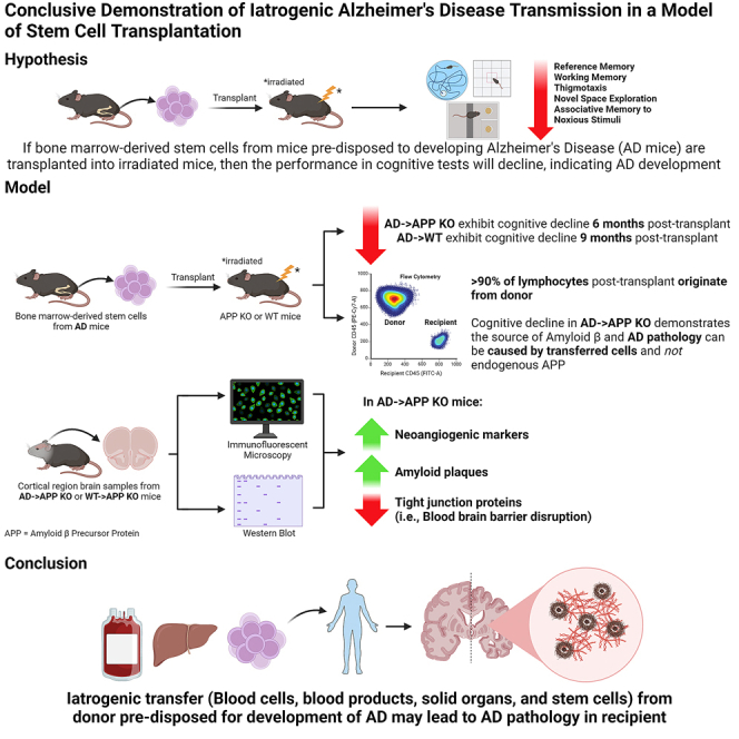
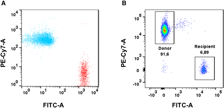
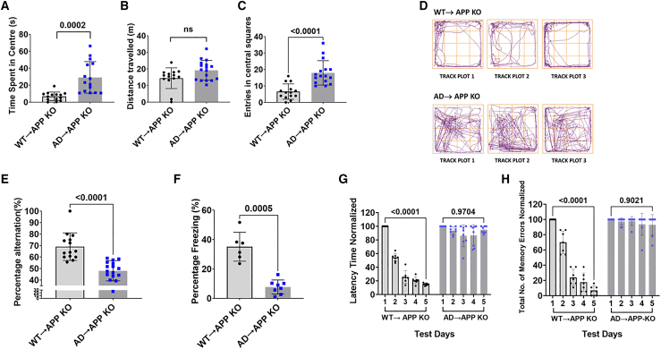
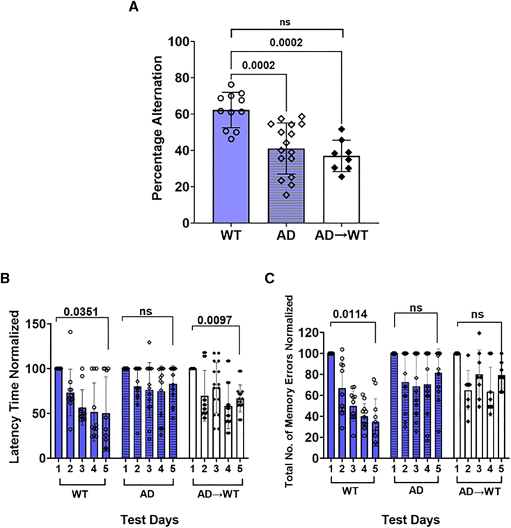
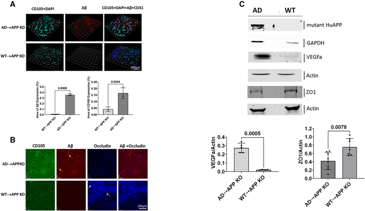
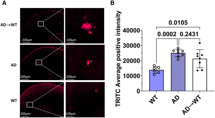
The risk of iatrogenic disease is often underestimated as a concern in contemporary medical procedures, encompassing tissue and organ transplantation, stem cell therapies, blood transfusions, and the administration of blood-derived products. In this context, despite the prevailing belief that Alzheimer's disease (AD) manifests primarily in familial and sporadic forms, our investigation reveals an unexpected transplantable variant of AD in a preclinical context, potentially indicating iatrogenic transmission in AD patients. Through adoptive transplantation of donor bone marrow stem cells carrying a mutant human amyloid precursor protein (APP) transgene into either APP-deficient knockout or normal recipient animals, we observed rapid development of AD pathological hallmarks. These pathological features were significantly accelerated and emerged within 6-9 months post transplantation and included compromised blood-brain barrier integrity, heightened cerebral vascular neoangiogenesis, elevated brain-associated β-amyloid levels, and cognitive impairment. Furthermore, our findings underscore the contribution of β-amyloid burden originating outside of the central nervous system to AD pathogenesis within the brain. We conclude that stem cell transplantation from donors harboring a pathogenic mutant allele can effectively transfer central nervous system diseases to healthy recipients, mirroring the pathogenesis observed in the donor. Consequently, our observations advocate for genomic sequencing of donor specimens prior to tissue, organ, or stem cell transplantation therapies, as well as blood transfusions and blood-derived product administration, to mitigate the risk of iatrogenic diseases.
Keywords: Alzheimer’s disease; CNS disorders; blood transfusion; iatrogenic diseases; megakaryocytes derived amyloid beta; mouse behaviour analysis; organ transplant; prion diseases; protein misfolding; stem-cell transplant.
Copyright © 2024 The Author(s). Published by Elsevier Inc. All rights reserved.
Conflict of interest statement
Declaration of interests Authors hold equity in the start-up company, Cava Healthcare, which possesses intellectual property related to these findings. This had no role in the study design, data collection, analysis or interpretation of data, or in the writing of the paper.
Figures
Similar articles
-
Alzheimer's disease amyloid-β pathology in the lens of the eye.Exp Eye Res. 2022 Aug;221:108974. doi: 10.1016/j.exer.2022.108974. Epub 2022 Feb 21. Exp Eye Res. 2022. PMID: 35202705 Free PMC article.
-
Neurons derived from sporadic Alzheimer's disease iPSCs reveal elevated TAU hyperphosphorylation, increased amyloid levels, and GSK3B activation.Alzheimers Res Ther. 2017 Dec 1;9(1):90. doi: 10.1186/s13195-017-0317-z. Alzheimers Res Ther. 2017. PMID: 29191219 Free PMC article.
-
Transplantation of in vitro cultured endothelial progenitor cells repairs the blood-brain barrier and improves cognitive function of APP/PS1 transgenic AD mice.J Neurol Sci. 2018 Apr 15;387:6-15. doi: 10.1016/j.jns.2018.01.019. Epub 2018 Feb 3. J Neurol Sci. 2018. PMID: 29571873
-
APP transgenic modeling of Alzheimer's disease: mechanisms of neurodegeneration and aberrant neurogenesis.Brain Struct Funct. 2010 Mar;214(2-3):111-26. doi: 10.1007/s00429-009-0232-6. Epub 2009 Nov 29. Brain Struct Funct. 2010. PMID: 20091183 Free PMC article. Review.
-
Insights on the Use of Transgenic Mice Models in Alzheimer's Disease Research.Int J Mol Sci. 2024 Feb 28;25(5):2805. doi: 10.3390/ijms25052805. Int J Mol Sci. 2024. PMID: 38474051 Free PMC article. Review.
Cited by
-
Rapid and sensitive determination of residual prion infectivity from prion-decontaminated surfaces.mSphere. 2024 Sep 25;9(9):e0050424. doi: 10.1128/msphere.00504-24. Epub 2024 Aug 27. mSphere. 2024. PMID: 39189773 Free PMC article.
-
Could Blood Transfusion Increase the Risk of Alzheimer's Disease? A Narrative Review.Healthcare (Basel). 2025 Feb 20;13(5):452. doi: 10.3390/healthcare13050452. Healthcare (Basel). 2025. PMID: 40077014 Free PMC article. Review.
-
Iatrogenic Dementia: Providing Insight into Transmissible Subtype of Alzheimer's Disease, Creutzfeldt-Jakob Disease and Cerebral Amyloid Angiopathy.Biomolecules. 2025 Apr 3;15(4):522. doi: 10.3390/biom15040522. Biomolecules. 2025. PMID: 40305264 Free PMC article. Review.
References
-
- PRNP Gene - Prion Protein . In: GeneCards, editor. 2023. (GeneCards the Human Gene Database)).
-
- Asante E.A., Gowland I., Linehan J.M., Mahal S.P., Collinge J. Expression Pattern of a Mini Human PrP Gene Promoter in Transgenic Mice. Neurobiol. Dis. 2002;10:1–7. - PubMed
-
- Asante E.A., Linehan J.M., Desbruslais M., Joiner S., Gowland I., Wood A.L., Welch J., Hill A.F., Lloyd S.E., Wadsworth J.D.F., Collinge J. BSE prions propagate as either variant CJD-like or sporadic CJD-like prion strains in transgenic mice expressing human prion protein. EMBO J. 2002;21:6358–6366. - PMC - PubMed
Publication types
MeSH terms
Substances
LinkOut - more resources
Full Text Sources
Medical
