. 2024 May;300(5):107237.
doi: 10.1016/j.jbc.2024.107237. Epub 2024 Mar 27.

Tau regulates Arc stability in neuronal dendrites via a proteasome-sensitive but ubiquitin-independent pathway

Affiliations

Tau regulates Arc stability in neuronal dendrites via a proteasome-sensitive but ubiquitin-independent pathway

Dina W Yakout et al. J Biol Chem. 2024 May.

Abstract

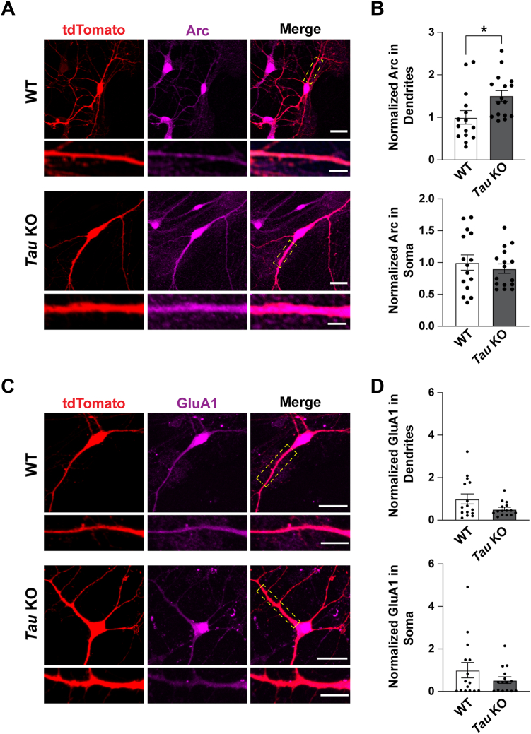
Tauopathies are neurodegenerative disorders characterized by the deposition of aggregates of the microtubule-associated protein tau, a main component of neurofibrillary tangles. Alzheimer's disease (AD) is the most common type of tauopathy and dementia, with amyloid-beta pathology as an additional hallmark feature of the disease. Besides its role in stabilizing microtubules, tau is localized at postsynaptic sites and can regulate synaptic plasticity. The activity-regulated cytoskeleton-associated protein (Arc) is an immediate early gene that plays a key role in synaptic plasticity, learning, and memory. Arc has been implicated in AD pathogenesis and regulates the release of amyloid-beta. We found that decreased Arc levels correlate with AD status and disease severity. Importantly, Arc protein was upregulated in the hippocampus of Tau KO mice and dendrites of Tau KO primary hippocampal neurons. Overexpression of tau decreased Arc stability in an activity-dependent manner, exclusively in neuronal dendrites, which was coupled to an increase in the expression of dendritic and somatic surface GluA1-containing α-amino-3-hydroxy-5-methyl-4-isoxazolepropionic acid receptors. The tau-dependent decrease in Arc was found to be proteasome-sensitive, yet independent of Arc ubiquitination and required the endophilin-binding domain of Arc. Importantly, these effects on Arc stability and GluA1 localization were not observed in the commonly studied tau mutant, P301L. These observations provide a potential molecular basis for synaptic dysfunction mediated through the accumulation of tau in dendrites. Our findings confirm that Arc is misregulated in AD and further show a physiological role for tau in regulating Arc stability and AMPA receptor targeting.

Keywords: AMPA receptors; Alzheimer’s disease; arc; proteasome; tau; ubiquitination.


Conflict of interest statement

Conflict of interest The authors declare that they have no conflicts of interests with the contents of this article.

Figures

Figure 1
Figure 1
Arc is increased in the hippocampus of Tau KO mice.A, Arc protein levels positively correlate with cognitive performance score and negatively correlate with AD progression scores. The mini-mental state examination (MMSE) indicates cognitive performance score, whereas the Consortium to Establish a Registry for Alzheimer’s Disease (CERAD) and Braak scores indicate AD progression. B, biochemical fractionation scheme. C, representative western blots showing Arc, GluA1, PSD-95, and Actin in the cytosol and light membrane fraction (S2). D, representative western blots showing Arc, GluA1, PSD-95, and Actin in the crude synaptosomal fraction (P2). Arc was significantly higher in the P2 fraction. Unpaired t test, t = 2.217, df = 12, p = 0.0467. E, representative western blots showing Arc, GluA1, PSD-95, and Actin in the synaptic vesicle fraction (S3). F, representative western blots showing Arc, GluA1, PSD-95, and Actin in the lysed synaptosomal membrane fraction (P3). No differences were found in GluA1 levels within any of the fractions. N = 6 animals per genotype, balanced for sex. AD, Alzheimer's disease; Arc, activity-regulated cytoskeleton-associated protein; PSD, postsynaptic density protein. ∗p < 0.05.
Figure 2
Figure 2
Activity-dependent increase of Arc in dendrites in Tau KO neurons is not associated with significant changes in surface GluA1 levels.A, representative images of primary hippocampal neurons from WT and Tau KO littermates transfected at DIV 9 with tdTomato to outline neuron morphology, treated with 2 μM TTX for 4 h, and fixed at DIV 10. Scale bar represents 20 μm. Scale bar in selected dendrites represents 5 μm. B, quantification of Arc showing significantly higher levels in dendrites but not the soma. Unpaired t test for Arc in dendrites, t = 2.517, df = 29, p = 0.0176; unpaired t test for Arc in soma, t = 0.677, df = 29, p = 0.504. n = 15 to 16 neurons from two independent biological replicates. C, representative images of primary hippocampal neurons from WT and Tau KO littermates transfected at DIV 12 with tdTomato to outline neuron morphology, treated with 2 μM TTX for 4 h, and fixed at DIV 13. Scale bar represents 20 μm. Scale bar in selected dendrites represents 10 μm. D, quantification of GluA1 showing no significant differences between WT and Tau KO in soma nor dendrites. Mann Whitney test for GluA1 in dendrites, p = 0.32; Mann Whitney test for GluA1 in soma p = 0.65, n = 15 neurons from two independent biological replicates. Arc, activity-regulated cytoskeleton-associated protein; TTX, tetrodotoxin. ∗p < 0.05.
Figure 3
Figure 3
GFP-tau but not GFP-P301L tau selectively reduces Arc in dendrites and correspondingly increases surface GluA1 in primary hippocampal neurons.A, primary hippocampal neurons transfected with GFP, GFP-tau, or GFP-P301L tau and treated with 2 μM TTX for 4 h. Tdtomato was used to outline neuron morphology. Scale bar represents 20 μm. Scale bar in selected dendrites represents 5 μm. B, quantification of Arc protein (magenta) in soma and dendrites showing a decrease in Arc selectively in dendrites with GFP-tau but not with GFP-P301L-tau. One-way ANOVA in dendrites F(2,55) = 5.885, p = 0.0048, Tukey’s post-hoc GFP versus GFP-Tau p = 0.0023; one-way ANOVA in soma F (2, 62) = 0.62, p = 0.54. n = 16 to 21 neurons from three independent biological replicates. C, primary hippocampal neurons overexpressing GFP, GFP-tau, and GFP-P301L tau and treated with 2 μM TTX for 4 h. TdTomato was used to outline neuron morphology. Neurons were fixed and then immunostained with an anti-GluA1 antibody. Scale bar represents 20 μm. Scale bar for selected dendrites represents 10 μm. D, quantification of surface GluA1 (magenta) in the soma and apical dendrites. Kruskal–Wallis test for dendrites, p = 0.0458; Kruskal–Wallis test for soma, p = 0.0034. n = 17 to 19 neurons from three independent biological replicates. Arc, activity-regulated cytoskeleton-associated protein; GFP-tau, GFP-tagged tau; TTX, tetrodotoxin. ∗p < 0.05, ∗∗p < 0.005.
Figure 4
Figure 4
Tau-induced decreases in dendritic Arc are proteasome-sensitive.A, primary hippocampal neurons overexpressing GFP or GFP-tau. tdTomato was used to outline neuron morphology. Cells were treated with TTX and then treated with either Vehicle (DMSO) or MG-132 (10 μM) for 4 h. Scale bar represents 20 μm. Scale bar in selected dendrites represents 5 μm. B, quantification of Arc protein (magenta) in the soma and apical dendrites showing a decrease in Arc selectively in dendrites in cultures treated with Vehicle but not in cultures treated with MG-132. Unpaired t test DMSO in dendrites t = 2.72, df = 27, p = 0.011; unpaired t test MG-132 in dendrites t = 0.3814, df = 27, p = 0.706; unpaired t test DMSO in soma t = 1.044, df = 27, p = 0.306; unpaired t test MG-132 in soma t = 0.16, df = 27, p = 0.874. n = 14 to 15 neurons from two independent biological replicates. Arc, activity-regulated cytoskeleton-associated protein; GFP-tau, GFP-tagged tau; TTX, tetrodotoxin. ∗p < 0.05.
Figure 5
Figure 5
Tau modulation of Arc in HEK293 cells is proteasome-sensitive but does not increase Arc ubiquitination.A, top, Titration of GFP-tau but not GFP reduces myc-Arc expression in HEK293 cells. HEK 293 cells were transfected with myc-Arc and increasing amounts of GFP or increasing amounts of GFP-tau. pcDNA3.1 was used as a DNA filler to keep the amount of transfected DNA between titration conditions identical. Cells were fixed, permeabilized, and then stained using anti-GFP (left) and anti-myc (right) antibodies. Bottom, quantification of GFP and myc-Arc in cells with increasing concentrations of GFP or GFP-tau. One-way ANOVA for GFP F (8, 18) = 0.6185, p = 0.7516; GFP-tau F (8, 27) = 5.560, p = 0.0003. Dunnett’s multiple comparisons test ∗∗p < 0.005, ∗∗∗∗p < 0.0001. n = 3 to 4. B, following transfection, HEK293 cells were treated with either Vehicle (DMSO) or MG-132 (10 μM) for 4 h. Left, representative western blots showing myc-Arc with increasing concentrations of GFP-tau in DMSO-treated HEK293 cells (0, 0.25, 0.5, 0.75, 1, and 1.5 μg). Actin was used as a loading control. Right, representative western blots showing myc-Arc with increasing concentrations of GFP-tau in MG-132–treated HEK293 cells (0, 0.25, 0.5, 0.75, 1, and 1.5 μg). Actin was used as a loading control. pcDNA3.1 was used as a DNA filler to keep the amount of transfected DNA between titration conditions identical. C, quantification of myc-Arc normalized to Actin showing a significant decrease in Arc with increasing tau in vehicle-treated cells (left) but not in the MG-132–treated cells (right). One-way ANOVA for DMSO control, F (5, 12) = 4.03, p = 0.022; one-way ANOVA for MG-132, F (5, 12) = 1.9, p = 0.15. n = 3. D, top, ubiquitin assay showing no enhancement in Arc ubiquitination when co-expressed with tau or P301L tau. RNF216 was used as a positive control. Bottom, input showing expression of myc-Arc, GFP-RNF216, GFP-tau, and GFP-P301L tau. E, coimmunoprecipitation assay showing pulldown of myc-Arc or myc-Arc ΔEB with an anti-myc antibody and immunoblotting with an anti-GFP antibody. GFP-RNF216 was used as a positive control. Arc, activity-regulated cytoskeleton-associated protein; EB, endophilin-binding; GFP-tau, GFP-tagged tau. ∗p < 0.05.
Figure 6
Figure 6
Tau-induced Arc removal does not depend on GSK3α/β-dependent Arc phosphorylation.A, left, representative western blots showing myc-Arc expressed alone or with GFP-tau. Cells were treated with Vehicle (Water) or CH98 (1–2 μM) for 4 h. Actin was used as a loading control. Right, quantification of myc-Arc showing a significant decrease with co-expression of GFP-tau after treatment with Vehicle (unpaired t test, t = 3.450, df = 18, p = 0.0029) or CH98 (unpaired t test, t = 3.417, df = 18, p = 0.0031). n = 9. B, schematic showing the structure of Arc with the location of mapped GSK3α/β phosphorylation sites S170, T175, T368, and T380. CE, left, representative western blots showing myc-Arc S170A/T175A, myc-Arc T368A, or myc-Arc T380A with Arc phosphorylation sites mutated to Alanine expressed alone or with GFP-tau. Actin was used as a loading control. Right, Quantification of myc-Arc S170A/T175A, myc-Arc T368A, or myc-Arc T380A showing a significant decrease when co-expressed with tau. Unpaired t test for Arc S170A/T175A, t = 4.913, df = 16, p = 0.0002; unpaired t test for Arc T368A, t = 8.714, df = 4, p = 0.001; unpaired t test for Arc T380A, t = 11.59, df = 4, p = 0.0003. n = 9 for S170A/T175A, n = 3 for T368A and T380A. Arc, activity-regulated cytoskeleton-associated protein; GFP-tau, GFP-tagged tau. ∗∗p < 0.05, ∗∗∗p < 0.001.
Figure 7
Figure 7
The endophilin-binding domain of Arc is essential for its reduction by tau.A, schematic showing the structure of Arc, highlighting the coiled-coil (CC) domain and the endophilin-binding (EB) domain on the N terminus and the N- and C-lobe on the C terminus. The location of the K92 acetylation site is shown. B, left, Representative Western blot showing WT myc-Arc, myc-Arc ΔC-terminal (lacking the C-terminal domain), myc-Arc ΔCC (lacking the coiled-coil motif on the N-terminal domain), and myc-Arc ΔEB (lacking the endophilin-binding domain on the N-terminus) expressed alone or with GFP-tau. Actin was used as a loading control. Right, Quantification of WT myc-Arc, myc-Arc ΔC-terminal, myc-Arc ΔCC, and myc-Arc ΔEB. Only Arc ΔEB does not show a decrease with tau overexpression (Unpaired t test for WT Arc t = 8.857, df = 10, ∗∗∗∗p < 0.0001; unpaired t test for Arc ΔC-terminal t = 6.471, df =1 0, ∗∗∗∗p < 0.0001; unpaired t test for Arc ΔCC t = 7.076, df = 10, ∗∗∗∗p < 0.0001; unpaired t test Arc ΔEB t = 0.2956, df = 10, p = 0.774). n = 6. C, left, Representative western blots showing myc-Arc K92Q with the acetylation site K92 mutated to glutamine expressed alone or with GFP-tau. Actin was used as a loading control. Right, Quantification of myc-Arc K92Q showing a significant decrease when co-expressed with tau. Unpaired t test t = 5.54, df = 10, p = 0.0002. n = 6. Arc, activity-regulated cytoskeleton-associated protein; GFP-tau, GFP-tagged tau. ∗∗∗p < 0.001, ∗∗∗∗p < 0.0001.
Supporting Figure S1

References

    1. Williams D.R. Tauopathies: classification and clinical update on neurodegenerative diseases associated with microtubule-associated protein tau. Intern. Med. J. 2006;36:652–660. - PubMed
    1. Kosik K.S., Joachim C.L., Selkoe D.J. Microtubule-associated protein tau (tau) is a major antigenic component of paired helical filaments in Alzheimer disease. Proc. Natl. Acad. Sci. U. S. A. 1986;83:4044–4048. - PMC - PubMed
    1. Wood J.G., Mirra S.S., Pollock N.J., Binder L.I. Neurofibrillary tangles of Alzheimer disease share antigenic determinants with the axonal microtubule-associated protein tau (tau) Proc. Natl. Acad. Sci. U. S. A. 1986;83:4040–4043. - PMC - PubMed
    1. Braak H., Braak E. Staging of Alzheimer's disease-related neurofibrillary changes. Neurobiol. Aging. 1995;16:271–278. Discussion 278–284. - PubMed
    1. Andreadis A., Brown W.M., Kosik K.S. Structure and novel exons of the human tau gene. Biochemistry. 1992;31:10626–10633. - PubMed

Publication types

MeSH terms