. 2024 Mar 29;14(1):7473.
doi: 10.1038/s41598-024-58075-x.

Control of breathing by orexinergic signaling in the nucleus tractus solitarii

Affiliations

Control of breathing by orexinergic signaling in the nucleus tractus solitarii

Yakun Wang et al. Sci Rep. .

Abstract

Orexin signaling plays a facilitatory role in respiration. Abnormalities in orexin levels correlate with disordered breathing patterns and impaired central respiratory chemoreception. Nucleus tractus solitarii (NTS) neurons expressing the transcription factor Phox2b contribute to the chemoreceptive regulation of respiration. However, the extent to which orexinergic signaling modulates respiratory activity in these Phox2b-expressing NTS neurons remains unclear. In the present study, the injection of orexin A into the NTS significantly increased the firing rate of the phrenic nerve. Further analysis using fluorescence in situ hybridization and immunohistochemistry revealed that orexin 1 receptors (OX1Rs) were primarily located in the ventrolateral subdivision of the NTS and expressed in 25% of Phox2b-expressing neurons. Additionally, electrophysiological recordings showed that exposure to orexin A increased the spontaneous firing rate of Phox2b-expressing neurons. Immunostaining experiments with cFos revealed that the OX1R-residing Phox2b-expressing neurons were activated by an 8% CO2 stimulus. Crucially, OX1R knockdown in these NTS neurons notably blunted the ventilatory response to 8% CO2, alongside an increase in sigh-related apneas. In conclusion, orexinergic signaling in the NTS facilitates breathing through the activation of OX1Rs, which induces the depolarization of Phox2b-expressing neurons. OX1Rs are essential for the involvement of Phox2b-expressing NTS neurons in the hypercapnic ventilatory response.


Conflict of interest statement

The authors declare no competing interests.

Figures
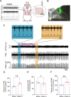
Figure 1
Figure 1
Injection of orexin A in the NTS potentiates PND activity. (A) Schematic of the setup showing recordings of PND in anesthetized mice. Top: ETCO2 levels; middle: raw PND waveforms; bottom: integrated PND signal, achieved through rectification and smoothing (time constant, 0.05 s). (B) Image of the unilateral injection site (green) within the NTS using fluorescent green beads. Scale bar, 50 μm. (C) Representative traces showing changes in the PND in response to unilateral injection of orexin A (1 μM, 100 nl per injection) in the NTS. The duration of orexin A injection is marked by a purple line. The blue and orange shadow regions show PND activity measured before and after injection of orexin A, with expanded views shown above for clarity. (DF) Quantification of normalized PND frequency, amplitude and minute output in response to injection of either saline or orexin A in the NTS. n = 5 mice for saline, n = 7 mice for orexin A, **P < 0.01 by two-tailed unpaired ttest. AP, area postrema, CC, central canal, DMNV, dorsal motor nucleus of vagus.
Figure 2
Figure 2
Expression pattern of OX1R-RNA and Phox2b in the NTS. (A) OX1R-RNA and Phox2b were identified using RNAscope in situ hybridization and immunohistochemical detection. Images showing the expression of OX1R-RNA (red, top), Phox2b (green, middle) and merged view (bottom) in the NTS. Scale bar, 100 μm. (B) Enlarged view of the square in (A). The arrows indicate OX1R-RNAPhox2b+ (green), OX1R-RNA+Phox2b+ (yellow), OX1R-RNA+Phox2b (red) neurons, respectively. Scale bar, 20 μm. (C,D) Quantitative analysis of expression of Phox2b and OX1R-RNA. Cells were manually counted in three sections from each mouse (n = 3 mice). sol, solitary tract, vlNTS, ventrolateral NTS, CC, central canal.
Figure 3
Figure 3
Orexin A regulates electrophysiological activity of NTSPhox2b neurons. (A) Schematic of viral injection strategy in Phox2b-Flp mice. (B) Images showing a fluorescent NTSPhox2b neurons in a brain slice using infrared differential interference contrast optics (left) and fluorescence microscopy (right). (C) Proportion of responsive (30%) and unresponsive (70%) NTSPhox2b neurons. n = 20 neurons from four mice. (D) Firing activity of a fluorescent NTSPhox2b neuron during bath application of orexin A (100 nM). Firing rate histograms (top traces; bin size, 10 s) were derived from cell-attached voltage-clamp recordings (bottom traces). (E) Quantitative analysis of firing rate in responsive NTSPhox2b neurons (n = 6, **P < 0.01, two-tailed paired t test).
Figure 4
Figure 4
Identification of CO2-activated OX1R-RNA-residing NTSPhox2b neurons. (A) Schematic diagram illustrating experimental protocols before histological experiments. (B,C) Combined application of RNAscope in situ hybridization and immunofluorescence staining showed co-expression of OX1R-RNA+ (red), cFos+ (purple) and Phox2b+ (green) neurons in the NTS. CO2-activated neurons were indicated by immunoreactivity to cFos. Square area-indicated views were enlarged in the right images. The arrows indicate co-expressing OX1R-RNA, cFos and Phox2b in NTS neurons. Scale bars, 100 μm (B,C, left), 20 µm (B,C, right). (D) Quantification of the number of OX1R-RNA+cFos+Phox2b+ neurons from mice exposing to either 100% O2 and 8% CO2. Cells were counted from seven sections (n = 3 mice) for 100% O2 and eight sections (n = 3 mice) for 8% CO2. ****P < 0.0001, 100% O2 vs. 8% CO2, unpaired t test. (E) Cell count analysis showing that 56% of CO2-activated NTSPhox2b neurons and 58% of CO2-activated Phox2b neurons expressed OX1R-RNA.
Figure 5
Figure 5
Knockdown of OX1R-RNA in NTSPhox2b neurons blunted the HCVR. (A) Schematic diagram illustrating a genetic strategy to knockdown OX1R-RNA through bilateral injections of the FRT-inducible virus AAV-FRT-OX1R-shRNA-mCherry (AAV-FRT-Scramble-shRNA-mCherry as control group) into the NTS from Phox2b-Flp mice. (B) Immunohistochemical validation of mCherry-expressing neurons. Scale bar, 200 µm. (C) Rostrocaudal distribution of mCherry-expressing neurons. Cells were counted in eight coronal sections (bregma: − 7.2 to − 7.9 mm; thickness: 25 µm; each separated by 75 µm) from each mouse (n = 3 mice). (D) qPCR to verify knockdown effectiveness. Following knockdown, ~ 64% of OX1R-RNA in NTSPhox2b neurons was retained (***P < 0.001, three technical replicates for each of three mice for both groups, two-tailed unpaired t test). (E) Schematic of monitoring respiratory function with the whole body plethysmography. (F) Typical traces showing eupnea, spontaneous apnea and sigh-related apnea. (G) Knockdown of OX1R-RNA in NTSPhox2b neurons increases sigh-related apnea, rather than spontaneous apnea (n = 7 mice for scramble group, n = 8 mice for knockdown group, ****P < 0.0001, two-tailed unpaired t test). (H) Knockdown of OX1R-RNA in NTSPhox2b neurons caused no significant change in the number of sighs. n = 5 mice for scramble group, n = 7 mice for knockdown group. Two-tailed unpaired t test. (I) The original airflow waveforms illustrating the HCVR in both Scramble-shRNA and OX1R-shRNA groups. (JL) Effect of knockdown of OX1R-RNA in NTSPhox2b neurons on the HCVR. Knockdown of OX1R-RNA in NTSPhox2b neurons caused an insignificant change in baseline breathing parameters but significantly blunted the HCVR during exposure to 8% CO2 (n = 9 mice for each group, **P < 0.01, ***P < 0.001, ****P < 0.0001, two-way ANOVA with Sidak’s multiple comparisons test). ns, not significant.

Similar articles

Cited by

References

    1. Korkalainen H, et al. Review and perspective on sleep-disordered breathing research and translation to clinics. Sleep Med. Rev. 2023;73:101874. doi: 10.1016/j.smrv.2023.101874. - DOI - PubMed
    1. Peppard PE, Hagen EW. The last 25 years of obstructive sleep apnea epidemiology-and the next 25? Am. J. Respir. Crit. Care Med. 2018;197:310–312. doi: 10.1164/rccm.201708-1614PP. - DOI - PubMed
    1. Javaheri S, Badr MS. Central sleep apnea: Pathophysiologic classification. Sleep. 2023;46(3):zsac113. doi: 10.1093/sleep/zsac113. - DOI - PMC - PubMed
    1. Barnett S, Li A. Orexin in respiratory and autonomic regulation. Health Dis. Compr. Physiol. 2020;10:345–363. doi: 10.1002/cphy.c190013. - DOI - PubMed
    1. Levy P, et al. Obstructive sleep apnoea syndrome. Nat. Rev. Dis. Prim. 2015;1:15015. doi: 10.1038/nrdp.2015.15. - DOI - PubMed