. 2024 Mar 30;7(1):388.
doi: 10.1038/s42003-024-06059-y.

Day length regulates gonadotrope proliferation and reproduction via an intra-pituitary pathway in the model vertebrate Oryzias latipes

Affiliations

Day length regulates gonadotrope proliferation and reproduction via an intra-pituitary pathway in the model vertebrate Oryzias latipes

Muhammad Rahmad Royan et al. Commun Biol. .

Abstract

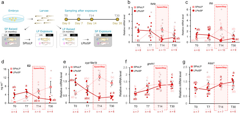
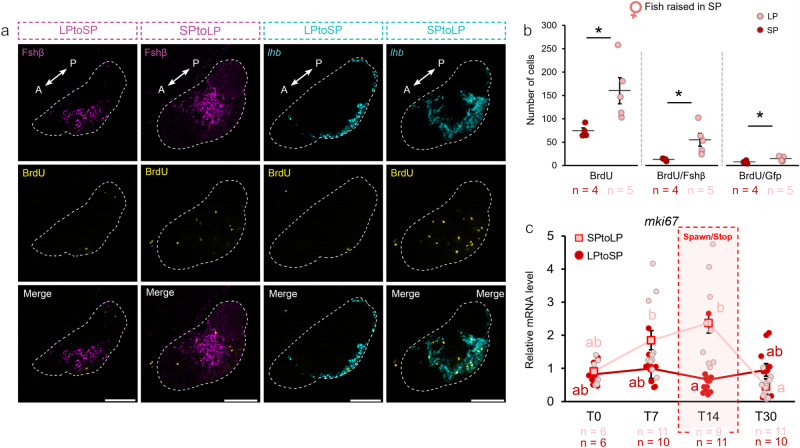
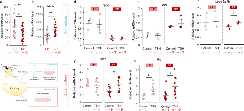
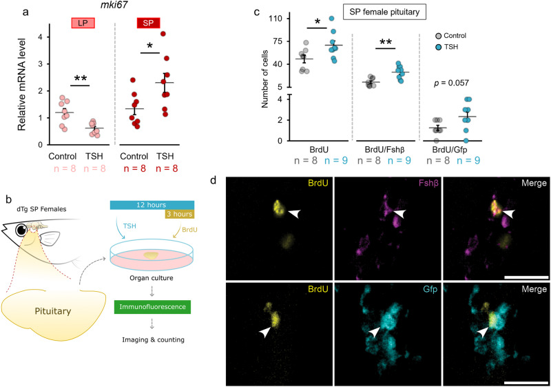
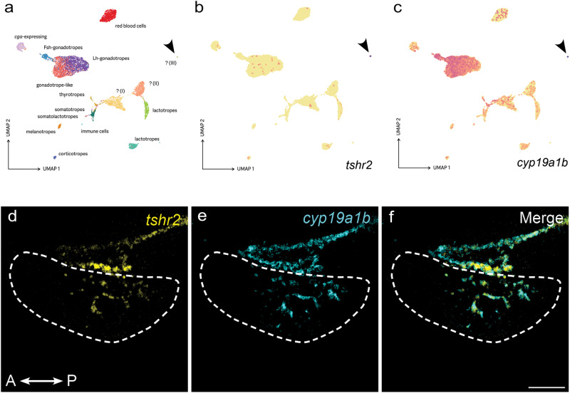
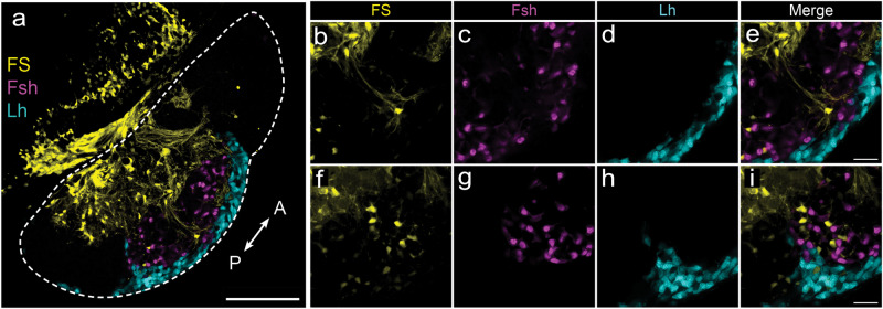
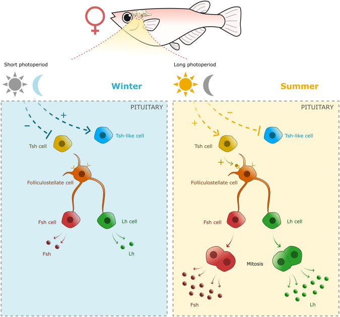
In seasonally breeding mammals and birds, the production of the hormones that regulate reproduction (gonadotropins) is controlled by a complex pituitary-brain-pituitary pathway. Indeed, the pituitary thyroid-stimulating hormone (TSH) regulates gonadotropin expression in pituitary gonadotropes, via dio2-expressing tanycytes, hypothalamic Kisspeptin, RFamide-related peptide, and gonadotropin-releasing hormone neurons. However, in fish, how seasonal environmental signals influence gonadotropins remains unclear. In addition, the seasonal regulation of gonadotrope (gonadotropin-producing cell) proliferation in the pituitary is, to the best of our knowledge, not elucidated in any vertebrate group. Here, we show that in the vertebrate model Japanese medaka (Oryzias latipes), a long day seasonally breeding fish, photoperiod (daylength) not only regulates hormone production by the gonadotropes but also their proliferation. We also reveal an intra-pituitary pathway that regulates gonadotrope cell number and hormone production. In this pathway, Tsh regulates gonadotropes via folliculostellate cells within the pituitary. This study suggests the existence of an alternative regulatory mechanism of seasonal gonadotropin production in fish.


Conflict of interest statement

The authors declare no competing interests.

Figures

Fig. 1
Fig. 1. Long photoperiod induces an increase in gene expression and circulating sex steroids.
a Illustration showing the experimental design. Fish were raised from hatching in LP or SP for 4 months before the photoperiod was changed. Fish were sampled on day 0 (just before the photoperiod change), 7, 14, and 30. bd The fluctuation of pituitary fshb and lhb expression as well as blood E2 levels observed in females at different time points following the photoperiod change. eg The fluctuation of cyp19a1b levels in the pituitary as well as gnrh1 and kiss1 in the brain at different time points following the photoperiod change. The letters (ac) show statistically differences between each time points and group. The statistical analyses were performed using non-parametric tests, Scheirer–Ray–Hare Test, followed by Dunn’s Test. All graphs are represented as mean ± SEM. Individual numbers (n) are annotated throughout the figure.
Fig. 2
Fig. 2. Long photoperiod induces an increase in gonadotrope cell mitosis in SP females.
a Confocal planes illustrating Fsh (magenta) and Lh (cyan) cell mitosis using BrdU marker (yellow) 5 days after photoperiod change (LPtoSP and SPtoLP). Scale bar: 100 µm. The dashed line indicates the pituitary (A: anterior, P: posterior). b Number of mitotic cells in SP females after exposing them to LP for 5 days or kept in SP for control. c Relative expression levels of a mitotic cell marker, mki67, in females before and 7, 14 or 30 days following the photoperiod change as described in Fig. 1A. The statistical analyses were performed using two-sample independent t-test (b) and the non-parametric test Scheirer–Ray–Hare Test followed by Dunn’s Test (c). The graph represents mean ± SEM. In b and c, the jittered dots represent each individual. In b, p-values < 0.05 are represented by *. In c, the letters (a, b) show statistically differences between each time points and group. Individual numbers (n) are annotated throughout the figure.
Fig. 3
Fig. 3. Tsh indirectly stimulates gene expression of gonadotrope cells in SP females.
tshba (a) and tshbb (b) mRNA levels in LP and SP condition in female medaka. c Experimental scheme illustrating the treatment of 0.5 µM of bovine TSH pituitary extract to dissociated cell culture and organ culture of medaka pituitary to evaluate direct and indirect effect of Tsh on gene expression of gonadotrope cells. df Effect of TSH or vehicle (control) on fshb, lhb and aromatase (cyp19a1b) mRNA levels in dispersed pituitary cell cultures in female medaka from LP and SP condition (n = 3; in which each replicate represents 4 pooled pituitaries), the bar represents mean ± SEM. gh Effect of TSH or vehicle (control) on fshb and lhb mRNA levels in ex vivo medaka pituitary organ cultures of female medaka from LP and SP condition. The statistical analyses were performed using two-sample independent t-test (b, df) or Mann–Whitney U test (a, g, h). All graphs (unless otherwise stated) are represented as mean ± SEM with the jitter dots representing each individual (p-values: *< 0.05; **< 0.01). Individual numbers (n) are annotated throughout the figure.
Fig. 4
Fig. 4. Tsh stimulates mitotic activity in the pituitary of SP females.
a Effect of 0.5 µM of bovine TSH pituitary extract for 24 h on mki67 levels in the female medaka pituitary organ cultures in LP and SP condition. b Schematic illustration of ex vivo medaka pituitary organ culture with bovine TSH pituitary extract and BrdU treatment to evaluate gonadotrope mitosis. c Number of mitotic cells in SP females exposed to TSH for 12 h. d Visual representation from the pituitary of dTg fish with Gfp signal for Lh cell (cyan), Fshβ for Fsh cells (magenta), and BrdU for mitotic cells (yellow) in the pituitary of SP females treated with TSH, as taken by Thunder fluorescence microscope. The statistical analyses were performed using two-sample independent t-test, in which the graph represents mean ± SEM while the jittered dots represent each individual (p-values: *< 0.05; **< 0.01). Individual numbers (n) are annotated throughout the figure. Arrows point to the mitotic cells. Scale bar: 20 µm.
Fig. 5
Fig. 5. Tsh regulation of gonadotrope cells is mediated via folliculostellate cells.
a The pituitary cell clusters identified by single-cell sequencing, showing three uncharacterized cell types (I–III). b, c tshr2 and cyp19a1b are specifically expressed in one of these uncharacterized cell types (III), with cyp19a1b also showing modest expression in other pituitary cell types (e.g. gonadotropes). Figures are UMAP projections generated using Seurat. Color scale: log-transformed relative expression level (maximized per gene). df Confocal planes showing co-expression of tshr2 and cyp19a1b in the same pituitary cells. Parasagittal section of brain and pituitary with a dashed line indicating the pituitary (A: anterior, P: posterior). Scale bar: 100 µm.
Fig. 6
Fig. 6. Folliculostellate cells connect to gonadotropes.
a Labeling of folliculostellate cells using the dipeptide β-Ala-Lys-Nε-AMCA in double transgenic (lhb-GfpII/fshb-DsRed2) medaka fish. be High magnification of confocal planes showing the projection of folliculostellate cell extensions (yellow) to gonadotrope cells (Lh: cyan; Fsh: magenta; scale bar: 100 µm). fi High magnification of confocal planes showing folliculostellate cells that border with gonadotropes. Dashed line indicates the whole parasagittal plane of the pituitary (A: anterior, P: posterior; scale bar: 20 µm).
Fig. 7
Fig. 7. Schema of the proposed hypothesis on the photoperiodic regulation of gonadotrope cell mitosis via melatoinin, Tsh, and folliculostellate cells.
In summer photoperiod (right panel), melatonin levels are suppressed, allowing Tsh cells to stimulate gene expression and mitosis of gonadotrope cells, via folliculostellate cells. As a result, an increasing number of gonadotrope cell due to mitosis participates in the increase in gonadotropin production necessary for gametogenesis and steroidogenesis, which allow the fish to reproduce. In winter photoperiod condition (left panel), high melatonin levels indirectly suppress tshba expression. In absence of Tsh stimulation, gene expression and mitosis of gonadotrope cells remain low. By contrast, melatonin indirectly upregulates tshbb expression that might inhibit Fsh and Lh, but this needs to be confirmed.

Similar articles

Cited by

References

    1. Shinomiya, A., Shimmura, T., Nishiwaki-Ohkawa, T. & Yoshimura, T. Regulation of Seasonal Reproduction by Hypothalamic Activation of Thyroid Hormone. Front. Endocrinol.5, 10.3389/fendo.2014.00012 (2014) - PMC - PubMed
    1. Bronson FH. Climate change and seasonal reproduction in mammals. Philos. Trans. R. Soc. Lond. B Biol. Sci. 2009;364:3331–3340. - PMC - PubMed
    1. Migaud H, Davie A, Taylor JF. Current knowledge on the photoneuroendocrine regulation of reproduction in temperate fish species. J. Fish. Biol. 2010;76:27–68. - PubMed
    1. Kanda S. Evolution of the regulatory mechanisms for the hypothalamic-pituitary-gonadal axis in vertebrates–hypothesis from a comparative view. Gen. Comp. Endocrinol. 2019;284:113075. - PubMed
    1. Yaron, Z. & Levavi-Sivan, B. in Encyclopedia of Fish Physiology Vol. 2 (ed A. P. Farrell) 1500–1508 (Academic Press, 2011). 10.1016/B978-0-12-374553-8.00058-7.

Publication types