. 2024 Mar 29;8(1):81.
doi: 10.1038/s41698-024-00572-3.

Tumor heterogeneity and clinically invisible micrometastases in metastatic breast cancer-a call for enhanced surveillance strategies

Affiliations

Tumor heterogeneity and clinically invisible micrometastases in metastatic breast cancer-a call for enhanced surveillance strategies

Eliza R Bacon et al. NPJ Precis Oncol. .

Abstract

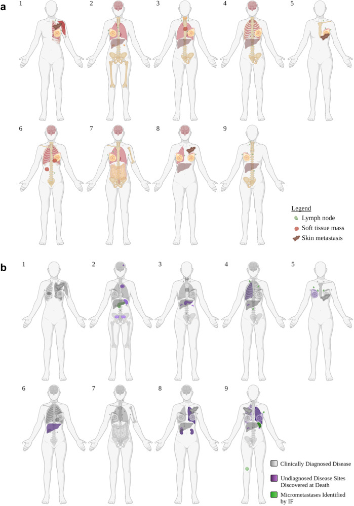
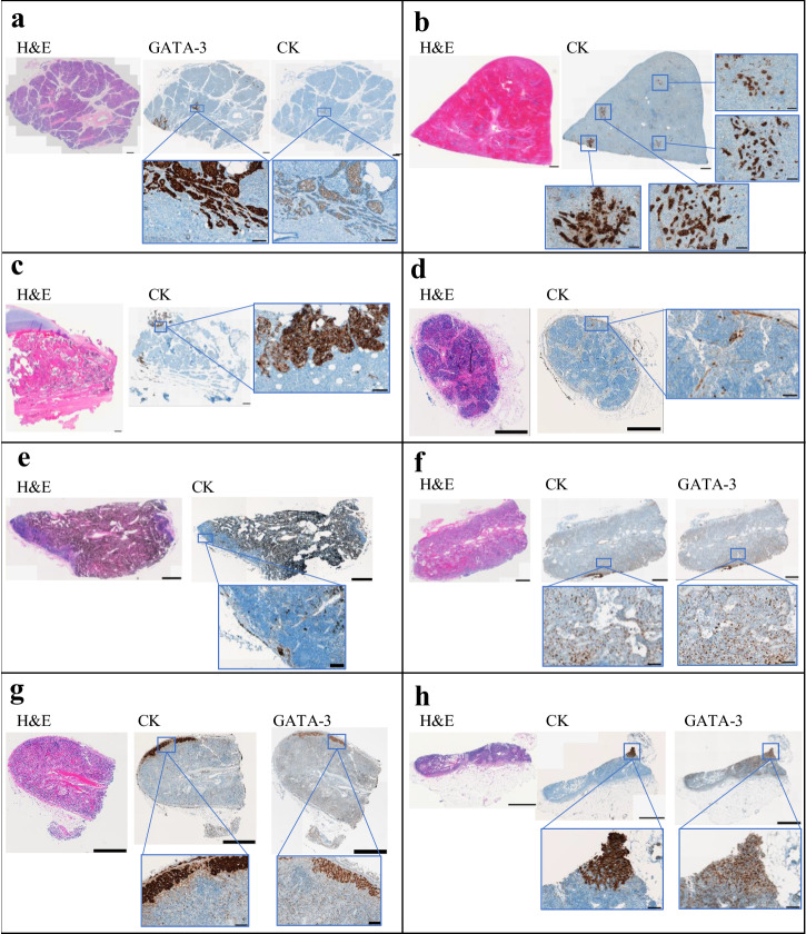
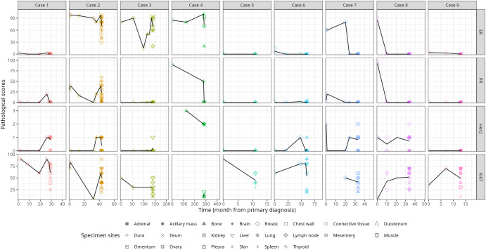
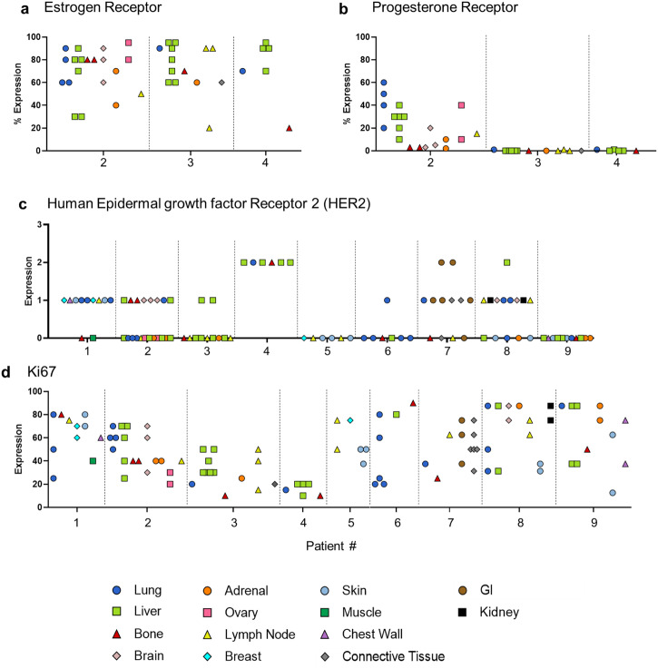
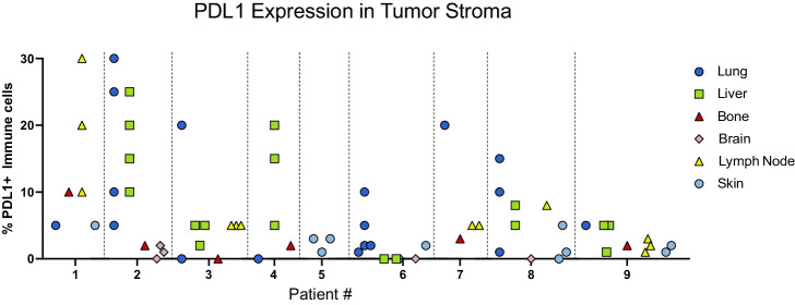
The biology of metastatic breast cancer (MBC) is understudied, primarily due to the difficulty of procuring multiple samples from patients with oligometastatic breast cancer. We developed a rapid postmortem tissue procurement program that allows the collection and analysis of numerous metastatic lesions, subclinical locations, and potential pre-metastatic niches that fall within this scope. We conducted a rapid postmortem tissue collection study on 9 patients with MBC. Patients and their families consented to donate tissues immediately after death in an IRB-approved study. Various disease subtypes, progression histories, organ involvement, and final causes of death are reported. In patients with hormone receptor-positive (HR+) disease, estrogen receptor (ER), progesterone receptor (PR), HER2, and Ki-67 expression were heterogeneous across metastatic lesions within individual patients. Disease phenotype at the end of life trended toward complete loss of HR expression. Nearly all (n = 7) patients exhibited extensive tumor involvement of additional organs that had not been previously diagnosed clinically and were not retrospectively visible on recent imaging. Of these seven individuals, three included organs uncommonly associated with MBC: kidney, spleen, pancreas, and ovary. Finally, we identified clinically undetectable micrometastases in several organs uncommonly involved in MBC. Our findings raise several clinically relevant questions regarding the mechanisms of metastatic progression. Insights from this study argue for better surveillance strategies for monitoring MBC. We highlight the need to capture more accurate biomarker information in the context of heterogeneous disease and urge the consideration of treatment strategies that combine multiple targeted therapies.


Conflict of interest statement

The authors declare no competing interests.

Figures

Fig. 1
Fig. 1. Diagnosed and unexpected disease.
a Schematic of all clinically diagnosed sites of disease throughout each patient’s history up until death. b Clinically diagnosed disease sites shaded in gray. Purple shading indicates diseases identified at postmortem that were not previously diagnosed or suspected while the patient was alive. Green shading indicates the presence of micrometastases that were identified in “disease-free” tissues via immunofluorescence (IF) Figures created with BioRender.com.
Fig. 2
Fig. 2. Identification of micrometastases in organs that rarely develop metastasis and in non-tumor-draining lymph nodes.
Whole-slide scale bars have been standardized to 1000 μm. Magnified images scales have all been standardized to 100 μm. a Patient 2: pancreas. No clinical history of disease in pancreas or surrounding lymph nodes. At postmortem: grossly normal no adjacent positive lymph nodes. H&E: negative; GATA-3: positive; ER: positive. b Patient 9: spleen. No clinical history of disease in spleen; adjacent lymph nodes activity shortly before death. At postmortem: grossly normal, no adjacent positive tissues. H&E: negative; CK: positive. c Patient 9: Patella (bone). No clinical history of bone metastasis. At postmortem: grossly normal and no adjacent positive tissues. H&E: negative; CK: positive. d Patient 4: right supraclavicular lymph node. No history of disease; did not directly drain any cancer sites. At postmortem: grossly normal and no adjacent grossly positive tissues. H&E: negative; CK: positive; GATA-3: negative (data not shown). e Patient 4: left interlobular lymph node. No history of disease in lymph node or left lung. At postmortem: grossly normal and no adjacent positive tissues. H&E: negative; CK: positive; GATA-3: negative (data not shown). f Patient 5: right hilar lymph node. No history of disease to lymph nodes or adjacent tissues. At postmortem: grossly normal and no adjacent positive tissues. H&E: negative; CK: positive; GATA-3: positive. g Patient 5: right interlobular lymph node. No history of disease to lymph nodes or adjacent tissues. At postmortem: grossly normal and no adjacent positive tissues. H&E: negative; CK: positive; GATA-3: positive. h Patient 9: left axillary lymph node. No history of disease in lymph nodes. At postmortem: grossly normal and no adjacent positive tissues. H&E: negative; CK: positive; GATA-3: positive. (CK: cytokeratin).
Fig. 3
Fig. 3. Hormone receptor (ER, PR, Her2) and tumor cell proliferation (Ki-67) changes during metastasis.
Data variables from clinical biopsies were combined with post-mortem evaluations to create a visual depiction of changes in tumor marker expressions overtime, where 0 months represents a patient’s first biopsy. When multiple values are present at a single timepoint, the black trend line corresponds to the average of all values. Due to discrepancies in standard-of-care practices, not all historical data variables were available for all patients.
Fig. 4
Fig. 4. Hormone receptor and tumor cell proliferation (Ki-67) heterogeneity at postmortem.
a Estrogen receptor (ER) expression in patients who maintained HR+ status at death. b Progesterone receptor expression in patients who maintained HR+ status at death. c Her2 expression across various metastatic sites across all patients. d Ki-67 expression across various metastatic sites across all patients.
Fig. 5
Fig. 5. PD-L1 expression.
The proportion of PD-L1 positive immune cells in the tumor stroma within tumor tissues collect at postmortem.
Fig. 6
Fig. 6. Tumor involved pancreas from patient 3.
a Although not visible on clinical imaging, this large pancreatic tumor was grossly identifiable at post-mortem. This tumor (5.7 × 2.5 × 1.3 cm3) was unlikely to have developed during the one-month span of time between the last imaging study and death (Table 3). b H&E confirmed the specimen to be 100% tumor (as scored by a clinical, board-certified breast cancer pathologist. Scale bar: 600 μm. c ×20 magnification of H&E.

Similar articles

Cited by

References

    1. Siegel RL, Miller KD, Fuchs HE, Jemal A. Cancer statistics, 2022. CA Cancer J. Clin. 2022;72:7–33. doi: 10.3322/caac.21708. - DOI - PubMed
    1. Bacon ER, Ihle K, Lee PP, Waisman JR. Building a rapid autopsy program – a step-by-step logistics guide. Transl. Med. Commun. 2020;5:23. doi: 10.1186/s41231-020-00074-x. - DOI
    1. Hooper JE. Rapid autopsy programs and research support: the pre- and post-COVID-19 environments. AJSP Rev. Rep. 2021;26:100–107. - PMC - PubMed
    1. Piñeiro R, Martínez-Pena I, López-López R. Relevance of CTC clusters in breast cancer metastasis. Adv. Exp. Med. Biol. 2020;1220:93–115. doi: 10.1007/978-3-030-35805-1_7. - DOI - PubMed
    1. Karlsson MC, Gonzalez SF, Welin J, Fuxe J. Epithelial-mesenchymal transition in cancer metastasis through the lymphatic system. Mol. Oncol. 2017;11:781–791. doi: 10.1002/1878-0261.12092. - DOI - PMC - PubMed