. 2024 Jul;31(7):1070-1080.
doi: 10.1038/s41417-024-00767-7. Epub 2024 Mar 29.

Generation of glioblastoma in mice engrafted with human cytomegalovirus-infected astrocytes

Affiliations

Generation of glioblastoma in mice engrafted with human cytomegalovirus-infected astrocytes

Joris Guyon et al. Cancer Gene Ther. 2024 Jul.

Abstract

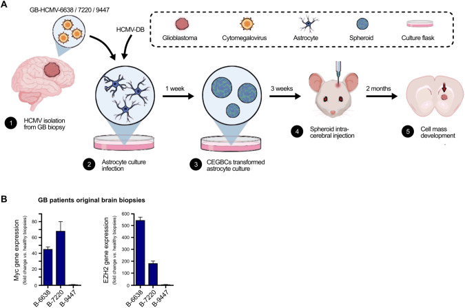
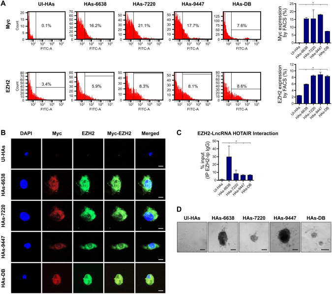
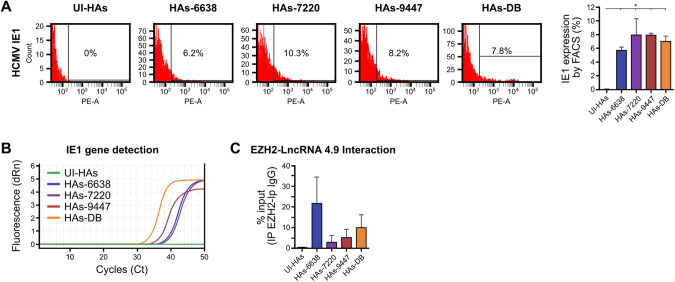
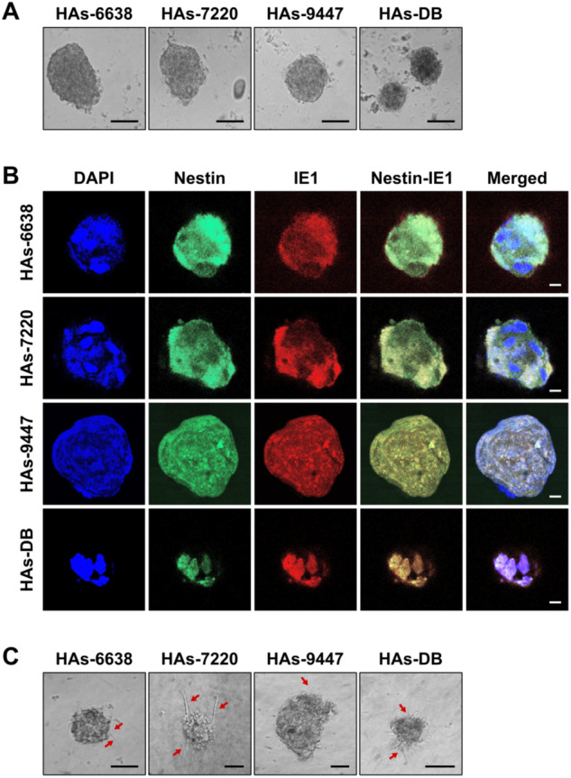
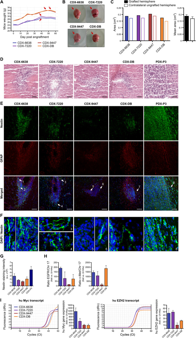
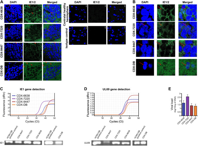
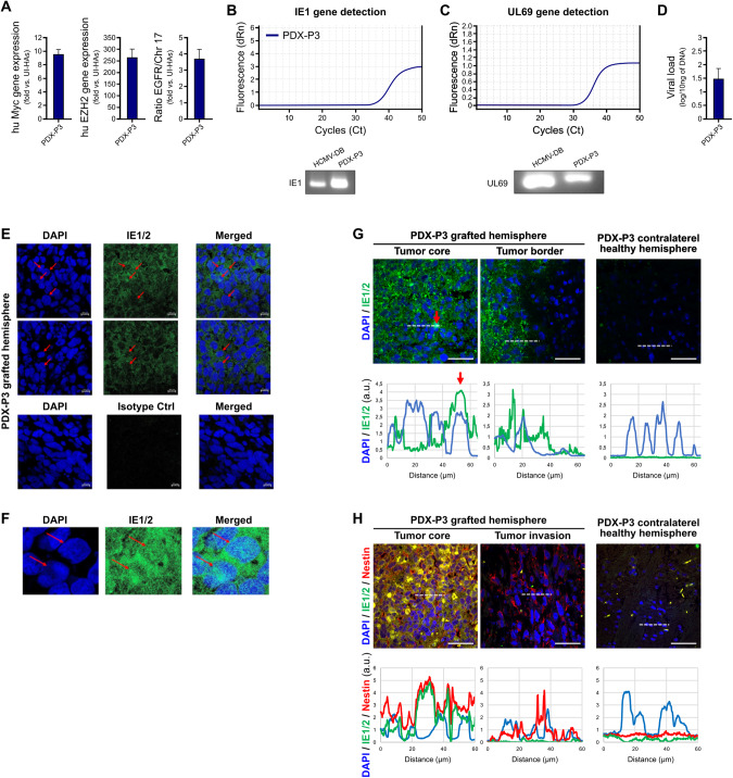
Mounting evidence is identifying human cytomegalovirus (HCMV) as a potential oncogenic virus. HCMV has been detected in glioblastoma multiforme (GB). Herewith, we present the first experimental evidence for the generation of CMV-Elicited Glioblastoma Cells (CEGBCs) possessing glioblastoma-like traits that lead to the formation of glioblastoma in orthotopically xenografted mice. In addition to the already reported oncogenic HCMV-DB strain, we isolated three HCMV clinical strains from GB tissues that transformed HAs toward CEGBCs and generated spheroids from CEGBCs that resulted in the appearance of glioblastoma-like tumors in xenografted mice. These tumors were nestin-positive mostly in the invasive part surrounded by GFAP-positive reactive astrocytes. The glioblastoma immunohistochemistry phenotype was confirmed by EGFR and cMet gene amplification in the tumor parallel to the detection of HCMV IE and UL69 genes and proteins. Our results fit with an HCMV-induced glioblastoma model of oncogenesis in vivo which will open the door to new therapeutic approaches and assess the anti-HCMV treatment as well as immunotherapy in fighting GB which is characterized by poor prognosis.


Conflict of interest statement

The authors declare no competing interests.

Figures

Fig. 1
Fig. 1. Protocol for the generation of glioblastoma tumors in mice engrafted with HCMV-infected astrocytes.
A A scheme of the experimental model used where HAs infected with HCMV GB-6638, GB-7220, GB-9447, and DB generated CEGBCs spheroids. After three weeks, the latter were stereotactically xenografted into the brains of randomly chosen Ragγ2C−/− mice generating a cell mass development day 40 to 50 post-xenografting. B Tumor landscape of the three (oncogenic) HCMV strains isolated from GB patients based on Myc and EZH2 expression as measured by RT-PCR.
Fig. 2
Fig. 2. Activation of Myc-EZH2 oncogenic pathway and colony formation in soft agar in HAs infected with the GB-HCMV strains.
A Myc and EZH2 protein expression as measured by FACS at day 3 post-infection in uninfected HAs and HAs infected with HCMV-GB and HCMV-DB strains. Histogram representing Myc and EZH2 expression in uninfected HAs and the HCMV-GB strains and HCMV-DB as measured by FACS. Data are represented as mean ± SD of two independent experiments. *p value ≤ 0.05. B Confocal microscopic images of Myc and EZH2 staining in HAs infected with the isolated HCMV-GB strains. Nuclei were counterstained with DAPI; magnification ×63, scale bar: 10 μm. C Histograms representing the lncRNA HOTAIR transcript detection in the EZH2 IP samples of HAs infected with HCMV-GB and DB strains, as measured by RT-qPCR. Mouse anti-IgG was used as an isotype control. Data are represented as mean ± SD of two independent experiments. *p value ≤ 0.05. D Colony formation in soft agar seeded with HCMV-GB and HCMV-DB -infected HAs; UI HAs were used as a control. Formed colonies were observed under an inverted light microscope. Magnification x200, scale bar: 100 µm.
Fig. 3
Fig. 3. Detection of HCMV and identification of the lncRNA4.9/EZH2 complex in HAs infected with the GB-HCMV strains.
A IE1 protein detection in HAs infected with the GB-HCMV strains and HCMV-DB as measured by FACS; UI HAs were used as a control. Histogram representing IE1 expression in uninfected HAs and the HCMV-GB strains and HCMV-DB as measured by FACS. B IE1 gene detection in HAs infected with the GB-HCMV strains and HCMV-DB as measured by qPCR. C lncRNA 4.9 transcript detection in the EZH2 IP samples of HAs infected with HCMV-GB and DB strains, as measured by RT-qPCR. Mouse anti-IgG was used as an isotype control. Data are represented as mean ± SD of two independent experiments.
Fig. 4
Fig. 4. Spheroid forming and invasion potentials of the HCMV-GB and HCMV-DB strains used for xenografts.
A Microscopic images of the spheroids generated from the isolated GB HCMV and DB strains; magnification ×100, scale bar: 20 μm. B Confocal microscopic images of concomitant Nestin/IE1 staining in spheroids generated from the isolated HCMV-GB and DB strains. Nuclei were counterstained with DAPI; magnification ×63, scale bar: 10 μm. C Microscopic images showing the invasion potential of CEBGCs-derived spheroids through protrusions (red arrows); magnification ×200, scale bar: 20 μm.
Fig. 5
Fig. 5. Generation of CMV-Derived Xenografts (CDX) and CDX tumors which displayed glioblastoma-like immunohistochemical and molecular phenotypes.
A Loss of weight in mice grafted with CEGBC-derived spheroids. B Brain photography after mice sacrifice; the left hemisphere (red arrows) corresponds to the one that has been implanted. C Histogram indicates the surface area of each cerebral hemisphere: grafted left and ungrafted control right. D Presence of glial proliferation combining increased cellularity and nuclear atypia with enlarged hyperchromatic nuclei in engrafted hemispheres. Tissue was stained using HES. Scale bar: 50 µm. EG Using immunohistochemistry staining, CDX tumor biopsies were positive (E, F) for nestin (CDX-6638 > CDX-7220 > CDX-9447); GFAP, a marker of reactive astrocytes upon viral infection was also detected. F Zoom in on the areas indicated by the arrows in (E) showing nestin filament proteins. Scale bar: 100 µm. G Histogram indicates the staining intensity of nestin of images presented in (E). H EGFR and cMet gene amplification in CDX tumor biopsies (fold increase versus normal uninfected astrocytes). I Human Myc and EZH2 gene expression in CDX tumor biopsies. Left panel: RT-qPCR curves of human Myc and EZH2 gene expression in CDX tumor biopsies. Right panel: Histograms represent the human Myc and EZH2 gene expression in CDX tumor biopsies (fold increase versus uninfected HAs). Data are represented as mean ± SD of three independent experiments.
Fig. 6
Fig. 6. HCMV detection in CDX tumors.
A Using immunohistochemistry staining, CDX tumor biopsies were positive for IE1/2 protein (red arrows indicated nuclear staining). Nuclei were counterstained with DAPI. IE1/2 protein detection in CDX engrafted hemisphere (tumor tissue) but not in the ungrafted contralateral hemisphere. Magnification ×63, scale bar: 10 μm. B IE1/2 protein detection in the nucleus and cytoplasm of cells present in the CDX tumor (red arrows indicated nuclear staining). Magnification ×63, scale bar: 10 μm. C IE1 gene detection (117 bp amplicon) in CDX tumors as measured by qPCR and gel loading. D UL69 gene detection (199 bp amplicon) in CDX tumors as measured by qPCR and gel loading. E. Histogram represents HCMV viral load in CDX brain; mean ± SD of four independent experiments.
Fig. 7
Fig. 7. PDX-P3 harbors HCMV.
A Enhanced Myc and EZH2 expression parallels EGFR gene amplification in PDX-P3 tumor biopsies. Data are represented as mean ± SD of three independent experiments. B IE1 gene detection (117 bp amplicon) in PDX-P3 tumor as measured by qPCR and gel loading. C UL69 gene detection (199 bp amplicon) in PDX-P3 tumor as measured by qPCR and gel loading. D Histogram represents HCMV viral load in PDX-P3 tumor; mean ± SD of four independent experiments. E Using immunohistochemistry staining, PDX-P3 tumor biopsies were positive for IE1/2 protein (red arrows indicated nuclear staining). Nuclei were counterstained with DAPI. Magnification x63, scale bar 10 μm. F IE1/2 protein detection in the nucleus and cytoplasm of cells present in the PDX-P3 tumor (red arrows indicated nuclear staining). Magnification x63, scale bar: 10 μm. G Tissue distribution of IE1/2 protein in PDX-P3. Upper panel: Detection of IE1/2 protein (green) and nucleus (blue) in the tumor core, in the tumor border but not in the healthy contralateral area. Nuclei were counterstained with DAPI. Scale bars: 50 µm. Lower panel: Graphs represent the staining intensity profile (a protein expression indicator) of IE1/2 and DAPI, delineated by the white dashed line (in the corresponding upper panel). The red arrow shows an overlap between the two staining for a cell. H Tissue distribution of IE/nestin in PDX-P3. Upper panel: IE/nestin double staining indicates the concomitant presence of the two proteins in the tumor core, but only nestin is detected in the invasive area. Nuclei were counterstained with DAPI. Scale bars: 50 µm. Lower panel: Graphs represent the staining intensity profile (a protein expression indicator) of IE1/2, nestin and DAPI, delineated by the white dashed line (in the corresponding upper panel).

Similar articles

Cited by

References

    1. Lee JH, Lee JE, Kahng JY, Kim SH, Park JS, Yoon SJ, et al. Human glioblastoma arises from subventricular zone cells with low-level driver mutations. Nature. 2018;560:243–7. doi: 10.1038/s41586-018-0389-3. - DOI - PubMed
    1. Louis DN, Perry A, Wesseling P, Brat DJ, Cree IA, Figarella-Branger D, et al. The 2021 WHO classification of tumors of the central nervous system: a summary. Neuro-Oncol. 2021;23:1231–51. doi: 10.1093/neuonc/noab106. - DOI - PMC - PubMed
    1. Zur Hausen H. Cancers in humans: a lifelong search for contributions of infectious agents, autobiographic Notes. Annu Rev Virol. 2019;6:1–28. doi: 10.1146/annurev-virology-092818-015907. - DOI - PubMed
    1. El Baba R, Herbein G. Immune landscape of CMV infection in cancer patients: from “canonical” diseases toward virus-elicited oncomodulation. Front Immunol. 2021;12:730765. doi: 10.3389/fimmu.2021.730765. - DOI - PMC - PubMed
    1. Geder L, Sanford EJ, Rohner TJ, Rapp F. Cytomegalovirus and cancer of the prostate: in vitro transformation of human cells. Cancer Treat Rep. 1977;61:139–46. - PubMed

LinkOut - more resources