. 2024 Jun;31(6):903-909.
doi: 10.1038/s41594-024-01252-3. Epub 2024 Mar 29.

Cryo-EM structures of amyloid-β and tau filaments in Down syndrome

Affiliations

Cryo-EM structures of amyloid-β and tau filaments in Down syndrome

Anllely Fernandez et al. Nat Struct Mol Biol. 2024 Jun.

Abstract

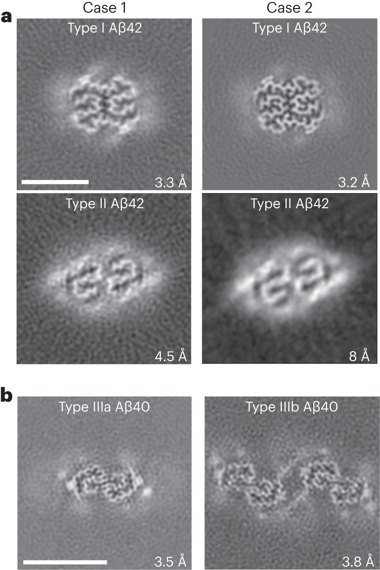
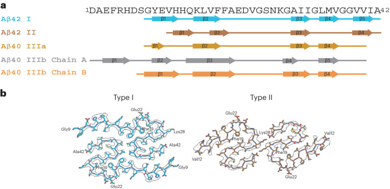
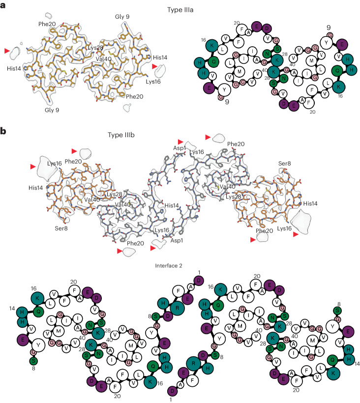
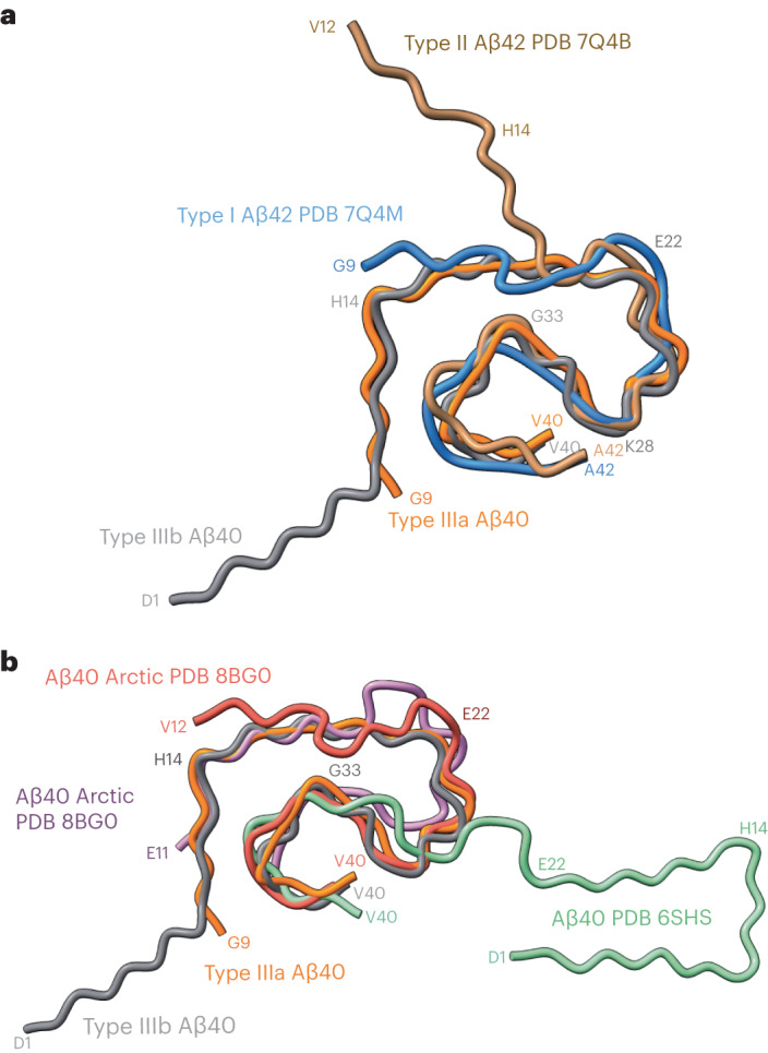
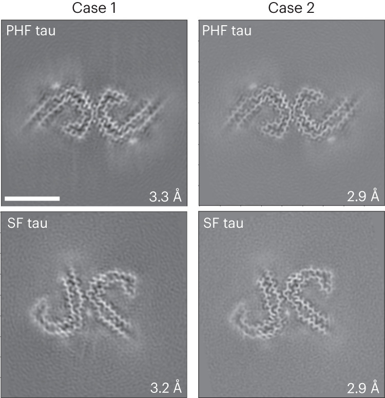
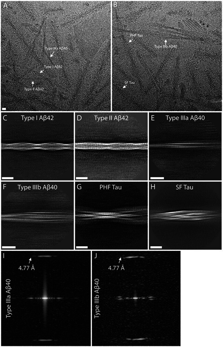
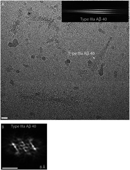
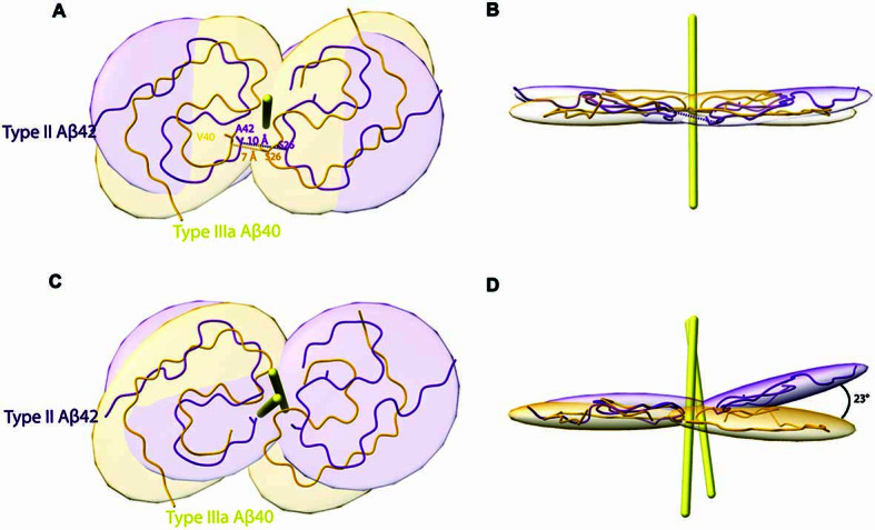
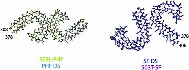
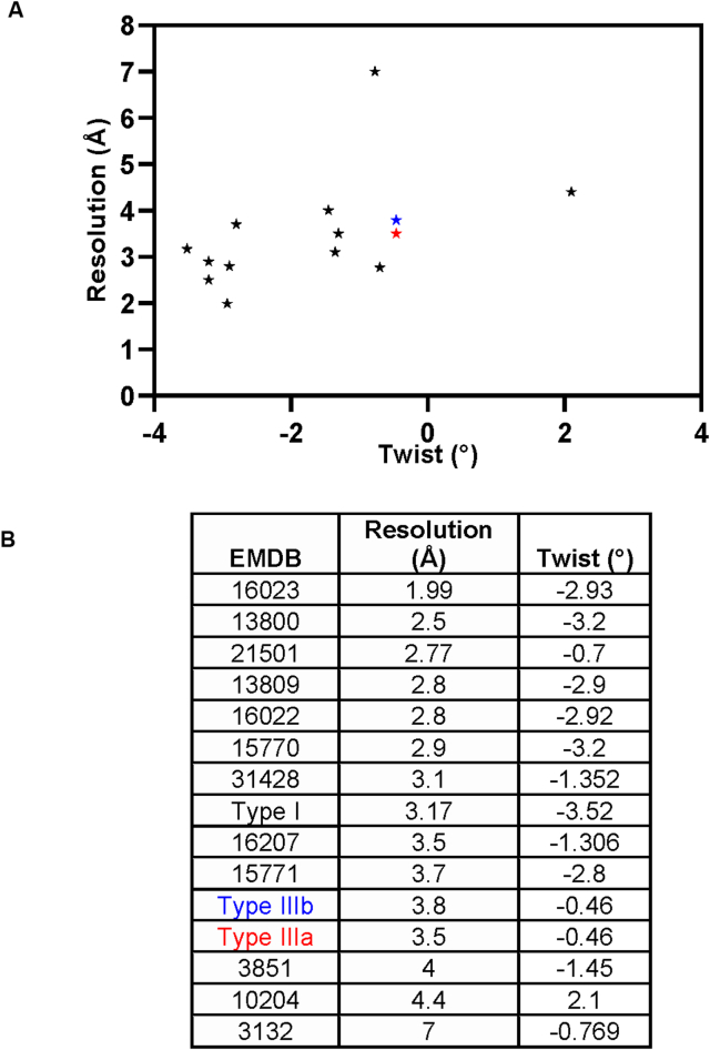
Adult individuals with Down syndrome (DS) develop Alzheimer disease (AD). Whether there is a difference between AD in DS and AD regarding the structure of amyloid-β (Aβ) and tau filaments is unknown. Here we report the structure of Aβ and tau filaments from two DS brains. We found two Aβ40 filaments (types IIIa and IIIb) that differ from those previously reported in sporadic AD and two types of Aβ42 filaments (I and II) identical to those found in sporadic and familial AD. Tau filaments (paired helical filaments and straight filaments) were identical to those in AD, supporting the notion of a common mechanism through which amyloids trigger aggregation of tau. This knowledge is important for understanding AD in DS and assessing whether adults with DS could be included in AD clinical trials.


Conflict of interest statement

The authors declare no competing interests.

Figures

Fig. 1
Fig. 1. Cryo-EM reconstructions of Aβ filaments.
a, Cryo-EM maps, depicted as the sum of ~5 Å central Z-slices, of Aβ42 filaments in DS cases 1 and 2. b, Case 2 type IIIa and IIIb Aβ40 filaments. The estimated resolution is shown on the bottom right. Scale bar, 5 nm (a and b). A total of 12,540 and 24,881 movies were collected for cases 1 and 2, respectively.
Fig. 2
Fig. 2. Aβ filaments in DS brain.
a, Aβ amino acid sequence showing the location of the β-strand regions in Aβ42 type I and II protofilaments and in Aβ40 type III protofilaments. Type I Aβ42 filaments and type II Aβ42 filaments comprise Gly9 to Ala42 and Val12 to Ala42, respectively. Type IIIa Aβ40 filaments are composed of two pairs of identical protofilaments that comprise residues Gly9 to Val40. Type IIIb Aβ40 filaments are organized as a dimer of dimers composed of two pairs of nonidentical protofilaments that comprise residues Ser8 to Val40 and Asp1 to Val40. b, Cryo-EM map in transparent gray, with an atomic model of a single molecule of type I and type II Aβ42 filaments in DS.
Fig. 3
Fig. 3. Aβ40 filaments in DS brain.
a,b, Atomic model of type IIIa Aβ40 (a) and type IIIb Aβ40 filaments (b). Type IIIa Aβ40 filaments are made of two identical protofilaments while type IIIb Aβ40 filaments are made of two nonidentical protofilaments. Type IIIa Aβ40 filaments extend from Gly9 to Val40, while type IIIb Aβ40 filaments extend from Ser8 to Val40 and from Asp1 to Val40. Red arrowheads indicate extra densities. Cartoon representation of amino acid residues: hydrophobic (white), positively charged (teal), polar (green), negatively charged (purple) and glycine (pink) residues.
Fig. 4
Fig. 4. Comparison of type III Aβ40 filaments with human Aβ40 and Aβ42 filaments.
a, Superposition of type I (blue) and II (brown) Aβ42 filaments, and type IIIa (orange) and IIIb (gray) Aβ40 filaments based on the central layer of their S-shaped domains. b, Superposition of the backbone structures of type IIIa (orange) and IIIb (gray) Aβ40 filaments with human Arctic chain A (red) and B (purple) Aβ40 filaments, and meningeal (green) human Aβ40 filaments.
Fig. 5
Fig. 5. Cryo-EM reconstructions of tau filaments.
Cryo-EM maps, depicted as central slices, of tau filaments from the two DS cases (1 and 2). The structures show identical pairs of C-shaped protofilaments and the symmetric inter-protofilament packing between PHFs and asymmetric packing for SFs. The estimated resolution is shown on the bottom right. Scale bar, 10 nm. A total of 12,540 and 24,881 movies were collected for cases 1 and 2, respectively.
Extended Data Fig. 1
Extended Data Fig. 1. Immunohistochemistry.
Immunohistochemical staining of sections of the frontal cortex (DS case #1, panels A, C, E) and temporal cortex (DS case #2, panels B, D, F), contralateral to the ones used for isolating amyloid fibrils, using antibodies specific for Aβ (a–d) and tau (E, F). A significant number of neuritic and diffused plaques can be observed using antibody 4G8 (A, B) and the C-terminal specific Aβ antibody against Aβ species ending at position 42 (C). Vascular amyloid deposition is seen using 4G8 (A) and the C-terminal specific Aβ antibody against Aβ species ending at position 40 (D). Presence of tau aggregates can be seen using the anti-tau antibody AT8 (E, F). Scale bars, 100 μm (A-C, F), 200 μm (D), and 50 μm (E).
Extended Data Fig. 2
Extended Data Fig. 2. Biochemical analysis of Aβ and tau in DS cases 1 and 2.
(a) Western blot shows the presence of Aβ peptides and tau isolated from the cerebral cortex from cases #1 and #2. Presence of Aβ was assessed using antibodies 4G8 and 6E10. Samples were treated with HFIP (+HFIP) to disaggregate Aβ fibrils before western blot using 4G8. Presence of tau was assessed using antibody HT7. Molecular masses are in kDa. This experiment was performed three times. (b) Representative images of immuno-TEM of sarkosyl insoluble Aβ filaments labeled by 6 nm immunogold particles and tau filaments labelled by 10 nm immunogold particles. Scale bar, 100 μm. This experiment was performed three times. (c) Mass Spectrometric analysis showing coverage of the Aβ peptide sequence after trypsin digestion. (d) Several proteins were identified in both DS cases (1,268), with 24 unique hits present in DS cases and in AD (32). This experiment was performed one time. Source data
Extended Data Fig. 3
Extended Data Fig. 3. Representative cryo-EM images of filaments from sarkosyl-insoluble fractions from case #2.
(a, b) Blue arrows indicate type I and purple arrows indicate type II Aβ42 filaments in two raw micrographs. Green arrows indicate type IIIa and red arrows indicate IIIb Aβ40 filaments. White and yellow arrows indicate PHF and SF tau filaments, respectively. (c–h) 2D class averages of type I, II, IIIa and IIIb Aβ filaments, and PHF and SF tau filaments. (i–j) Power spectra of IIIa and IIIb Aβ40 filaments 2D class averages. The scale bars represent 10 nm. A total of 24,881 movies were collected for case #2.
Extended Data Fig. 4
Extended Data Fig. 4. Cryo-EM analysis of Type IIIa Aβ40 filaments in DS-1.
(a) Representative type IIIa Aβ40 filaments, indicated in the cryo-EM micrograph with a white arrow, and corresponding 2D class average. (b) 3D reconstruction of type IIIa Aβ40 filaments. Scale bars represent 10 nm. A total of 12,540 movies were collected for case #1.
Extended Data Fig. 5
Extended Data Fig. 5. Type III Aβ40 is a distinct structure.
Superposition of two-fold rotation axes of type IIIa Aβ40 (yellow) with type II Aβ42 (purple) filaments reveals differences in the packing of residues (a, b). Superposition of a single chain of type IIIa Aβ40 with a single chain of type II Aβ42 shows that the two chains are similar, with the second monomer having a residual rotation of 23 ° (c, d).
Extended Data Fig. 6
Extended Data Fig. 6. Atomic model of tau in DS.
Map fit in the model for PHF and SF from DS compared to PHFs and SF in AD (33).
Extended Data Fig. 7
Extended Data Fig. 7. Twist values of Aβ filaments shown in a scatter plot (A) and table (B).
Data points for the DS type IIIa and IIIb Aβ40 filaments are colored in red and blue, respectively.
Extended Data Fig. 8
Extended Data Fig. 8. Fourier shell correlation curves.
Fourier shell correlation (FSC) curves for independently refined two half maps and map vs model for PHF tau (a), SF tau (b), type I (c), IIIa (d) and IIIb (e) Aβ filaments. The FSC threshold of 0.143 and 0.5 are shown as blue and orange line respectively.
Extended Data Fig. 9
Extended Data Fig. 9. Cryo-EM image analysis of Down syndrome amyloids.
The diagram shows the workflow of image analysis and 3D reconstruction of the two types of tau filaments, and four types of Aβ filaments using RELION 4.0.

Similar articles

Cited by

References

    1. Fortea J, et al. Alzheimer’s disease associated with Down syndrome: a genetic form of dementia. Lancet Neurol. 2021;20:930–942. doi: 10.1016/S1474-4422(21)00245-3. - DOI - PMC - PubMed
    1. Ballard C, Mobley W, Hardy J, Williams G, Corbett A. Dementia in Down syndrome. Lancet Neurol. 2016;15:622–636. doi: 10.1016/S1474-4422(16)00063-6. - DOI - PubMed
    1. Dierssen M. Down syndrome: the brain in trisomic mode. Nat. Rev. Neurosci. 2012;13:844–858. doi: 10.1038/nrn3314. - DOI - PubMed
    1. Hattori M, et al. Chromosome 21 mapping and sequencing consortium, the DNA sequence of human chromosome 21. Nature. 2000;405:311–319. doi: 10.1038/35012518. - DOI - PubMed
    1. Davidson YS, Robinson A, Prasher VP, Mann DMA. The age of onset and evolution of Braak tangle stage and Thal amyloid pathology of Alzheimer’s disease in individuals with Down syndrome. Acta Neuropathol. Commun. 2018;6:56. doi: 10.1186/s40478-018-0559-4. - DOI - PMC - PubMed