. 2024 Sep;26(9):2227-2239.
doi: 10.1007/s12094-024-03450-3. Epub 2024 Mar 29.

Personalizing non-small cell lung cancer treatment through patient-derived xenograft models: preclinical and clinical factors for consideration

Affiliations

Personalizing non-small cell lung cancer treatment through patient-derived xenograft models: preclinical and clinical factors for consideration

Vered Fuchs et al. Clin Transl Oncol. 2024 Sep.

Abstract

Purpose: In the pursuit of creating personalized and more effective treatment strategies for lung cancer patients, Patient-Derived Xenografts (PDXs) have been introduced as preclinical platforms that can recapitulate the specific patient's tumor in an in vivo model. We investigated how well PDX models can preserve the tumor's clinical and molecular characteristics across different generations.

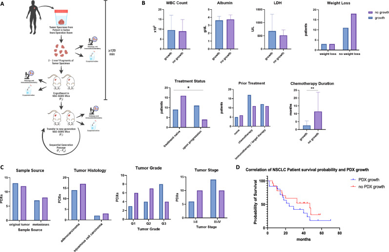
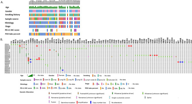
Methods: A Non-Small Cell Lung Cancer (NSCLC) PDX model was established in NSG-SGM3 mice and clinical and preclinical factors were assessed throughout subsequent passages. Our cohort consisted of 40 NSCLC patients, which were used to create 20 patient-specific PDX models in NSG-SGM3 mice. Histopathological staining and Whole Exome Sequencing (WES) analysis were preformed to understand tumor heterogeneity throughout serial passages.

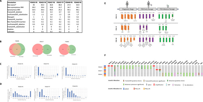
Results: The main factors that contributed to the growth of the engrafted PDX in mice were a higher grade or stage of disease, in contrast to the long duration of chemotherapy treatment, which was negatively correlated with PDX propagation. Successful PDX growth was also linked to poorer prognosis and overall survival, while growth pattern variability was affected by the tumor aggressiveness, primarily affecting the first passage. Pathology analysis showed preservation of the histological type and grade; however, WES analysis revealed genomic instability in advanced passages, leading to the inconsistencies in clinically relevant alterations between the PDXs and biopsies.

Conclusions: Our study highlights the impact of multiple clinical and preclinical factors on the engraftment success, growth kinetics, and tumor stability of patient-specific NSCLC PDXs, and underscores the importance of considering these factors when guiding and evaluating prolonged personalized treatment studies for NSCLC patients in these models, as well as signaling the imperative for additional investigations to determine the full clinical potential of this technique.

Keywords: NSG-SGM3; Non-small cell lung cancer; Patient-derived xenografts; Precision medicine; Preclinical models.


Conflict of interest statement

All authors declare no financial or non-financial competing interests.

Figures

Fig. 1
Fig. 1
Establishment of the NSCLC PDX model platform A Schematic presentation of PDX engraftment protocol in NSG-SGM3. Tumor tissues were collected from NSCLC donor patients undergoing surgical tumor removal after obtaining informed consent. Following resection, the tumor biopsy was kept in a saline solution and transferred to the pathology lab for examination. After pathology confirmation, within 120 min, the biopsy was dissected into smaller fragments of 2–3 mm3 and implanted subcutaneously into the tissue back or flank area of NSG-SGM3 mice, thus creating the first generation of Patient-Derived Xenograft (PDX), referred to as P0 (passage 0). The remaining tumor tissue was cryopreserved for genetic analysis and assessed for pathology and immunohistochemistry. Once a formed PDX reached the size of 1000mm3, mice were euthanized, and the tumor was dissected into smaller implant fragments of 2–3mm3. Subsets of these were as follows: (1) transferred into a new generation of NSG-SGM3 mice (P1–P10), (2) cryopreserved for genetic analysis and sample bank collection, and (3) assessed for pathology and immunohistochemistry. The process repeated sequentially throughout multiple passages of PDX mice. B Clinical and demographic parameters of NSCLC donor patients. The patient's demographic and clinical data were collected at the tumor excision and PDX engraftment point. PDX engraftment was analyzed against the following parameters: WBC count, albumin levels, LDH levels, weight loss > 10% from diagnosis of disease until engraftment of PDX, treatment history, types of treatments, and chemotherapy treatment duration. Data are shown as mean results. Error bars represent standard deviation (SD). C Pathological features of NSCLC PDXs. Several pathological features were analyzed during surgical tumor excision and correlated to PDX growth success and growth rate. These included sample source, tumors' histological type, tumors' histological differentiation grade, and tumors' histological staging according to TNM classification for NSCLC. D Correlation of NSCLC patient survival probability and PDX growth. PDX growth was correlated with the survival time of corresponding patients. Kaplan–Meier survival curves are shown. The tick marks represent censoring
Fig. 2
Fig. 2
Oncoplot presenting the clinical background and molecular alterations of each NSCLC PDX patient. A The plot displays the age, gender, smoking history, PDX sample source, tumor histology, tumor stage, PDL-1 immunohistochemistry score, and the PDX take percent within engrafted mice. The PDX take percent represents the percentage of PDXs successfully engrafted in the mice. PDL-1 immunohistochemistry score is indicated in percentage ranges. B The plot displays alterations found by clinical liquid or tissue sequencing panels for each patient. Alterations are color coded based on their type (e.g., amplifications, deletions, missense mutations). The alterations identified by clinical sequencing panels are shown on the upper side of the plot
Fig. 3
Fig. 3
Growth patterns of NSCLC PDX grafts in NSG-SGM3 mice. Following the engraftment of tumors in mice, PDX size was measured regularly, and once the xenograft reached the size of 1000 mm3, mice were euthanized. The PDX was serially transferred to the next generation of mice. A Passage patterns of 13 selected NSCLC PDXs engrafted in NSG-SGM3 mice over 80 weeks. Each bar represents the time from the initial engraftment of the PDX. Mouse icons represent the passage of the PDX to a new generation of NSG-SGM3 mice. The passage number is indicated inside the mouse icons. B Growth kinetics of 5 PDXs in NSG-SGM3 mice throughout 15 weeks from engraftment (P0) to first passage (P1). C Time intervals between engraftment of PDXs to first passage (P0–P1), first passage to second passage (P1–P2), and second passage to third passage (P2–P3). Lines represent the median results. D Correlation of first PDX passage duration time (P0–P1) and tumor histological differentiation grade. Lines represent median results. E Relationship between patient overall survival and corresponding PDX duration time to the first passage
Fig. 4
Fig. 4
Immuno-histological staining characterization of NSCLC PDXs. PDXs were stained for hematoxylin and eosin (H&E) and analyzed for tumor structure and pathological clinical markers, including TTF1, P40, Ki67, and PDL1. A Representative H&E, TTF1, and P40 staining results are shown for PDX14 in the patient tumor, early (P0), intermediate (P5), and late (P8) PDX passages. B Representative Ki67 and PD-L1 staining results are shown for PDX40 in early (P0), intermediate (P5), and late (P8) PDX passages. C Immuno-histological staining results of selected NSCLC PDXs. Histopathology images were acquired at a magnification of X100 for the hematoxylin & eosin, TTF-1, p40, and PD-L1 slides and at a magnification of X40 for Ki67 slides
Fig. 5
Fig. 5
Whole exome sequencing (WES) analysis. Selected NSCLC PDXs from paired early and late corresponding passages were analyzed by WES. A Annotation distribution (%) of passed filter variants according to WES analysis for selected NSCLC PDXs from early and corresponding late passages. B Venn diagram representing shared variants across early and late passages of paired PDXs. C The distribution of frequencies of passed filter variants occurred only in early PDX passages. D The distribution of passed filter variants occurred both in the early and in the corresponding late passages of NSCLC PDXs. E Comparison of clinically relevant variants found in diagnostic NSG performed for NSCLC patients before tumor resection and WES analysis results of the corresponding early and late PDX passages. Clinically relevant genes are schematically represented as rectangles; gene names are presented above each rectangle; gene alterations are described as stars; and the specific mutation is noted below each rectangle. A double rectangle represents gene amplification; a shortened rectangle represents gene deletion or copy number loss. F Oncoplot showing the molecular alterations of three NSCLC PDXs throughout sequential passages. Each row represents a single PDX sample, with the patient ID on the left. The columns indicate different molecular alterations detected by clinical sequencing panels at the original tumor and by whole exome sequencing in the first passage (P0) and advanced passages (P6–P8). Alterations are color coded based on their type (e.g., amplifications, deletions, missense mutations)

Similar articles

Cited by

References

    1. Sung H, Ferlay J, Siegel RL, Laversanne M, Soerjomataram I, Jemal A, et al. Global cancer statistics 2020: GLOBOCAN estimates of incidence and mortality worldwide for 36 cancers in 185 countries. CA Cancer J Clin. 2021;71(3):209–49. 10.3322/caac.21660 - DOI - PubMed
    1. Zhou L, Wang XL, Deng QL, Du YQ, Zhao NQ. The efficacy and safety of immunotherapy in patients with advanced NSCLC: A systematic review and meta-analysis. Sci Rep. 2016;6: 32020. 10.1038/srep32020 - DOI - PMC - PubMed
    1. Wang M, Herbst RS, Boshoff C. Toward personalized treatment approaches for non-small-cell lung cancer. Nat Med 2021 278. 2021;27(8):1345–56. - PubMed
    1. Chen DS, Mellman I. Elements of cancer immunity and the cancer-immune set point. Nature. 2017;541:321–30. 10.1038/nature21349 - DOI - PubMed
    1. Champiat S, Ileana E, Giaccone G, Besse B, Mountzios G, Eggermont A, et al. Incorporating immune-checkpoint inhibitors into systemic therapy of NSCLC. J Thorac Oncol. 2014;9:144–53. 10.1097/JTO.0000000000000074 - DOI - PubMed

LinkOut - more resources