

Effect of chitosan and CMCS on dentin after Er:YAG laser irradiation: shear bond strength and surface morphology analysis
- PMID: 38553692
- PMCID: PMC10979601
- DOI: 10.1186/s12903-024-04097-w
Effect of chitosan and CMCS on dentin after Er:YAG laser irradiation: shear bond strength and surface morphology analysis
Abstract
Objectives: The aim of the present study was to evaluate the effect of chitosan and carboxymethyl chitosan (CMCS) on dentin surface morphology and bonding strength after irradiation of Er:YAG laser.
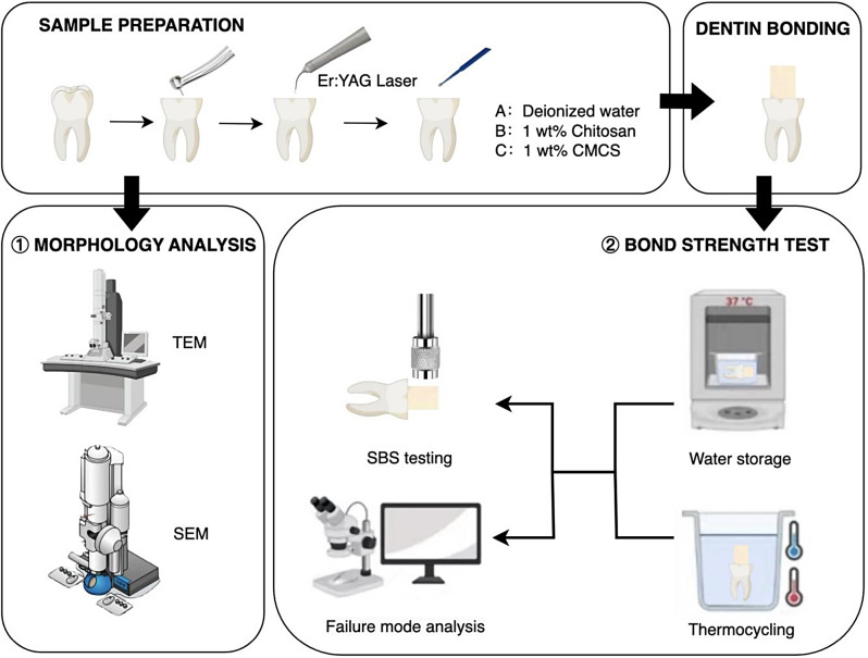
Methods: Eighty-four laser-irradiated dentin samples were randomly distributed into three groups (n = 28/group) according to different surface conditioning process: deionized water for 60s; 1wt% chitosan for 60s; or 1wt% CMCS for 60s. Two specimens from each group were subjected to TEM analysis to confirm the presence of extrafibrillar demineralization on dentin fibrils. Two specimens from each group were subjected to morphological analysis by SEM. Seventy-two specimens (n = 24/group) were prepared, with a composite resin cone adhered to the dentin surface, and were then randomly assigned to one of two aging processes: storage in deionized water for 24 h or a thermocycling stimulation. The shear bond strength of laser-irradiated dentin to the resin composite was determined by a universal testing machine. Data acquired in the shear bond strength test was analyzed by one-way ANOVA with the Tukey honestly significant difference post hoc test and Independent Samples t-test (α = 0.05).
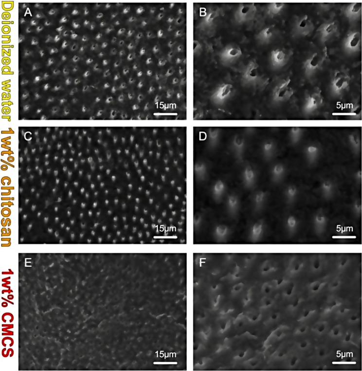
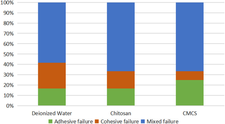
Results: CMCS group presented demineralized zone and a relatively smooth dentin surface morphology. CMCS group had significantly higher SBS value (6.08 ± 2.12) without aging (p < 0.05). After thermal cycling, both chitosan (5.26 ± 2.30) and CMCS group (5.82 ± 1.90) presented higher bonding strength compared to control group (3.19 ± 1.32) (p < 0.05). Chitosan and CMCS group preserved the bonding strength after aging process (p > 0.05).
Conclusions: CMCS has the potential to be applied in conjunction with Er:YAG laser in cavity preparation and resin restoration.
Keywords: Carboxymethyl chitosan; Chitosan; Dentin bonding; Er:YAG laser; Extrafibrillar.
© 2024. The Author(s).
Conflict of interest statement
The authors declare no competing interests.
Figures


Similar articles
-
Influence of water flow rate on shear bond strength of resin composite to Er:YAG cavity preparation.Am J Dent. 2008 Apr;21(2):124-8. Am J Dent. 2008. PMID: 18578182
-
Shear bond strength of different adhesives to Er:YAG laser-prepared dentin.J Adhes Dent. 2006 Oct;8(5):319-25. J Adhes Dent. 2006. PMID: 17080880 Clinical Trial.
-
Bond strengths of one-step self-etch adhesives to laser-irradiated and bur-cut dentin after water storage and thermocycling.Photomed Laser Surg. 2012 Apr;30(4):214-21. doi: 10.1089/pho.2011.3170. Epub 2012 Mar 15. Photomed Laser Surg. 2012. PMID: 22420776 Clinical Trial.
-
The effect of Er:YAG laser irradiation on the bond stability of self-etch adhesives at different dentin depths.Lasers Med Sci. 2017 Jul;32(5):967-974. doi: 10.1007/s10103-017-2194-x. Epub 2017 Mar 30. Lasers Med Sci. 2017. PMID: 28357598 Free PMC article.
-
The influence of Er:YAG laser treatment on the shear bond strength of enamel and dentin: a systematic review and meta-analysis.Quintessence Int. 2020;51(1):8-16. doi: 10.3290/j.qi.a43648. Quintessence Int. 2020. PMID: 31781691
Cited by
-
Effects of different energy levels of Er: YAG laser-assisted bleaching on teeth bleaching efficacy and immediate resin bonding: a study on safety and clinical applicability.Lasers Med Sci. 2025 May 22;40(1):238. doi: 10.1007/s10103-025-04489-6. Lasers Med Sci. 2025. PMID: 40402274
-
Effect of chitosan and chlorhexidine on demineraslised dentin to enhance adhesion.Bioinformation. 2025 Feb 28;21(2):185-188. doi: 10.6026/973206300210185. eCollection 2025. Bioinformation. 2025. PMID: 40322705 Free PMC article.
-
Antibacterial activity, cytotoxicity, and microshear bond strength of an experimental adhesive system containing chitosan-based silver oxide particles.Odontology. 2025 Jun 4. doi: 10.1007/s10266-025-01126-0. Online ahead of print. Odontology. 2025. PMID: 40467946
References
-
- de Azevedo CS, Carneiro PMA, Aranha ACC, Francisconi-dos-Rios LF, de Freitas PM, Matos AB. Long-term effect of Er:YAG laser on adhesion to caries-affected dentin. Lasers Dent Sci. 2018;2(1):19–28. doi: 10.1007/s41547-017-0013-0. - DOI
MeSH terms
Substances
Grants and funding
LinkOut - more resources
Full Text Sources
