. 2024 Jul 18;5(3):100287.
doi: 10.1016/j.xhgg.2024.100287. Epub 2024 Mar 29.

Menke-Hennekam syndrome; delineation of domain-specific subtypes with distinct clinical and DNA methylation profiles

Collaborators, Affiliations

Menke-Hennekam syndrome; delineation of domain-specific subtypes with distinct clinical and DNA methylation profiles

Sadegheh Haghshenas et al. HGG Adv. .

Erratum in

  • Menke-Hennekam syndrome; delineation of domain-specific subtypes with distinct clinical and DNA methylation profiles.
    Haghshenas S, Bout HJ, Schijns JM, Levy MA, Kerkhof J, Bhai P, McConkey H, Jenkins ZA, Williams EM, Halliday BJ, Huisman SA, Lauffer P, de Waard V, Witteveen L, Banka S, Brady AF, Galazzi E, van Gils J, Hurst ACE, Kaiser FJ, Lacombe D, Martinez-Monseny AF, Fergelot P, Monteiro FP, Parenti I, Persani L, Santos-Simarro F, Simpson BN; MKHK Research Consortium; Alders M, Robertson SP, Sadikovic B, Menke LA. Haghshenas S, et al. HGG Adv. 2024 Oct 10;5(4):100337. doi: 10.1016/j.xhgg.2024.100337. Epub 2024 Sep 21. HGG Adv. 2024. PMID: 39306848 Free PMC article. No abstract available.

Abstract

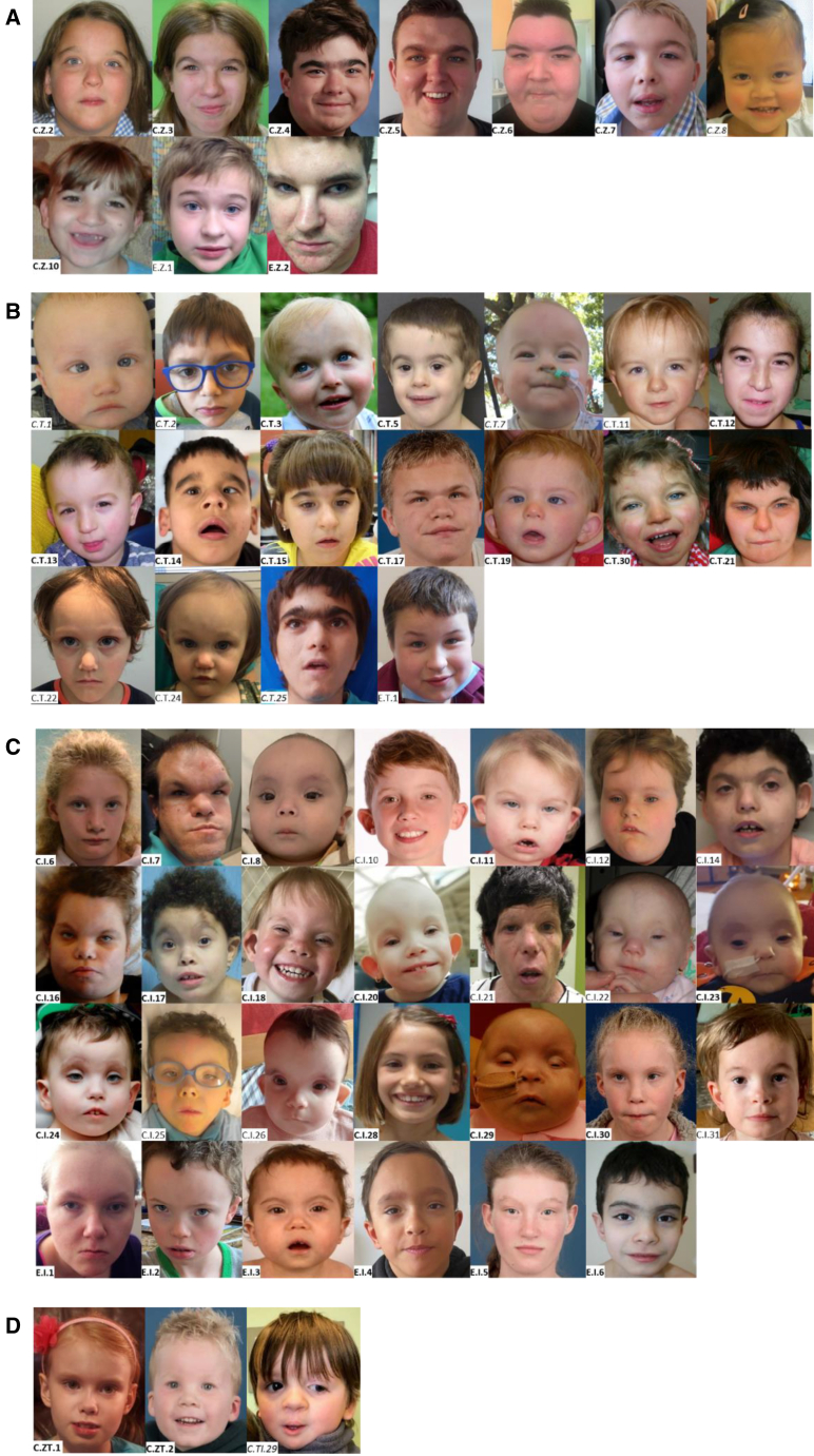
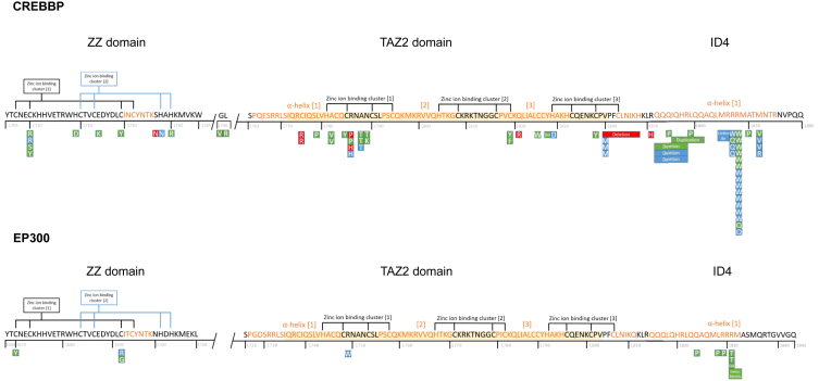
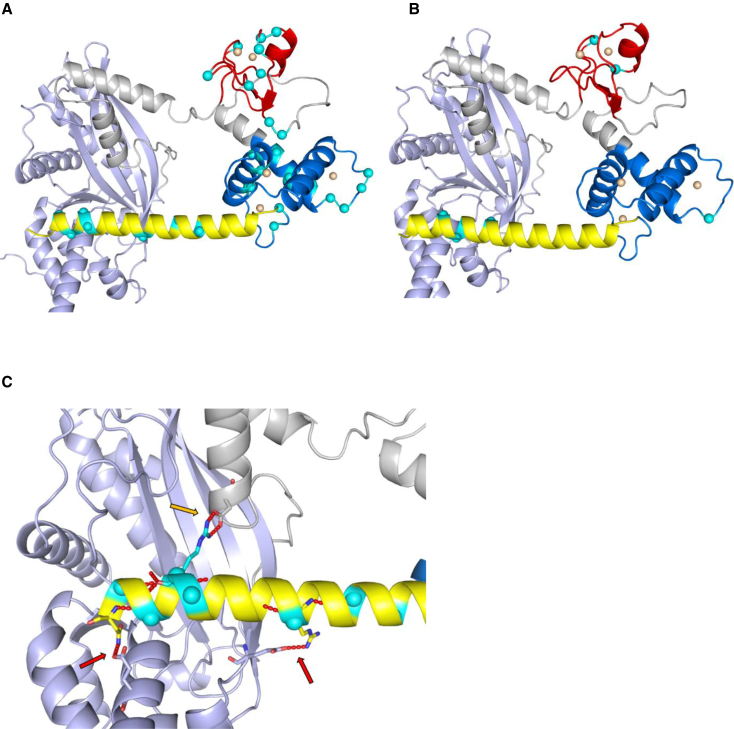
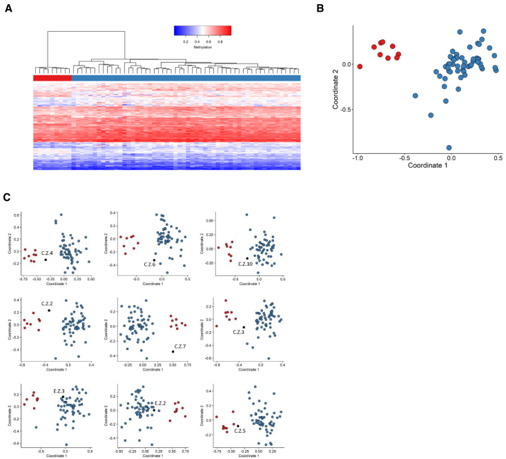
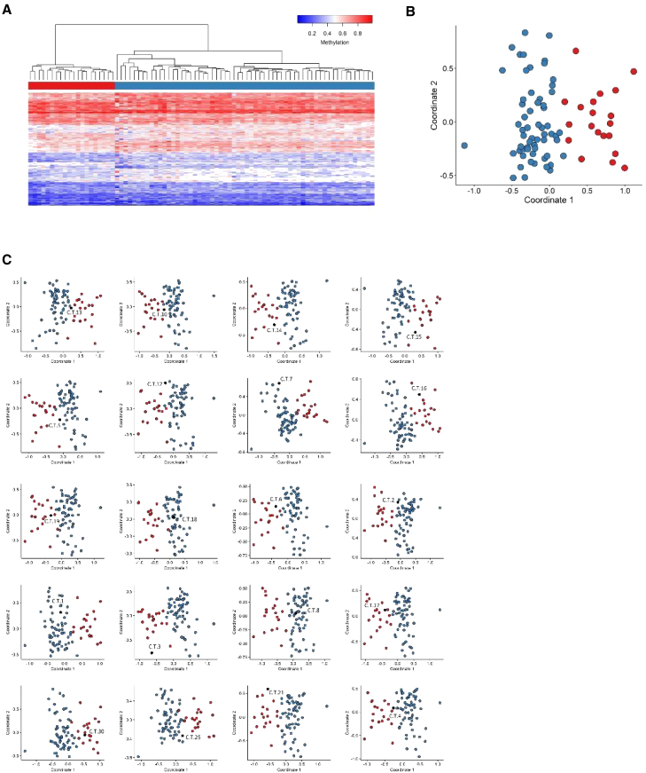
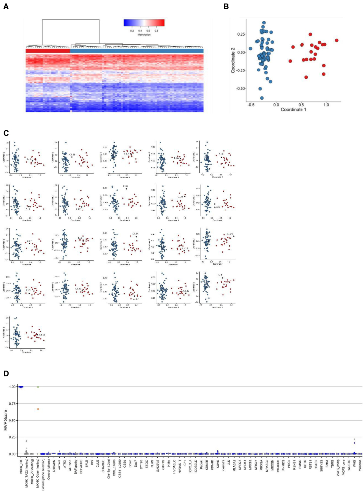
CREB-binding protein (CBP, encoded by CREBBP) and its paralog E1A-associated protein (p300, encoded by EP300) are involved in histone acetylation and transcriptional regulation. Variants that produce a null allele or disrupt the catalytic domain of either protein cause Rubinstein-Taybi syndrome (RSTS), while pathogenic missense and in-frame indel variants in parts of exons 30 and 31 cause phenotypes recently described as Menke-Hennekam syndrome (MKHK). To distinguish MKHK subtypes and define their characteristics, molecular and extended clinical data on 82 individuals (54 unpublished) with variants affecting CBP (n = 71) or p300 (n = 11) (NP_004371.2 residues 1,705-1,875 and NP_001420.2 residues 1,668-1,833, respectively) were summarized. Additionally, genome-wide DNA methylation profiles were assessed in DNA extracted from whole peripheral blood from 54 individuals. Most variants clustered closely around the zinc-binding residues of two zinc-finger domains (ZZ and TAZ2) and within the first α helix of the fourth intrinsically disordered linker (ID4) of CBP/p300. Domain-specific methylation profiles were discerned for the ZZ domain in CBP/p300 (found in nine out of 10 tested individuals) and TAZ2 domain in CBP (in 14 out of 20), while a domain-specific diagnostic episignature was refined for the ID4 domain in CBP/p300 (in 21 out of 21). Phenotypes including intellectual disability of varying degree and distinct physical features were defined for each of the regions. These findings demonstrate existence of at least three MKHK subtypes, which are domain specific (MKHK-ZZ, MKHK-TAZ2, and MKHK-ID4) rather than gene specific (CREBBP/EP300). DNA methylation episignatures enable stratification of molecular pathophysiologic entities within a gene or across a family of paralogous genes.

Keywords: CREB-binding protein; DNA methylation; E1A-associated protein p300; MKHK; Menke-Hennekam syndrome; Rubinstein-Taybi syndrome; episignatures; intellectual disability; intrinsically disordered linker; zinc-finger domain.


Conflict of interest statement

Declaration of interests B.S. is a shareholder in EpiSign Inc., a biotech firm involved in commercial application of EpiSign technology.

Figures

Figure 1
Figure 1
Facial morphology of the presently described individuals with a variant in the MKHK region of CREBBP and EP300 Facial features of individuals with a variant in (A) the ZZ domain, (B) TAZ2-domain, (C) ID4, and (D) in between the ZZ and TAZ2-domain (C.ZT.1 en C.ZT.2) and in between TAZ2 and ID4 (C.TI.29). Study numbers are depicted within each photograph reflecting the affected gene (C or E representing CREBBP and EP300) and domain (Z, T, I, ZT, and TI representing ZZ, TAZ2, ID4, the region between ZZ and TAZ2, and the region between TAZ2 and ID4, respectively) followed by a unique number. Study numbers of individuals with a confirmed MKHK episignature or methylation profile are displayed in bold, and those of individuals who were tested but in whom no MKHK episignature or methylation profile was found are in italics. Photographs of the lateral facial characteristics and of the hands and feet are shown in Figure S1. Detailed description of facial and distal limb morphology of all individuals can be found in Table S3.
Figure 2
Figure 2
Schematic overview of the predicted amino acid changes within the MKHK region of CBP (NP_004371.2) and p300 (NP_001420.2) ZZ, zinc-finger ZZ-type (CBP residues 1,705–1,745; p300 1,668–1,708); TAZ2, zinc-finger TAZ-type (CBP 1,772–1,840; p300 1,735–1,803); ID4, first α helix of the fourth intrinsically disordered region (CBP residues 1,852–1,875, p300 1,810–1,833). Yellow shaded residues in TAZ2 as defined in NCBI reference. Note that three variants are located outside these domains/region (two in between ZZ and TAZ2 and one in between TAZ2 and ID4). Green box represents confirmed methylation profile or episignature (in ID4). Red box, no methylation profile or episignature; blue, no result available. The ID4 episignature was also found in the two individuals with a variant in between ZZ and TAZ2 (C.ZT.1 and moderately in C.ZT.2).
Figure 3
Figure 3
3D predicted protein structures of CBP (NP_004371.2) and p300 (NP_001420.2) (A) 3D predicted protein structure of CBP, including HAT domain (1,342–1,649, in purple), ZZ domain (1,705–1,745, in red), TAZ2-domain (1,772–1,840, in blue) and first α helix of ID4 (1,852–1,875, in yellow). Gray structures not part of functional domain according to NCBI consensus (CDD:239077 and CDD:426615). Cyan spheres represent residue variants, duplications and deletions not included. Orange spheres represent zinc ions. (B) 3D predicted protein structure of p300, including HAT domain (1,306–1,612), ZZ domain (1,668–1,708), TAZ2-domain (1,735–1,803), and first α helix of ID4 (1,810–1,833). (C) ID4 to HAT domain relation in CBP. Red arrows indicate hydrogen bonds (in red) between ID4 and HAT residues: (ID4 + HAT) Arg1857 + Glu1370, Asn1873 + Glu1551. Golden arrow indicates hydrogen bond Arg1868 + Asp1665.
Figure 4
Figure 4
Assessment of the strength of the identified MKHK-ZZ methylation profile and cross-validation Using the selected set of probes, unsupervised and supervised models were applied in order to verify the robustness, sensitivity, and specificity of the selected probes in distinguishing ZZ samples from matched control samples. (A) Hierarchical clustering, where rows represent probes and columns represent samples. The heatmap color scale demonstrates the methylation levels from blue (no methylation) to red (full methylation). On the heatmap pane, red represents ZZ case samples and blue represents control individuals. (B) MDS, where red and blue circles depict case and control samples, respectively. Plots A and B demonstrate clear separation of the case and control groups. (C) In order to inspect the sensitivity of the identified ZZ methylation profile, rounds of leave-one-out cross-validation were performed, using all but one ZZ case sample for probe selection at each trial. In each MDS plot, the blue circles represent the matched control samples, red circles indicate ZZ case samples that were used for probe selection, and the black circle depicts the ZZ case sample that was left out from the probe selection process. It was observed that three ZZ samples clustered with control samples when used for testing (C.Z.6, E.Z.3, and E.Z.2) and two samples fell between case and control groups (C.Z.10 and C.Z.3), demonstrating that the selected set of probes are not sensitive enough to classify all ZZ samples correctly. Blue circles represent training samples, while gray circles depict testing samples.
Figure 5
Figure 5
Assessment of the strength of the identified MKHK-TAZ2 methylation profile and cross-validation (A) Hierarchical clustering, with red representing TAZ2 case samples and blue depicting matched control samples in the heatmap panel. (B) MDS, where red and blue circles represent case and control samples, respectively. A mild difference is observed between the methylation patterns of the case and control groups in both plots (A and B). (C) Cross-validation results for the TAZ2 episignature. In six iterations, the TAZ2 sample that was not used for probe selection, indicated with black, clustered with control individuals, demonstrated by blue circles (C.T.18, C.T.1, C.T.8, C.T.2, C.T.7, and C.T.25) and, in five rounds, the testing TAZ2 sample clustered between the training TAZ2 samples, red circles, and the control individuals (C.T.13, C.T.10, C.T.5, C.T.12, C.T.4), indicating that the selected probes are not sensitive enough to classify all TAZ2 samples correctly.
Figure 6
Figure 6
Assessment of the strength of the identified MKHK-ID4 episignature, cross-validation, and MVP scores (A) Hierarchical clustering, with red representing ID4 case samples and blue depicting matched control samples in the heatmap panel. (B) MDS, where red and blue circles represent case and control samples, respectively. Clear separation of the ID4 case samples and control samples is observed in plots A and B. (C) Rounds of leave-one-out cross-validation were performed, using all but one ID4 case sample for probe selection at each trial. It was observed that, at all iterations, the black circle (ID4 sample not used for probe selection) clustered with the red ones (ID4 samples used for probe selection) and far from the blue circles (control individuals), demonstrating the sensitivity of the episignature. (D) MVP scores created by the SVM constructed using the ID4 selected probes. All the ID4 case samples have received MVP scores near 1 and all the control samples and case samples from other disorders have received scores near 0, indicating full specificity of the model. All the other MKHK samples, other than individuals C.ZT.1 and C.ZT.2 (demonstrated by green and orange circle, respectively), have received low MVP scores. Blue circles represent training samples, while gray circles depict testing samples.

References

    1. Petrij F., Giles R.H., Dauwerse H.G., Saris J.J., Hennekam R.C., Masuno M., Tommerup N., van Ommen G.J., Goodman R.H., Peters D.J., et al. Rubinstein-Taybi syndrome caused by mutations in the transcriptional co-activator CBP. Nature. 1995;376:348–351. doi: 10.1038/376348a0. - DOI - PubMed
    1. Roelfsema J.H., White S.J., Ariyürek Y., Bartholdi D., Niedrist D., Papadia F., Bacino C.A., den Dunnen J.T., van Ommen G.J.B., Breuning M.H., et al. Genetic heterogeneity in Rubinstein-Taybi syndrome: mutations in both the CBP and EP300 genes cause disease. Am. J. Hum. Genet. 2005;76:572–580. doi: 10.1086/429130. - DOI - PMC - PubMed
    1. Cross E., Duncan-Flavell P.J., Howarth R.J., Hobbs J.I., Thomas N.S., Bunyan D.J. Screening of a large Rubinstein-Taybi cohort identified many novel variants and emphasizes the importance of the CREBBP histone acetyltransferase domain. Am. J. Med. Genet. 2020;182:2508–2520. doi: 10.1002/ajmg.a.61813. - DOI - PubMed
    1. Hennekam R.C.M. Rubinstein-Taybi syndrome. Eur. J. Hum. Genet. 2006;14:981–985. doi: 10.1038/sj.ejhg.5201594. - DOI - PubMed
    1. Menke L.A., van Belzen M.J., Alders M., Cristofoli F., DDD Study. Ehmke N., Fergelot P., Foster A., Gerkes E.H., Hoffer M.J.V., et al. CREBBP mutations in individuals without Rubinstein-Taybi syndrome phenotype. Am. J. Med. Genet. 2016;170:2681–2693. doi: 10.1002/ajmg.a.37800. - DOI - PubMed