[Ga68] DOTATATE PET/MRI-guided radiosurgical treatment planning and response assessment in meningiomas
- PMID: 38553990
- PMCID: PMC11300004
- DOI: 10.1093/neuonc/noae067
[Ga68] DOTATATE PET/MRI-guided radiosurgical treatment planning and response assessment in meningiomas
Abstract
Background: Our purpose was to determine the utility of [68Ga]-DOTATATE PET/MRI in meningioma response assessment following radiosurgery.
Methods: Patients with meningioma prospectively underwent postoperative DOTATATE PET/MRI. Co-registered PET and gadolinium-enhanced T1-weighted MRI were employed for radiosurgery planning. Follow-up DOTATATE PET/MRI was performed at 6-12 months post-radiosurgery. Maximum absolute standardized uptake value (SUV) and SUV ratio (SUVRSSS) referencing superior sagittal sinus (SSS) blood pool were obtained. Size change was determined by Response Assessment in Neuro-Oncology (RANO) criteria. Association of SUVRSSS change magnitude and progression-free survival (PFS) was evaluated using Cox regression.
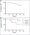
Results: Twenty-seven patients with 64 tumors (26% World Health Organization [WHO]-1, 41% WHO-2, 26% WHO-3, and 7% WHO-unknown) were prospectively followed post stereotactic radiosurgery (SRS) or stereotactic body radiotherapy (SBRT; mean dose: 30 Gy, modal dose 35 Gy, mean of 5 fractions). Post-irradiation SUV and SUVRSSS decreased by 37.4% and 44.4%, respectively (P < .0001). Size product decreased by 8.9%, thus failing to reach the 25% significance threshold as determined by RANO guidelines. Mean follow-up time was 26 months (range: 6-44). Overall mean PFS was 83% and 100%/100%/54% in WHO-1/-2/-3 subcohorts, respectively, at 34 months. At maximum follow-up (42-44 months), PFS was 100%/83%/54% in WHO-1/-2/-3 subcohorts, respectively. Cox regression analyses revealed a hazard ratio of 0.48 for 10-unit reduction in SUVRSSS in the SRS cohort.
Conclusions: DOTATATE PET SUV and SUVRSSS demonstrated marked, significant decrease post-radiosurgery. Lesion size decrease was statistically significant; however, it was not clinically significant by RANO criteria. DOTATATE PET/MR thus represents a promising imaging biomarker for response assessment in meningiomas treated with radiosurgery.
Clinicaltrials.gov identifier: NCT04081701.
Keywords: DOTATATE; PET/MRI; meningioma; outcomes; radiosurgery.
© The Author(s) 2024. Published by Oxford University Press on behalf of the Society for Neuro-Oncology. All rights reserved. For commercial re-use, please contact reprints@oup.com for reprints and translation rights for reprints. All other permissions can be obtained through our RightsLink service via the Permissions link on the article page on our site—for further information please contact journals.permissions@oup.com.
Figures


Similar articles
-
Evaluating diagnostic accuracy and determining optimal diagnostic thresholds of different approaches to [68Ga]-DOTATATE PET/MRI analysis in patients with meningioma.Sci Rep. 2022 Jun 3;12(1):9256. doi: 10.1038/s41598-022-13467-9. Sci Rep. 2022. PMID: 35661809 Free PMC article.
-
68Ga-DOTATATE PET-CT as a tool for radiation planning and evaluating treatment responses in the clinical management of meningiomas.Radiat Oncol. 2021 Aug 16;16(1):151. doi: 10.1186/s13014-021-01875-6. Radiat Oncol. 2021. PMID: 34399805 Free PMC article.
-
Gallium-68 DOTATATE PET in the Evaluation of Intracranial Meningiomas.J Neuroimaging. 2019 Sep;29(5):650-656. doi: 10.1111/jon.12632. Epub 2019 May 20. J Neuroimaging. 2019. PMID: 31107591
-
PET imaging in patients with meningioma-report of the RANO/PET Group.Neuro Oncol. 2017 Nov 29;19(12):1576-1587. doi: 10.1093/neuonc/nox112. Neuro Oncol. 2017. PMID: 28605532 Free PMC article. Review.
-
Semi-automated segmentation methods of SSTR PET for dosimetry prediction in refractory meningioma patients treated by SSTR-targeted peptide receptor radionuclide therapy.Eur Radiol. 2023 Oct;33(10):7089-7098. doi: 10.1007/s00330-023-09697-8. Epub 2023 May 6. Eur Radiol. 2023. PMID: 37148355 Review.
Cited by
-
Preoperative PET/MRI and radio-guided surgery using [Cu64]DOTATATE in meningioma: a feasibility study. Illustrative case.J Neurosurg Case Lessons. 2025 Mar 24;9(12):CASE24867. doi: 10.3171/CASE24867. Print 2025 Mar 24. J Neurosurg Case Lessons. 2025. PMID: 40127474 Free PMC article.
-
The role of nuclear medicine in central nervous system evaluation.Neuroradiol J. 2025 Jun 25:19714009251345100. doi: 10.1177/19714009251345100. Online ahead of print. Neuroradiol J. 2025. PMID: 40557769 Free PMC article. Review.
-
An Update on DOTA-Peptides PET Imaging and Potential Advancements of Radioligand Therapy in Intracranial Meningiomas.Life (Basel). 2025 Apr 7;15(4):617. doi: 10.3390/life15040617. Life (Basel). 2025. PMID: 40283171 Free PMC article. Review.
-
Meningioma: Novel Diagnostic and Therapeutic Approaches.Biomedicines. 2025 Mar 7;13(3):659. doi: 10.3390/biomedicines13030659. Biomedicines. 2025. PMID: 40149634 Free PMC article. Review.
-
Meningioma: Molecular Updates from the 2021 World Health Organization Classification of CNS Tumors and Imaging Correlates.AJNR Am J Neuroradiol. 2025 Feb 3;46(2):240-250. doi: 10.3174/ajnr.A8368. AJNR Am J Neuroradiol. 2025. PMID: 38844366 Review.
References
-
- Ivanidze J, Roytman M, Sasson A, et al.. Molecular imaging and therapy of somatostatin receptor positive tumors. Clin Imaging. 2019;56(July-Aug):146–154. - PubMed
-
- Mirimanoff RO, Dosoretz DE, Linggood RM, Ojemann RG, Martuza RL.. Meningioma: Analysis of recurrence and progression following neurosurgical resection. J Neurosurg. 1985;62(1):18–24. - PubMed
-
- Ehresman JS, Garzon-Muvdi T, Rogers D, et al.. The relevance of simpson grade resections in modern neurosurgical treatment of world health organization grade I, II, and III Meningiomas. World Neurosurg. 2018;109(Jan):e588–e593. - PubMed
-
- Ivanidze J, Roytman M, Lin E, et al.. Gallium-68 DOTATATE PET in the evaluation of intracranial meningiomas. J Neuroimaging. 2019;29(5):650–656. - PubMed
Publication types
MeSH terms
Substances
Associated data
Grants and funding
LinkOut - more resources
Full Text Sources
Medical

