. 2024 May 8;52(8):4659-4675.
doi: 10.1093/nar/gkae212.

The crystal structure of bacteriophage λ RexA provides novel insights into the DNA binding properties of Rex-like phage exclusion proteins

Affiliations

The crystal structure of bacteriophage λ RexA provides novel insights into the DNA binding properties of Rex-like phage exclusion proteins

Myfanwy C Adams et al. Nucleic Acids Res. .

Abstract

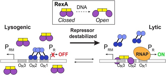
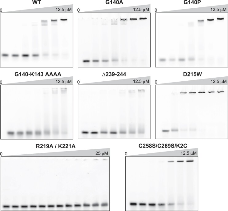
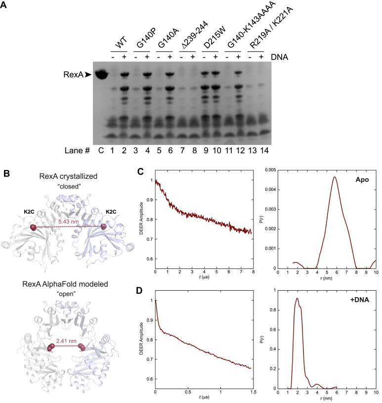
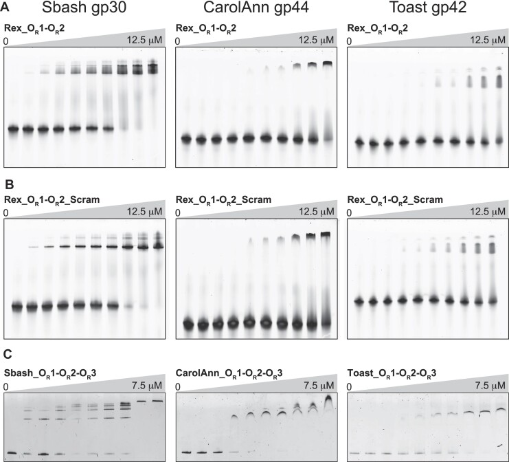
RexA and RexB function as an exclusion system that prevents bacteriophage T4rII mutants from growing on Escherichia coli λ phage lysogens. Recent data established that RexA is a non-specific DNA binding protein that can act independently of RexB to bias the λ bistable switch toward the lytic state, preventing conversion back to lysogeny. The molecular interactions underlying these activities are unknown, owing in part to a dearth of structural information. Here, we present the 2.05-Å crystal structure of the λ RexA dimer, which reveals a two-domain architecture with unexpected structural homology to the recombination-associated protein RdgC. Modelling suggests that our structure adopts a closed conformation and would require significant domain rearrangements to facilitate DNA binding. Mutagenesis coupled with electromobility shift assays, limited proteolysis, and double electron-electron spin resonance spectroscopy support a DNA-dependent conformational change. In vivo phenotypes of RexA mutants suggest that DNA binding is not a strict requirement for phage exclusion but may directly contribute to modulation of the bistable switch. We further demonstrate that RexA homologs from other temperate phages also dimerize and bind DNA in vitro. Collectively, these findings advance our mechanistic understanding of Rex functions and provide new evolutionary insights into different aspects of phage biology.


Figures

Graphical Abstract
Graphical Abstract
Figure 1.
Figure 1.
Structure of RexA. (A) Structure of λ RexA dimer. Split globular domain and dimerization domain are labeled in one monomer are colored green and blue respectively. Second monomer is colored yellow for contrast. Dashed box highlights flexible loop connections (pink and red) between domains (see panel C). (B) Topology diagram of RexA monomer. Pink circle indicates putative G140 hinge and asterisk denotes position of the kink present in the C-terminal α8 helix. (C) Zoomed view of box in A showing conformations of the hinge loop (pink) and swivel loop (red). Pink sphere shows position of putative G140 hinge. (D) Dimerization interface of RexA. Interdigitating helices are labeled. (E, F) Hydrophobic (E) and hydrogen bonding (F) interactions stabilize the RexA dimer interface. Hydrophobic side chains are shown as spheres and hydrogen bonds are depicted as dashed black lines. Participating side chains are labeled, with ‘O’ superscript denoting backbone carbonyl oxygen.
Figure 2.
Figure 2.
RexA shares structural homology with RdgC. (A) Superposition of RexA split globular domain (green) and RdgC center domain (beige). (B) Superposition of RexA dimerization domains (blue) and RdgC base domains (pink). (C) Superposition of crystallized RexA dimer with crystallized RdgC dimer. Structures aligned via the dimerization/base domains and individual domains are colored as in (A) and (B). (D) Structural alignment of individual RexA domains onto RdgC dimer yields a modeled ‘open’ conformation.
Figure 3.
Figure 3.
Putative RexA conformational change suggested by RdgC and AlphaFold modeling. (A–D) Electrostatic surfaces of (left to right) crystallized RexA dimer (A), modeled open RexA dimer based on RdgC homology (B), modeled RexA dimer predicted via AlphaFold-Multimer (78) (C) and RdgC (PDB: 2OWL) (D). Scale bar indicates electrostatic surface coloring from –3 KbT/ec to + 3 KbT/ec. (E–H) Domain organization of crystallized RexA dimer (E), modeled open RexA dimer based on RdgC homology (F), modeled RexA dimer predicted from AlphaFold-Multimer (78) (G) and RdgC (H). Structurally analogous domains are similarly colored to highlight their relative positions in each structure. Dashed circle shows the relative position of the RdgC central pore in each structure based on superposition (see Figure 2C and D) through which DNA is thought to pass (71). (I–K) Distribution of conserved residues on the crystallized (I), RdgC-modeled (J), and AlphaFold-modeled (K) RexA dimer structures. Coloring generated using the ConSurf server (50) and the sequence alignment in Supplementary Figure S6.
Figure 4.
Figure 4.
RexA mutants alter DNA binding activity. EMSA analysis of DNA binding by wildtype and mutant RexA proteins. Binding was performed at 25°C for 30 min in a 20 μl reaction containing 500 nM of Rex_OR1-OR2 annealed double-stranded DNA with increasing concentrations of each RexA protein construct (0, 0.1, 0.2, 0.5, 2, 8 and 12.5 μM; for R291A/K221A double mutant: 0, 0.1, 0.2, 0.5, 2, 8, 12.5, 15, 18.5, 20, 22.5 and 25 μM). Gels were stained with SYBR Gold in 1× TAE buffer for 20 min at 25°C to visualize. See Supplementary Table S2 for substrate oligonucleotide sequences. EMSA experiments were carried out a minimum of three times, each with independently purified batches of protein. Calculated Kd values can be found in Supplementary table S3.
Figure 5.
Figure 5.
Biochemical and biophysical analyses of RexA mutants support a DNA-dependent conformational change. (A) Limited proteolysis of RexA constructs in the absence (–) and presence (+) of 25 μM EMSA_02 (unlabeled) annealed double-stranded DNA. Samples were incubated with 50 μg/ml of trypsin for 30 min at room temperature and then subjected to SDS-PAGE and Coomassie staining to visualize. Lanes are numbered below with ‘C’ denoting the untreated control sample (no protease, no DNA). Proteolysis experiments were carried out three times independently, each using a different batch of purified protein. Representative experiment is shown. (B) Predicted distances between the K2C substitutions (red spheres) in the crystallized (closed) and AlphaFold modeled (open) RexA dimer structures. Individual monomers are colored gray and light blue. (C, D) Time domain signals and distance distributions from DEER spectroscopy of K2C RexA in the absence (C) or presence (D) of 20 μM EMSA_02 (unlabeled) annealed double-stranded DNA.
Figure 6.
Figure 6.
Effects DNA binding and conformational mutants on RexA functions in vivo. (A) Genetic map of the PLPR dual reporter constructed by the insertion of the λ immunity region into the E. coli lac operon (21). Reporter strains contain the temperature-sensitive cI857 repressor with the PR lytic promoter driving expression lacZ and the PL lytic promoter driving expression of the firefly luciferase gene luc. (B, C) Plaque assays testing exclusion of T4, T4rII, and λimm phages (top, middle, and bottom rows on each plate, respectively) by PLPR dual reporter strains containing either wildype rexA (B) or rexA mutants (C). Exclusion phenotypes of E. coli strains that either lack the dual reporter insertion (MG1655) or with the rex genes replaced with a chloramphenicol resistance cassette (rexAB <> cat) are shown for comparison in (B). Note that wildtype λ forms turbid plaques on MG1655 since bacterial lysogens arise within these plaques whereas clear plaques are formed by the purely lytic phages T4 and T4rII. Strains are labeled as follows (see Supplementary Table S4 for full details): MG1655, LT351, rexA+rexB+, LT732; rexAB <> cat, LT772; R219A/K221A, LT2294; Δ239–244, LT2302; D215W, LT2298. (D) Representative papillation from dual reporter strains containing either wildtype rexA (WT) or rexA mutants Δ239–244 and D215W, respectively, in the context of either cro+ (top row) or cro27 (bottom) alleles. Strains are as follows (see Supplementary Table S4 for full details): LT1886, rexA+ cro+; LT1055, rexA+ cro27; LT2302, rexA(Δ239–244) cro+; LT2303, rexA(Δ239–244) cro27; LT2298, rexA(D215W) cro+; LT2299, rexA(D215W) cro27. (E) Quantitation of red papillae in individual colonies for the six genotypes shown in (D). The data are plotted as scatterplots, with each small vertical line indicating the number of papillae found in a single colony. At least 100 hundred colonies were scored for each genotype (WT cro+, n = 100; Δ239–244 cro+, n = 109; D215W cro+, n = 104, WT cro27, n = 100; Δ239–244 cro27, n = 106; D215W cro27, n = 107). The error bars show the SD. See Supplementary statistical analysis of papillation data for t-test results. In all cases, the number of papillae per colony observed with the rexA mutants was significantly different from that found with the wildtype RexA strains.
Figure 7.
Figure 7.
RexA homologs bind DNA non-specifically. EMSA analysis of Sbash gp30, CarolAnn gp44, and Toast gp42 binding to DNA substrates containing λ phage OR1-OR2 operator sites (A), scrambled sequences for λ OR1 and OR2 operators (B) and analogous OR1-OR2-OR3 operator regions specific to each phage (C). Binding was performed at 25°C for 30 min in a 20 μl reaction containing 500 nM of each DNA substrate with increasing concentrations of each phage protein (0, 0.1, 0.2, 0.5, 2, 8 and 12.5 μM for experiments with OR1-OR2 substrates; 0, 0.1, 0.25, 0.5, 0.75, 1, 1.25, 2.5, 3.75, 7.5 μM for experiments with OR1–OR2–OR3 substrates). Gels were stained with SYBR Gold in 1× TAE buffer for 20 min at 25°C to visualize. See Supplementary Table S2 and Supplementary Figure S12 for oligonucleotide sequences and detailed descriptions of each substrate. EMSA experiments were carried out a minimum of three times, each with independently purified batches of protein. Calculated Kd values can be found in Supplementary Table S3.

Similar articles

Cited by

References

    1. Lederberg E. Lysogenicity in E. coli K12. Genetics. 1951; 36:560.
    1. Gottesman M.E., Weisberg R.A.. Little Lambda, who made thee?. Microbiol. Mol. Biol. R. 2004; 68:796–813. - PMC - PubMed
    1. Campbell A. The future of bacteriophage biology. Nat. Rev. Genet. 2003; 4:471–477. - PMC - PubMed
    1. Ptashne M., Hopkins N.. The operators controlled by the lambda phage repressor. Proc. Natl. Acad. Sci. U.S.A. 1968; 60:1282–1287. - PMC - PubMed
    1. Dodd I.B., Shearwin K.E., Egan J.B.. Revisited gene regulation in bacteriophage λ. Curr. Opin. Genet. Dev. 2005; 15:145–152. - PubMed

Publication types

MeSH terms