Review
. 2024 May 8;52(8):4111-4123.
doi: 10.1093/nar/gkae199.

Critical steps in the assembly process of the bacterial 50S ribosomal subunit

Affiliations
Review

Critical steps in the assembly process of the bacterial 50S ribosomal subunit

Amal Seffouh et al. Nucleic Acids Res. .

Abstract

During assembly, ribosomal particles in bacteria fold according to energy landscapes comprised of multiple parallel pathways. Cryo-electron microscopy studies have identified a critical maturation step that occurs during the late assembly stages of the 50S subunit in Bacillus subtilis. This step acts as a point of convergency for all the parallel assembly pathways of the subunit, where an assembly intermediate accumulates in a 'locked' state, causing maturation to pause. Assembly factors then act on this critical step to 'unlock' the last maturation steps involving the functional sites. Without these factors, the 50S subunit fails to complete its assembly, causing cells to die due to a lack of functional ribosomes to synthesize proteins. In this review, we analyze these findings in B. subtilis and examine other cryo-EM studies that have visualized assembly intermediates in different bacterial species, to determine if convergency points in the ribosome assembly process are a common theme among bacteria. There are still gaps in our knowledge, as these methodologies have not yet been applied to diverse species. However, identifying and characterizing these convergency points can reveal how different bacterial species implement unique mechanisms to regulate critical steps in the ribosome assembly process.


Figures

Graphical Abstract
Graphical Abstract
Figure 1.
Figure 1.
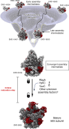
Overview of the 50S subunit assembly process in B. subtilis. The 50S ribosomal particles at the early stages of assembly follow parallel assembly pathways that converge into a critical maturation stage. The assembly intermediate that accumulates is ‘locked’, and its maturation is paused. RbgA, YphC and YsxC act on this intermediate to ‘unlock’ it and complete the maturation of the ribosomal particle. The rRNA in the cryo-EM maps is colored in light gray, and the r-proteins are shown in red. This figure was prepared from EMD entries 16 497, 16 500, 16 509, 16 506, 16 503, 16 504 (30), 20 435 (17) and 6306 (56).
Figure 2.
Figure 2.
Convergency points in the 50S assembly pathway of B. subtilis. (A) The late stages of assembly in the 50S subunit involve the maturation of the central protuberance (CP), the L1 and L7/12 stalks and the functional core (A, P and E sites). Panel A shows these landmarks overlaid in the structure of the mature 50S subunit. The rRNA helices comprising the A, P and E site are indicated. The components of the central protuberance, 5S rRNA and H80-88 are also indicated. (B) The assembly intermediates that accumulate under depletion of assembly factors RbgA (45SRbgA), YphC (45SYphC) and YsxC (44.5SYsxC) present depletion of r-proteins uL16, bL27, bL28, b33, b35 and b36. The panel shows the location of these proteins in the mature 50S subunit structure. Panels (A) and (B) were produced from PDB 3J9W (56). (C) Cryo-EM structures of the 45SRbgA, 45SYphC and 44.5SYsxC particles showing that these assembly intermediates are structurally similar. The rRNA in the cryo-EM maps is colored in light gray, and the r-proteins are shown in red. Structure representations were prepared from EMD entries 20435 (25) (45SRbgA) and (57) (45SYphC and 44.5SYsxC).
Figure 3.
Figure 3.
The essential role of RbgA at the critical maturation steps of the 50S subunit in B. subtilis. (A) This panel shows how RbgA binding to the P site in the 45SRbgA assembly intermediate triggers the folding of critical rRNA helices in the A site (H91 and H92) and P site (H93), all colored in cyan. RbgA binding also stabilizes the L1 stalk (colored in dark blue), H38 (colored in purple) and the binding of r-protein uL6. The remaining rRNA helices are shown in light gray and all r-proteins are shown in red. Structure representations were prepared from EMD entries 20 435 and 20 441 (25). (B) The rRNA helices H97 and H42 in the 45SRbgA particle are latched together through the atomic interactions shown in the panel, locking the maturation of the intermediate and making it dependent on RbgA to continue its maturation process. (C) This panel shows how H97 and H42 are linked in the cryo-EM structure of the 45SRbgA particle. The binding of r-protein uL6 stabilizes this interaction. The lack of uL6 incorporation in the case of the R70P uL6 variant or the incorporation of the uL6 R3C variant into the corresponding 44S particles causes H97 and H42 to remain unlatched, making the 44S intermediates less dependent on RbgA to continue their maturation. Structure representations were prepared from EMD entries 29 437 (44SR3C), 24 950 (44SR70P) (17) and 20 435 (45SRbgA) (25). (D) The panel shows the canonical maturation pathway of the 45SRbgA particle where maturation of the functional A, P and E sites occurs after the central protuberance (CP). Binding of uL6 and the dependency of RbgA for this maturation steps ensure this specific order for the maturation of this critical structural motifs of the 50S subunit. Structure representations were prepared from EMD entries 20 491, 20 435 and 20 441 (25) and 6306 (56). (E) In the 44SR3C particles, incorporating the R3C uL6 variant allows maturation to evolve through an alternative pathway. In this case, the maturation of these intermediates is less dependent on RbgA and the maturation of the functional sites is possible before the central protuberance develops. The 44SR70P particle matures following a similar pathway but it is not shown in the panel. Structure representations were prepared from EMD entries 24 940, 24 960, 24 962 (17) and 6306 (56). In panels (C)–(E), the rRNA in the cryo-EM maps is colored in light gray, and the r-proteins are shown in red.
Figure 4.
Figure 4.
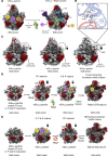
Assembly intermediates from an in vitro 50S reconstitution reaction from purified E. coli components. (A) Schematic of the 50S in vitro assembly assay, consisting of two steps. Under conditions indicated 33S, 41S and 48S precursors form during step 1. In step 2, the 48S precursor is converted into an active 50S subunit under the influence of heat (55°C) and 20 mM Mg2+. (B) 50S assembly map based on the Nierhaus map (58), refined by Williamson (59). In contrast to the classical assembly map showcasing the order in which proteins bind to the RNA and the protein′s interdependencies, this map orders the precursor states obtained by cryo-EM analysis from material subjected to step 1 of the 50S reconstitution protocol (30). From left to right 5′of the 23S rRNA to 3′; from top to bottom order in which proteins were found to bind to the LSU in vivo (time). Particle densities in the cryo-EM maps are colored according to the architectural domains of the 23S rRNA they belong to. A 23S rRNA 2D map shown as insert appears in the same color code. All ribosomal proteins on the y-axis appear in the same color code. Structure representations were prepared from EMD entries 16 509 (d1), 16 508 (d1_L4), 16 507 (d1_L4/L23), 16 506 (d12), 16 505 (d12_L23), 16 502 (d12-CP), 16 503 (d126), 16 501 (d126-CP), 16 504 (d136), 16 499 (C), 16 498 (C_L2), 16 497 (C-CP), 16 496 (C-CP_L28), 16 494 (C-CP-H68) and 16 495 (C-CP_L28/L2) (30). (C) Nomenclature of the precursors. Early intermediates in this reaction are named according to the 23S rRNA domains whose cryo-EM densities were exhibited. Later, these intermediates extend their names by adding the name of the additional structural elements which density appears, such as L28, L2 or H68. Late maturation stages containing all domains of the core of the LSU (I, II, III and VI) are called C particles. The formation of the core demarcates the end of early 50S assembly. From there, particles that exhibit the central protuberance, add CP to their names. Different routes lead from state C to state C-CP-H68 (48S), the end point of step1 of 50S in vitro assembly. * Proteins not resolved in the cryo-EM maps.
Figure 5.
Figure 5.
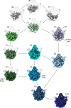
Assembly intermediates from an in vitro 50S reconstitution the iSAT reaction reaction from purified E. coli components. Cryo-EM maps obtained from the 50S assembly intermediates that accumulate in the iSAT reaction (36). Intermediates are named in this study using a subsequent combination of letters and numbers. The name of each intermediate is indicated in their upper left side. Key rRNA helices forming the P (H69) and E sites (H68) are indicated in intermediates where they become first apparent. The location of the A site, still partially immature is indicated in the most mature intermediate (E6). Structure representations were prepared from EMD entries 29 056 (B1), 29 042 (B2), 29 058 (C1), 29 059 (C2), 29 060 (C3), 29 061 (C4), 29 062 (E1), 29 063 (E2), 29 064 (E3), 29 065 (E4), 29 066 (E5), 29 067 (E6) and 29 057 (G1) (36).
Figure 6.
Figure 6.
Convergency intermediates observed in B. subtilis and E. coli using various experimental methodologies. Structurally similar intermediates representing convergency points in the assembly process isolated using a RbgA-depletion B. subtilis strain (17,25) (A), an in vitro reconstitution assays with E. coli purified components (30) (B) and iSAT also with E. coli elements (36) (C). The name of the intermediate using the original terminology in each study is indicated below. The lock besides the name indicates that these three intermediates may represent a locked convergency point, requiring the interaction with assembly factors to progress into the mature 50S subunit. The assembly factors regulating the locked assembly point are still unknown in E. coli likely different from those described for RbgA in B. subtilis. In the three panels, the rRNA in the cryo-EM maps is colored in light gray, and the r-proteins are shown in red. This figure was prepared from EMD entries 20 435 (25) (45SRbgA), 16 495 (C-CP-L28-L2) (30) and 29 063 (E2) (36).
Figure 7.
Figure 7.
50S assembly intermediates from an E. coli ΔrlmE strain. Cryo-EM structures of three 50S assembly intermediates purified from an E. coli strain lacking the rlmE gene. The panel in the right shows the cryo-EM structure of the mature 50S subunit purified from the same strain. The rRNA helices forming the A, P and E functional sites that are still not formed in state I are indicated. These helices adopt a mature conformation and show density in the cryo-EM maps for state II, III and 50S subunits. H42 and H97 forming the L7/L12 stalk are also indicated in each class. These two helices are unlatched in state I but latched in states 2 and 3 and the mature subunit. The rRNA in the cryo-EM maps is colored in light gray, and the r-proteins are shown in red. This figure was prepared from EMD entries 30 214 (state I), 30 213 (state II), 30 212 (state III) and 30 215 (mature 50S) (49).
Figure 8.
Figure 8.
Assembly factors assisting convergent assembly intermediates in bacteria may act in conjuction. Left panel: Cryo-EM map of a mitoribosome large subunit assembly intermediate from Trypanosome brucei (EMD 11000) (left panel) (54) with assembly factors Mtg1 (RbgA homologue), mt-EngA (YphC homologue) and mt-EngB (YsxC homologue) bound simultaneously to the interface area of the subunit. Middle panel: Cryo-EM map of the B. subtilis 45SRbgA particle with RbgA, colored in yellow, bound to the P site (EMD 20441) (25). The binding site of RbgA in the subunit is equivalent to the binding site of Mtg1 in T. Brucei. Right panel: Cryo-EM map of the E. coli 50S subunit with EngA bound in the E site (EMD 6149) (60). The binding site of EngA (YphC homologue) in the subunit is equivalent to the binding site of mt-EngA in T. Brucei. There are no structures from protein homologues of mt-EngB bound to bacterial ribosomal subunits or assembly intermediates. In the three structures r-proteins are colored in red and rRNA is colored in light gray. Assembly factor mt-EngA and EngA are colored in dark blue and mt-EngB is colored in cyan.

Similar articles

Cited by

References

    1. Gutgsell N.S., Jain C.. Gateway role for rRNA precursors in ribosome assembly. J. Bacteriol. 2012; 194:6875–6882. - PMC - PubMed
    1. Traub P., Nomura M.. Studies on the assembly of ribosomes in vitro. Cold Spring Harb. Symp. Quant. Biol. 1969; 34:63–67. - PubMed
    1. Rohl R., Nierhaus K.H.. Assembly map of the large subunit (50S) of Escherichia coli ribosomes. Proc. Natl. Acad. Sci. U.S.A. 1982; 79:729–733. - PMC - PubMed
    1. Woodson S.A. RNA folding and ribosome assembly. Curr. Opin. Chem. Biol. 2008; 12:667–673. - PMC - PubMed
    1. Woodson S.A. RNA folding pathways and the self-assembly of ribosomes. Acc. Chem. Res. 2011; 44:1312–1319. - PMC - PubMed

Publication types

MeSH terms