. 2024 Mar 30;73(5):78.
doi: 10.1007/s00262-024-03659-9.

Lipid droplets-related Perilipin-3: potential immune checkpoint and oncogene in oral squamous cell carcinoma

Affiliations

Lipid droplets-related Perilipin-3: potential immune checkpoint and oncogene in oral squamous cell carcinoma

Yijia He et al. Cancer Immunol Immunother. .

Abstract

Background: Lipid droplets (LDs) as major lipid storage organelles are recently reported to be innate immune hubs. Perilipin-3 (PLIN3) is indispensable for the formation and accumulation of LDs. Since cancer patients show dysregulated lipid metabolism, we aimed to elaborate the role of LDs-related PLIN3 in oral squamous cell carcinoma (OSCC).

Methods: PLIN3 expression patterns (n = 87), its immune-related landscape (n = 74) and association with B7-H2 (n = 51) were assessed by immunohistochemistry and flow cytometry. Real-time PCR, Western blot, Oil Red O assay, immunofluorescence, migration assay, spheroid-forming assay and flow cytometry were performed for function analysis.

Results: Spotted LDs-like PLIN3 staining was dominantly enriched in tumor cells than other cell types. PLIN3high tumor showed high proliferation index with metastasis potential, accompanied with less CD3+CD8+ T cells in peripheral blood and in situ tissue, conferring immunosuppressive microenvironment and shorter postoperative survival. Consistently, PLIN3 knockdown in tumor cells not only reduced LD deposits and tumor migration, but benefited for CD8+ T cells activation in co-culture system with decreased B7-H2. An OSCC subpopulation harbored PLIN3highB7-H2high tumor showed more T cells exhaustion, rendering higher risk of cancer-related death (95% CI 1.285-6.851).

Conclusions: LDs marker PLIN3 may be a novel immunotherapeutic target in OSCC.

Keywords: B7-H2; Lipid droplets; OSCC; PD-L1; Perilipin-3.


Conflict of interest statement

The authors have no relevant financial or non-financial interests to disclose.

Figures

Fig. 1
Fig. 1
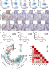
Expression pattern of spotted LDs-like PLIN3 and association with proliferation index and postoperative status. a Representative image of IHC (the bar above is 50 µm and below is 10 µm) and Oil Red O (Scale bars for TCs, FLCs, and TILs indicate 8, 10, and 4 µm, respectively) for low and high expression of PLIN3 in normal epithelial, TCs, FLCs, and TILs in OSCC. b The IHC score of PLIN3 in normal, TCs, FLCs, and TILs from OSCC patients (n = 87). c, d Expression of PLIN3 in HNSCC was analyzed by single-cell RNA sequencing (GSE103322, GSE164690). e Single-cell RNA-seq results of PLIN3 in tongue tissues (https://www.proteinatlas.org). f–h Correlation between Ki-67 staining and PLIN3 in TCs, TILs and FLCs. i, j The effect of PLIN3 on postoperative recurrence and metastasis status in OSCC patients were shown by two-way ANOVA (mixed model). Results are shown as mean ± SEM. p = two-tailed t test
Fig. 2
Fig. 2
PLIN3 is associated with the immunosuppressive microenvironment of tumor tissue in situ. a–e Correlation between PLIN3 RNA expression and CD8B, CD4, CD56, CD68, as well as CD19 in HNSCC from cBioportal database. f–k Relationship between PLIN3 expression and CD8+ T cell, CD4+ T cell, CD56+ NK cell, CD68+ TAM, and CD19+ B cell infiltration in serial sections of OSCC followed by IHC (n = 10). l–n Quantitative statistics on the correlation between PLIN3 and CD8, CD4 and CD4/CD8 ratio in situ of OSCC patients. o Correlation analysis between PLIN3 and immune cell infiltrations in HNSCC samples using TIMER. p Heatmap illustrating the correlation between PLIN3 and immune checkpoints from cBioportal database. Results are shown by Pearson correlation analysis
Fig. 3
Fig. 3
The change of lymphocytes subset in PBMC of OSCC patients according to PLIN3 level. a Flow cytometry contour plots showing the strategy for gating lymphocytes. b–g The ratio and absolute number of human CD3+T cells, CD3+CD4+ helper/inducer T cells, CD3+CD8+ cytotoxic T cells, CD3CD16+CD56+ NK cells and CD3CD19+ B cells in blood were analyzed in PLIN3low and PLIN3high groups of TCs, TILs, and FLCs by BD Multitest™ reagent in OSCC patients (n = 74). Results are shown by two-way ANOVA. p = Sidak’s multiple comparison test
Fig. 4
Fig. 4
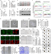
PLIN3 heterogeneity in tumor microenvironment leads to different clinical outcomes. a–i Kaplan–Meier survival analyses for overall survival time (OS), metastasis-free survival time (MFS), and disease-free survival time (DFS) of OSCC patients according to the protein expression of PLIN3 in TCs, TILs, and FLCs. j–l The effects of PLIN3 expression on the prognosis of OS in cervical squamous cell carcinoma, esophageal squamous cell carcinoma and liver hepatocellular carcinoma patients were shown by Kaplan–Meier plotter database. m Cox regression models for OS and DFS in OSCC patients to determine the independent risk factors, adjusted hazard ratio (HR), and 95% confidence interval (CI) of OSCC. Survival analyses including OS, MFS, and DFS were evaluated by Kaplan–Meier and log-rank test
Fig. 5
Fig. 5
Knockdown of PLIN3 inhibits abnormal lipid accumulation and biological behavior in OSCC cells. a PLIN3 expression in human normal keratinocytes (HACAT), Cal27, Cal33, and HN6 cell lines. b, c Knockdown expression of PLIN3 in HN6 cells with three independent siRNA, confirmed using qPCR and WB. d, e PLIN3-knockdown HN6 or PLIN3-overexpressing Cal 33 cell lines were generated by transfecting lentivirus and validation by qPCR and WB. f GSEA of PLIN3 mRNA and OSCC signaling pathways. g Oil Red O staining was conducted in HN6 and Cal33 cells. h–k Immunofluorescent and Oil Red O were performed to detect LDs content in HN6 cells treated with OA and knocked down with PLIN3. The bar is 8 μm. l–n Wound healing and transwell assay; 3D spheroid models were performed to detect HN6 cell migration and proliferation. The results represent the mean ± SEM from three independent experiments. p = two-tailed t test
Fig. 6
Fig. 6
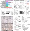
PLIN3 upregulates B7-H2/PD-L1 level and induces CD8+T cells exhaustion in OSCC patients. a KEGG pathway classification of PLIN3 in OSCC cells. b GO annotated positive regulation of lymphocyte migration and myeloid leukocyte migration. c Schematic diagram of human tumor cells/immunocytes co-culture system. d After co-cultured with PLIN3 knockdown HN6, the percentage of CD3+ CD8+ T cells in PBMC were analyzed. e, f qPCR and WB analysis showed the mRNA and protein expression of PD-L1 and B7-H2 in HN6 cells. g, h Correlation between PLIN3 and B7-H2 shown by immunohistochemistry and quantitative analysis (n = 51). i, j Kaplan–Meier survival curves for overall survival time (OS) and disease-free survival time (DFS) of OSCC patients according to the expression of PLIN3 and B7-H2 in tumor cells. k, l The ratio and absolute count of lymphocytes subset of PBMCs in OSCC patients with distinct PLIN3 and B7-H2 expression in tumor cells
Fig. 7
Fig. 7
Schematic of the study. Patients with PLIN3high tumors had a poor prognosis and displayed a more likely to occur distant metastasis, which exhibited as high intensity B7-H2 expression and less infiltrated CD8+ T cells. *p < 0.05, **p < 0.01, ***p < 0.001, ****p < 0.0001, ns, no significance

Similar articles

Cited by

References

    1. Pai S, Yadav VK, Kuo KT, et al. PDK1 inhibitor BX795 improves cisplatin and radio-efficacy in oral squamous cell carcinoma by downregulating the PDK1/CD47/Akt-mediated glycolysis signaling pathway. Int J Mol Sci. 2021 doi: 10.3390/ijms222111492. - DOI - PMC - PubMed
    1. Lindemann A, Takahashi H, Patel AA, Osman AA, Myers JN. Targeting the DNA damage response in OSCC with TP53 mutations. J Dent Res. 2018;97:635–644. doi: 10.1177/0022034518759068. - DOI - PMC - PubMed
    1. Colevas AD, Yom SS, Pfister DG, et al. NCCN guidelines insights: head and neck cancers, version 1.2018. J Natl Compr Cancer Netw. 2018;16:479–490. doi: 10.6004/jnccn.2018.0026. - DOI - PubMed
    1. Ding L, Ren J, Zhang D, et al. A novel stromal lncRNA signature reprograms fibroblasts to promote the growth of oral squamous cell carcinoma via LncRNA-CAF/interleukin-33. Carcinogenesis. 2018;39:397–406. doi: 10.1093/carcin/bgy006. - DOI - PubMed
    1. Zhao M, He Y, Zhu N, Song Y, Hu Q, Wang Z, Ni Y, Ding L. IL-33/ST2 signaling promotes constitutive and inductive PD-L1 expression and immune escape in oral squamous cell carcinoma. Br J Cancer. 2023;128:833–843. doi: 10.1038/s41416-022-02090-0. - DOI - PMC - PubMed

MeSH terms