. 2024 Mar 30;15(1):2802.
doi: 10.1038/s41467-024-47074-1.

Flexible large-area ultrasound arrays for medical applications made using embossed polymer structures

Affiliations

Flexible large-area ultrasound arrays for medical applications made using embossed polymer structures

Paul L M J van Neer et al. Nat Commun. .

Abstract

With the huge progress in micro-electronics and artificial intelligence, the ultrasound probe has become the bottleneck in further adoption of ultrasound beyond the clinical setting (e.g. home and monitoring applications). Today, ultrasound transducers have a small aperture, are bulky, contain lead and are expensive to fabricate. Furthermore, they are rigid, which limits their integration into flexible skin patches. New ways to fabricate flexible ultrasound patches have therefore attracted much attention recently. First prototypes typically use the same lead-containing piezo-electric materials, and are made using micro-assembly of rigid active components on plastic or rubber-like substrates. We present an ultrasound transducer-on-foil technology based on thermal embossing of a piezoelectric polymer. High-quality two-dimensional ultrasound images of a tissue mimicking phantom are obtained. Mechanical flexibility and effective area scalability of the transducer are demonstrated by functional integration into an endoscope probe with a small radius of 3 mm and a large area (91.2×14 mm2) non-invasive blood pressure sensor.


Conflict of interest statement

A patent application has been filed by the authors (L.C.J.M.P., J.-L.P.J.v.d.S., R.G.F.V., P.L.M.J.v.N., and G.H.G.) under the number EP3869575A1. This patent describes the technology and device structure of the ultrasound transducers used in this work. PillarWave is a trademark of TNO. The remaining authors declare no competing interests.

Figures

Fig. 1
Fig. 1. PillarWaveTM technology.
a Confocal microscope image of the P(VDF-TrFE) film directly after embossing. b Schematic cross section of the flexible ultrasound transducers. On top of a polyimide foil (thickness 14 μm) and a patterned molybdenum-aluminum electrode (500 nm thick) a ca. 40 μm P(VDF-TrFE) film is embossed resulting in 3D structures that are ca. 70 μm high with a residual layer of ca. 10 μm below. On top of the 3D structures, a 10 μm P(VDF-TrFE) film is laminated at elevated temperatures, whereafter a 500 nm molybdenum-aluminum top-electrode is deposited. The stack is finished with an isolating and flexible encapsulation film. c Side view of the complete transducer after lamination of the top electrode. The white areas are the P(VDF-TrFE) film. The orangish areas are air-filled gaps between the pillars. d Photograph of the finished ultrasound transducer foil, illustrating its thinness of 0.1 mm and mechanical flexibility. e Transducer foil wrapped around the inner wire of a dilatation catheter for percutaneous transluminal angioplasty (PTCA) (Blue Medical Force NC) that has a radius of 0.25 mm for intravascular ultrasound. f Ultrasound transducer foil placed in the neck on top of the carotid artery for blood pressure measurements.
Fig. 2
Fig. 2. Characteristics of a single element.
a Transmit efficiency and b receive sensitivity as a function of frequency in water. c Transmit efficiency at 8.93 MHz as a function of location at transducer surface measured in water. d Magnitude and e phase of the electrical impedance as a function of frequency for the condition in which the active surface area of the transducer is in contact with air or oil. Experimental results are indicated by solid lines. Modeled results use dotted lines.
Fig. 3
Fig. 3. Characteristics of 128-element flexible polymer array transducer.
a Photograph of the flexible array while slightly bent. The design consists of two 64-element arrays—their locations are indicated by the gold-colored top electrodes –, one used in transmission, one used in reception. b Photograph of the array integrated on a 6-mm EUS probe. c Pulse-echo signal of the array wrapped around the EUS probe measured in water.
Fig. 4
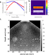
Fig. 4. High-resolution imaging of tissue-mimicking phantom using a 128-element flexible polymer array transducer.
a Measured transmit and receive transfer functions versus frequency. b Area uniformity of the peak transmit transfer at 8.2 MHz at the transducer surface. The color scale indicates the peak transmit transfer in Pa/V. c B-mode image captured with plane wave compounding. The gray scale indicates the intensity in dB.
Fig. 5
Fig. 5. Large-area flexible ultrasonic blood pressure sensor.
a Photograph of a large area of flexible ultrasonic blood pressure sensor while still on the support glass. See Fig. 1f for a photograph of the blood sensor placed in the neck. b Transmit transfer in Pa/V at the resonance frequency of 8.2 MHz of transmit elements, obtained using hydrophone measurements. c The pressure waveforms derived from the measured carotid phantom vessel diameters as a function of time. The diastolic pressure pd was taken from the reference blood pressure sensor, and the vessel stiffness α was fitted such that the obtained systolic peak pressure matched with the reference blood pressure. The red curve shows the blood pressure measured using a reference blood pressure meter. d Recorded in vivo ultrasound data of the carotid of a healthy volunteer of the optimally positioned array element.

References

    1. Cobbold, R. S. C. Foundations Of Biomedical Ultrasound (Oxford Univ. Press, 2007).
    1. Szabo, T. L. Diagnostic Ultrasound Imaging: Inside Out (Academic Press, 2014).
    1. Global Business Intelligence. Ultrasound systems market to 2019—Technological advancements, wide applications and device portability to drive future growth. https://www.gbiresearch.com/report-store/market-reports/medtech/ultrasou... growth (2013).
    1. Wang C, et al. Bioadhesive ultrasound for long-term continuous imaging of diverse organs. Science. 2022;377:517–523. doi: 10.1126/science.abo2542. - DOI - PubMed
    1. Omidvar A, Cretu E, Rohling R, Cresswell M, Hodgson AJ. Flexible polyCMUTs: fabrication and characterization of a flexible polymer-based capacitive micromachined ultrasonic array for conformal ultrasonography. Adv. Mater. Technol. 2023;8:2201316. doi: 10.1002/admt.202201316. - DOI