. 2024 Jun;98(6):1859-1875.
doi: 10.1007/s00204-024-03708-3. Epub 2024 Mar 30.

Effects of the nerve agent VX on hiPSC-derived motor neurons

Affiliations

Effects of the nerve agent VX on hiPSC-derived motor neurons

Catherine Schaefers et al. Arch Toxicol. 2024 Jun.

Abstract

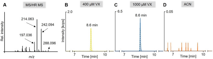
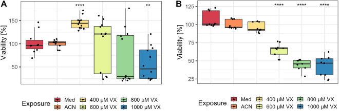
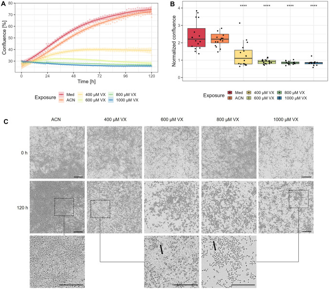
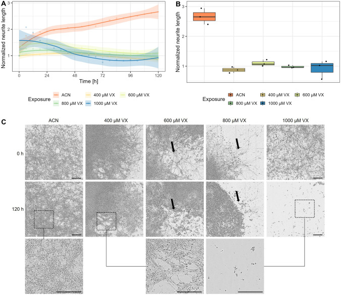
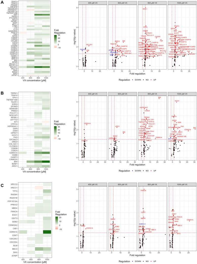
Poisoning with the organophosphorus nerve agent VX can be life-threatening due to limitations of the standard therapy with atropine and oximes. To date, the underlying pathomechanism of VX affecting the neuromuscular junction has not been fully elucidated structurally. Results of recent studies investigating the effects of VX were obtained from cells of animal origin or immortalized cell lines limiting their translation to humans. To overcome this limitation, motor neurons (MN) of this study were differentiated from in-house feeder- and integration-free-derived human-induced pluripotent stem cells (hiPSC) by application of standardized and antibiotic-free differentiation media with the aim to mimic human embryogenesis as closely as possible. For testing VX sensitivity, MN were initially exposed once to 400 µM, 600 µM, 800 µM, or 1000 µM VX and cultured for 5 days followed by analysis of changes in viability and neurite outgrowth as well as at the gene and protein level using µLC-ESI MS/HR MS, XTT, IncuCyte, qRT-PCR, and Western Blot. For the first time, VX was shown to trigger neuronal cell death and decline in neurite outgrowth in hiPSC-derived MN in a time- and concentration-dependent manner involving the activation of the intrinsic as well as the extrinsic pathway of apoptosis. Consistent with this, MN morphology and neurite network were altered time and concentration-dependently. Thus, MN represent a valuable tool for further investigation of the pathomechanism after VX exposure. These findings might set the course for the development of a promising human neuromuscular test model and patient-specific therapies in the future.

Keywords: Apoptosis; BCL2A1; CASP10; Nerve agent; Neurotoxicity; qRT-PCR.


Conflict of interest statement

The authors declare that they have no conflict of interest.

Figures

Fig. 1
Fig. 1
Mass spectrometric detection of VX-derived tyrosine adducts, Tyr(-EMP). The MS/HR MS spectrum of Tyr(-EMP) (m/z 288.096) (A) was extracted from the chromatographic peak of 1000 µM VX at 8.6 min (C). The most prominent structure confirming product ions are labelled. μLC-ESI MS/HR MS analysis of phosphonylated tyrosines Tyr(-EMP) derived from 400 µM VX (B), 1000 µM VX (C), and solvent control (ACN) (D) exposed MN after lysis and pronase-catalyzed proteolysis. Traces represent extracted ion chromatogram of the most intense product ion of Tyr(-EMP) (m/z 214.063 ± 0.005)
Fig. 2
Fig. 2
Changes in viability. After 5 days of exposure to 400 µM, 600 µM, 800 µM, 1000 µM VX, medium (Med) or solvent (ACN) control, the viability of mature neurons (A) and NPC (B) was tested using the XTT assay. The percentage of viable cells (n = triplicates per group from three independent experiments) and the significance of each group compared to corresponding solvent controls are illustrated. Data are represented as Tukey boxplots. **p < 0.01, ****p < 0.0001
Fig. 3
Fig. 3
Changes in confluence and morphology of MN. A After exposure to 400 µM, 600 µM, 800 µM, 1000 µM VX, medium or solvent (ACN) control, the percentage of confluence was determined by the area covered by MN using the IncuCyte® microscope every hour in a total period of 120 h. Results are illustrated as means and trend with 99% confidence interval (n = three independent experiments). B Confluence after 120 h of exposure to 400 µM, 600 µM, 800 µM, 1000 µM VX, medium (Med) or solvent (ACN) control normalized to confluence before exposure (0 h). The significance of each group compared to the corresponding solvent controls is presented (n = three independent experiments). Data are represented as Tukey box plots. ****p < 0.0001. C Representative images from MN before (0 h) and after (120 h) exposure to solvent control (ACN), 400 µM, 600 µM, 800 µM, or 1000 µM VX. For better illustration, images of MN after exposure to solvent control (ACN; left), 400 µM (middle) and 1000 µM (right) VX are additionally displayed with 2.6 × magnification. Black arrows indicate changes in cell-to-cell interactions. Scale bars, 200 µm
Fig. 4
Fig. 4
Changes in neurite network of MN. A The neurite length of MN, exposed to 400 µM, 600 µM, 800 µM, 1000 µM VX or solvent control (ACN), was determined every 6 h over a period of 120 h using the NeuroTrack software of the IncuCyte® microscope. Results are displayed as means ± SD and trend with 99% confidence interval (n = three biological replicates). B Illustration of changes in neurite length after 120 h of exposure to 400 µM, 600 µM, 800 µM, 1000 µM VX or solvent control (ACN) normalized to neurite length before exposure (0 h). Data are represented as Tukey box plots (n = three biological replicates). C Representative images of neurite network before (0 h) and after exposure (120 h) to solvent control (ACN), 400 µM, 600 µM, 800 µM, or 1000 µM VX. Black arrows indicate changes in neurite network. For better illustration, images of MN after exposure to solvent control (ACN; left), 400 µM (middle), or 1000 µM (right) VX with 2.6 × magnification are additionally displayed. Scale bars, 200 µm
Fig. 5
Fig. 5
Regulation of genes after VX exposure in MN. Genes associated with apoptosis (A), neurotoxicity (B) and DNA damage and repair (C) were tested 5 days after exposure to 400 µM, 600 µM, 800 µM or 1000 µM VX in comparison to solvent control (ACN) via qRT-PCR. Fold regulation of up- or downregulated genes (≥ 2.0 or ≤  − 2.0 in combination with p < 0.05) are shown as means in a heat map (left) and all genes in a volcano plot (right) (n = three independent experiments)
Fig. 6
Fig. 6
Changes on the protein level in MN. Upregulation of CASP10 in comparison to solvent control (ACN) was observed using Western Blot. Fold changes ± standard deviation and representative bands are shown (n = three independent experiments)

References

    1. Abou-Donia MB. Organophosphorus ester-induced chronic neurotoxicity. Arch Environ Health. 2003;58(8):484–497. doi: 10.3200/AEOH.58.8.484-497. - DOI - PubMed
    1. Benitez A, Liu W, Palovcak A, Wang G, Moon J, An K, Kim A, Zheng K, Zhang Y, Bai F, Mazin AV, Pei X-H, Yuan F, Zhang Y. FANCA promotes DNA double-strand break repair by catalyzing single-strand annealing and strand exchange. Mol Cell. 2018;71(4):621–628.e4. doi: 10.1016/j.molcel.2018.06.030. - DOI - PMC - PubMed
    1. Berridge MV, Herst PM, Tan AS. Tetrazolium dyes as tools in cell biology: New insights into their cellular reduction. Biotechnol Annu Rev. 2005;11:127–152. doi: 10.1016/S1387-2656(05)11004-7. - DOI - PubMed
    1. Bloch-Shilderman E, Rabinovitz I, Egoz I, Yacov G, Allon N, Nili U. Determining a threshold sub-acute dose leading to minimal physiological alterations following prolonged exposure to the nerve agent VX in rats. Arch Toxicol. 2018;92(2):873–892. doi: 10.1007/s00204-017-2108-5. - DOI - PubMed
    1. Bohush A, Bieganowski P, Filipek A. Hsp90 and Its Co-Chaperones in Neurodegenerative Diseases. Int J Mol Sci. 2019;20(20):4976. doi: 10.3390/ijms20204976. - DOI - PMC - PubMed

MeSH terms