. 2024 Mar 30;15(1):2788.
doi: 10.1038/s41467-024-47149-z.

Clinically used broad-spectrum antibiotics compromise inflammatory monocyte-dependent antibacterial defense in the lung

Affiliations

Clinically used broad-spectrum antibiotics compromise inflammatory monocyte-dependent antibacterial defense in the lung

Patrick J Dörner et al. Nat Commun. .

Abstract

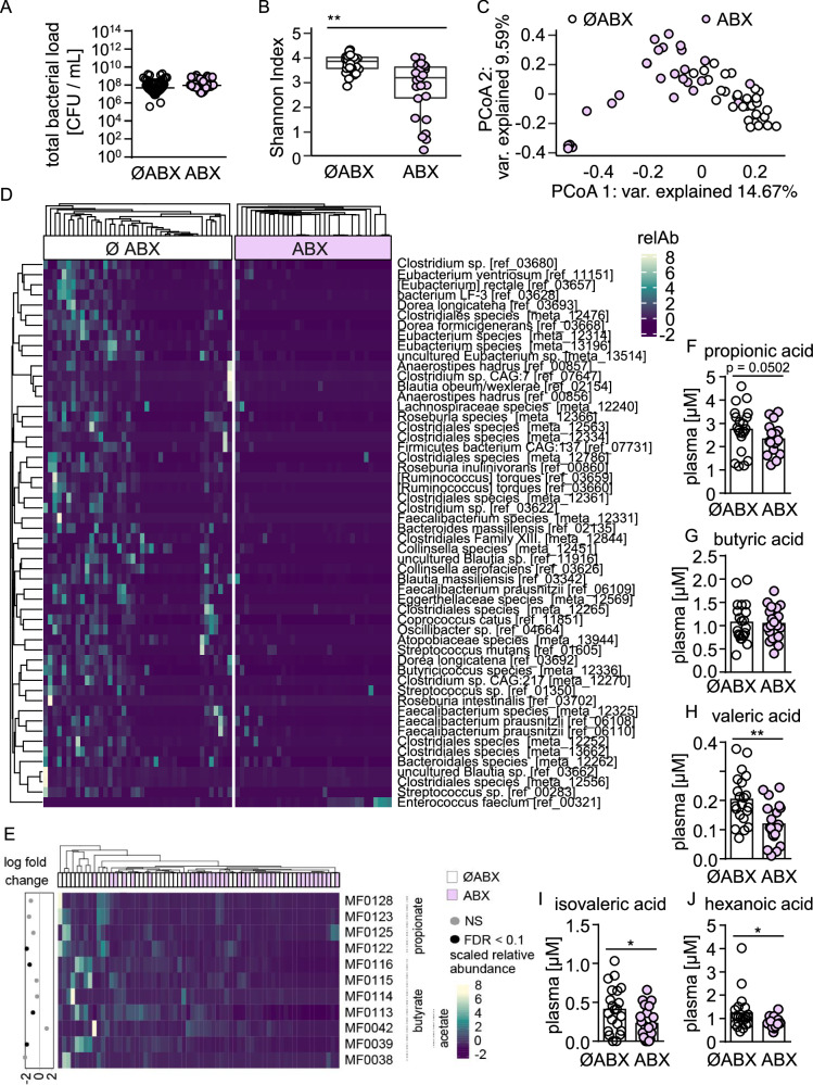
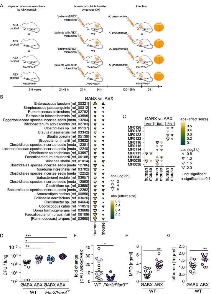
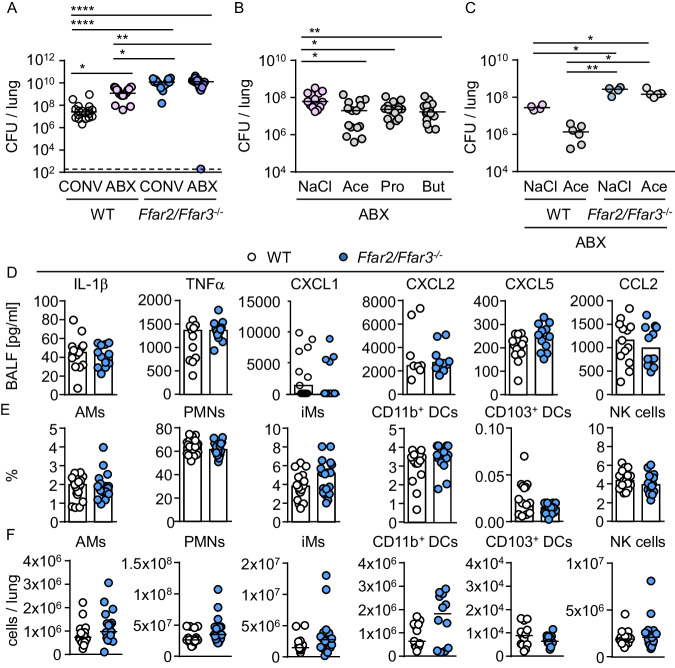
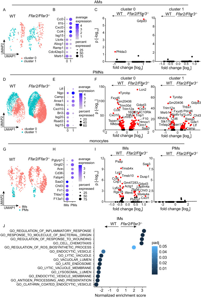
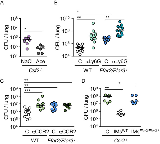
Hospital-acquired pneumonia (HAP) is associated with high mortality and costs, and frequently caused by multidrug-resistant (MDR) bacteria. Although prior antimicrobial therapy is a major risk factor for HAP, the underlying mechanism remains incompletely understood. Here, we demonstrate that antibiotic therapy in hospitalized patients is associated with decreased diversity of the gut microbiome and depletion of short-chain fatty acid (SCFA) producers. Infection experiments with mice transplanted with patient fecal material reveal that these antibiotic-induced microbiota perturbations impair pulmonary defense against MDR Klebsiella pneumoniae. This is dependent on inflammatory monocytes (IMs), whose fatty acid receptor (FFAR)2/3-controlled and phagolysosome-dependent antibacterial activity is compromized in mice transplanted with antibiotic-associated patient microbiota. Collectively, we characterize how clinically relevant antibiotics affect antimicrobial defense in the context of human microbiota, and reveal a critical impairment of IM´s antimicrobial activity. Our study provides additional arguments for the rational use of antibiotics and offers mechanistic insights for the development of novel prophylactic strategies to protect high-risk patients from HAP.


Conflict of interest statement

The authors declare no competing interests.

Figures

Fig. 1
Fig. 1. Antimicrobial therapy in patients is associated with reduced diversity of the gut microbiota and decreased abundance of SCFA producers.
A Total bacterial loads (CFU) in fecal samples of antibiotic-treated (ABX; n = 12) and untreated patients (ØABX; n = 17). BE Shotgun metagenomic sequencing was conducted on fecal samples of antibiotic-treated (ABX; n = 26) and untreated (ØABX; n = 29) patients. B α-diversity (Shannon index) and (C) PCoA of beta diversity were calculated on pairwise Bray-Curtis dissimilarities. D Heatmap indicating the scaled relative abundance of bacterial species altered in antibiotic-treated patients compared to non-antibiotic treated controls. E Abundances of gene modules associated with the production of SCFAs in fecal samples of antibiotic-treated and untreated patients. F–J SCFA concentrations in plasma samples of antibiotic-treated (ABX; n = 21) and untreated (ØABX; n = 21) patients. Mann-Whitney U test (two-tailed) was used for bacterial loads (A, B) and Wilcoxon-Mann-Whitney U test (two-tailed) was used for plasma SCFA concentrations (F–J) and values are shown as median with each dot representing the data from one patient. B The box plot represents median, 25th and 75th percentiles—interquartile range; IQR—and whiskers extend to maximum and minimum values *P < 0.05, **P < 0.01, ***P < 0.005.
Fig. 2
Fig. 2. Human microbiota alterations induced by antibiotics or FFAR2/FFAR3 deficiency compromise pulmonary antibacterial defense in mice.
A Experimental scheme of human microbiota transplantation and infection experiments for BE. Antibiotic-associated alterations in the relative abundance of bacterial species and functional modules in fecal samples of transplanted mice and patient samples, significantly altered species with similar directionality are shown (B), and variation in SCFA modules (C) (n = 26 for ØABX, n = 29 for ABX). D Bacterial loads (CFU) in lungs of human microbiota transplanted mice infected with K. pneumoniae for 24 h, and E fold changes in lung bacterial loads of mice transplanted with ABX-associated human microbiota vs. ABX-naïve human microbiota (n = 12 for ØABX WT; n = 12 for ABX WT; n = 11 for ØABX Ffar2/Ffar3-/-; n = 9 for ABX Ffar2/Ffar3-/-). F, G Myeloperoxidase and albumin levels in bronchoalveolar lavage fluid (BALF) of infected mice, transplanted with ABX-naïve human microbiota or ABX-associated human microbiota (n = 12 for ØABX WT; n = 12 for ABX WT). C FDR correction was done to account for multiple testing using the Benjamini-Hochberg method (two-sided). Kruskal-Wallis test followed by Dunn´s multiple comparison was applied to the bacterial load dataset (D). The fold change analysis (E) and myeloperoxidase (MPO) and albumin measurements (F–G) were analyzed by Mann-Whitney U test (two-tailed). Values are shown as median (D–G), each dot represents the data from a single mouse. The solid black border of triangles in C indicates q < 0.1 (FDR corrected in the human samples, two-sided), and x on the triangles indicates p < 0.05 (targeted testing only within the significant modules in human samples). *P < 0.05, **P < 0.01, ***P < 0.005. Downward facing triangle: increased in ØABX in comparison to ABX.
Fig. 3
Fig. 3. SCFA receptor deficiency does not influence pulmonary cytokine production and immune cell infiltration during K. pneumoniae infection.
A Conventionally colonized (CONV) or antibiotic (ABX)-treated WT and Ffar2/Ffar3-/- mice were infected with K. pneumoniae for 24 h and bacterial loads (CFU) in lungs were assessed (n = 18 for CONV WT; n = 18 for ABX WT; n = 17 for CONV Ffar2/Ffar3-/-; n = 17 for ABX Ffar2/Ffar3-/-). B WT mice treated with antibiotics (ABX) were additionally given SCFA or NaCl. Lung bacterial loads were assessed after 24 h (n = 13 for NaCl; n = 15 for Ace; n = 15 for Pro; n = 15 for But). C WT and Ffar2/Ffar3-/- mice treated with antibiotics (ABX) were additionally given acetate (200 mM, Ace) or NaCl, and lung bacterial loads were assessed (n = 4 for NaCl WT; n = 6 for Ace WT; n = 4 for NaCl Ffar2/Ffar3-/-; n = 5 for Ace Ffar2/Ffar3-/-). DF Conventionally housed mice were intranasally infected with K. pneumoniae for 12 h, cytokine levels in BALF were measured by multiplex ELISA (n = 12 for WT; n = 13 for Ffar2/Ffar3-/-) or AMs (alveolar macrophages), PMNs (pulmonary neutrophils), IMs (inflammatory monocytes), CD11b+ DCs (dendritic cells), CD103b+ DCs, and NK cells (natural killer cells) in lung tissue were analyzed by FACS (n = 19 for WT; n = 18 for Ffar2/Ffar3-/-). Kruskal-Wallis test followed by Dunn´s multiple comparison was applied to the bacterial load datasets (AC). Mann-Whitney U test (two-tailed) was applied for lung cell populations and cytokines (D–F). Values are shown as median, each dot represents the data from one mouse. *P < 0.05, **P < 0.01, ***P < 0.005.
Fig. 4
Fig. 4. FFAR2/FFAR3 deficiency affects gene expression in IMs (inflammatory monocytes) and PMNs (pulmonary neutrophils).
scRNAseq of lung cells from WT and Ffar2/Ffar3-/- mice infected with K. pneumoniae for 12 h (n = 4 for WT, n = 4 for Ffar2/Ffar3-/-). A, D, G UMAPs of AMs, PMNs, and monocytes subclusters. B, E, H Dot plots of the most differentially expressed genes for each cell subcluster. C, F, I Volcano plots of differentially expressed genes for each cell subclusters. J GSEA indicating significantly up- and downregulated pathways in IMs of infected WT and Ffar2/Ffar3-/- mice. Wilcoxon Rank Sum test was used and adjusted based on bonferroni correction using all genes in the dataset. J Bar plot indicating significantly up- and downregulated GSEA pathways in IMs of infected WT and Ffar2/Ffar3-/- mice. Calculation of p-value estimation is based on an adaptive multi-level split Monte-Carlo scheme followed by BH correction.
Fig. 5
Fig. 5. FFAR2/FFAR3 control antibacterial activity via IMs.
A Bacterial loads (CFU) in lung tissue of ABX-treated Csf2-/- mice receiving either NaCl or acetate (n = 8 for NaCl, n = 6 for Ace) 24 h after infection with K. pneumoniae. B Bacterial loads in WT and Ffar2/Ffar3-/- mice treated intraperitoneally with anti-Ly6G or control antibody (C) and infected for 24 h with K. pneumoniae (n = 14 for WT C, n = 13 for Ffar2/Ffar3-/- C, n = 13 for WT αLy6G; n = 14 for Ffar2/Ffar3-/- αLy6G). C Bacterial loads in WT and Ffar2/Ffar3-/- mice treated intraperitoneally with anti-CCR2 or control antibody (C) and infected for 24 h with K. pneumoniae (n = 12 for WT C, n = 11 for Ffar2/Ffar3-/- C, n = 12 for WT αCCR2; n = 11 for Ffar2/Ffar3-/- αCCR2). D) Bacterial loads in the lungs of Ccr2-/- mice transplanted intravenously with IM´s of WT or Ffar2/Ffar3-/- animals or treated with PBS, and infected with K. pneumoniae (n = 7 for PBS, n = 6 for WT; n = 6 for Ffar2/Ffar3-/-). Kruskal-Wallis test followed by Dunn´s multiple comparison was applied to the datasets. Values are shown as median, each dot represents the data from a single mouse. *P < 0.05, **P < 0.01, ***P < 0.005.
Fig. 6
Fig. 6. Antibiotic-induced microbiota alterations compromise FFAR2/FFAR3-controlled antibacterial activity of IMs (inflammatory monocytes).
A IMs of WT and Ffar2/Ffar3-/- mice were isolated from the bone marrow by FACS sorting, stimulated for 20 minutes with 1 mM acetate (Ace) or control medium prior to incubation with K. pneumoniae. WT IMs were also stimulated with 1 mM acetate in combination with 50 µM bafilomycin A1 (Baf), 0.75 µg cytochalasin D (Cyto), or 100 µg N,N´,N”-Triacetylchitotriose (Tri) prior to incubation with the bacteria. Values are shown as median, each dot represents the mean data from triplicates from cells of one mouse (n = 8 for K. pneumoniae without IMs; n = 12 for IMWT; n = 12 for IMWT + Ace; n = 9 for IMWT + Ace + Baf; n = 9 IMWT + Ace + Cyto; n = 9 for IMWT + Ace + Tri; n = 12 for IMFfar2/Ffar3-/-; n = 12 for IMFfar2/Ffar3-/- + Ace). Kruskal-Wallis test followed by Dunn´s multiple comparison was applied to the dataset. B WT and Ffar2/Ffar3-/- mice were transplanted with human microbiota from ABX-naïve or antibiotic-treated patients, IMs were isolated from the bone marrow by FACS sorting, incubated with K. pneumoniae, and CFUs were counted at the indicated time points. Values are shown as median, each dot represents the mean data from triplicates from a single mouse (n = 4 for K. pneumoniae without IMs; n = 6 for IMWT ØABX; n = 4 for IMWT ABX; n = 5 for IMFfar2/Ffar3-/- ØABX; n = 5 for IMFfar2/Ffar3-/- ABX). 2-way ANOVA test followed by Tukey´s multiple comparison between given time points and groups was applied to the datasets. *P < 0.05, **P < 0.01, ***P < 0.005.

References

    1. Torres, A. et al. International ERS/ESICM/ESCMID/ALAT guidelines for the management of hospital-acquired pneumonia and ventilator-associated pneumonia: Guidelines for the management of hospital-acquired pneumonia (HAP)/ventilator-associated pneumonia (VAP) of the European Respiratory Society (ERS), European Society of Intensive Care Medicine (ESICM), European Society of Clinical Microbiology and Infectious Diseases (ESCMID) and Asociacion Latinoamericana del Torax (ALAT). Eur. Respir. J. 5010.1183/13993003.00582-2017 (2017). - PubMed
    1. Cassini A, et al. Burden of six healthcare-associated infections on European population health: estimating incidence-based disability-adjusted life years through a population prevalence-based modelling study. PLoS Med. 2016;13:e1002150. doi: 10.1371/journal.pmed.1002150. - DOI - PMC - PubMed
    1. Melsen WG, et al. Attributable mortality of ventilator-associated pneumonia: a meta-analysis of individual patient data from randomised prevention studies. Lancet Infect. Dis. 2013;13:665–671. doi: 10.1016/S1473-3099(13)70081-1. - DOI - PubMed
    1. Kollef MH. Ventilator-associated pneumonia. A multivariate analysis. JAMA. 1993;270:1965–1970. doi: 10.1001/jama.1993.03510160083034. - DOI - PubMed
    1. Rello J, Ausina V, Ricart M, Castella J, Prats G. Impact of previous antimicrobial therapy on the etiology and outcome of ventilator-associated pneumonia. Chest. 1993;104:1230–1235. doi: 10.1378/chest.104.4.1230. - DOI - PubMed

Substances

LinkOut - more resources