

Influence of implant distribution on the biomechanical behaviors of mandibular implant-retained overdentures: a three-dimensional finite element analysis
- PMID: 38555452
- PMCID: PMC10981806
- DOI: 10.1186/s12903-024-04146-4
Influence of implant distribution on the biomechanical behaviors of mandibular implant-retained overdentures: a three-dimensional finite element analysis
Abstract
Objective: To assess stress distribution in peri-implant bone and attachments of mandibular overdentures retained by small diameter implants, and to explore the impact of implant distribution on denture stability.
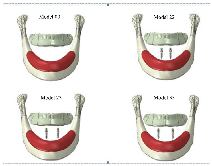
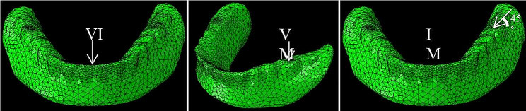
Methods: Through three-dimensional Finite Element Analysis (3D FEA), four models were established: three models of a two mandibular implants retained overdenture (IOD) and one model of a conventional complete denture (CD). The three IOD models consisted of one with two implants in the bilateral canine area, another with implants in the bilateral lateral incisor area, and the third with one implant in the canine area, and another in the lateral incisor area. Three types of loads were applied on the overdenture for each model: a 100 N vertical load and a inclined load on the left first molar, and a100N vertical load on the lower incisors. The stress distribution in the peri-implant bone, attachments, and the biomechanical behaviors of the overdentures were analyzed.
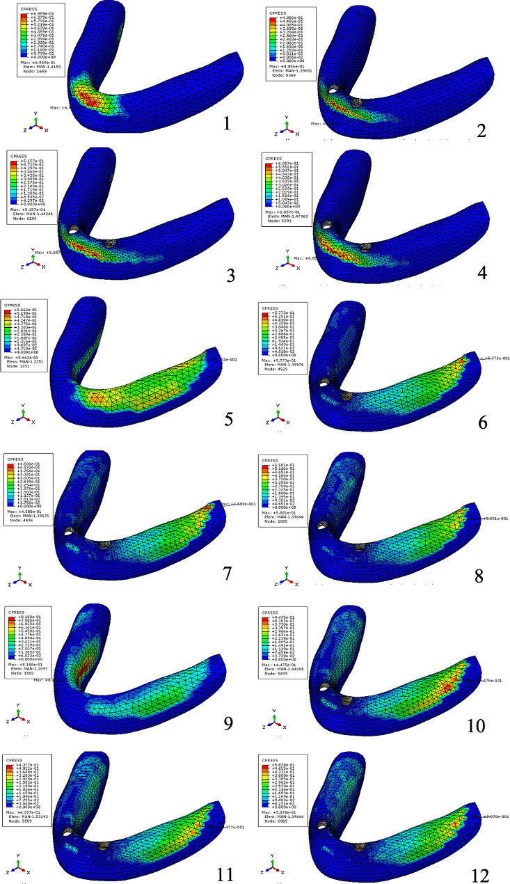
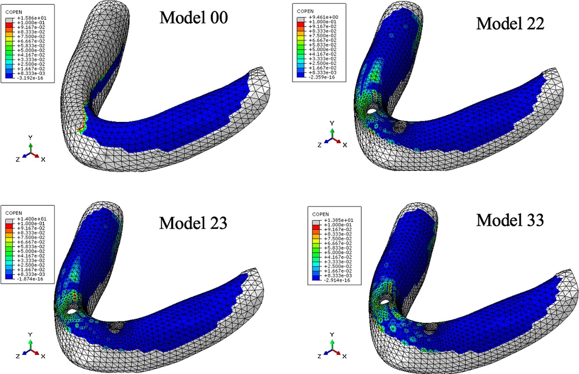
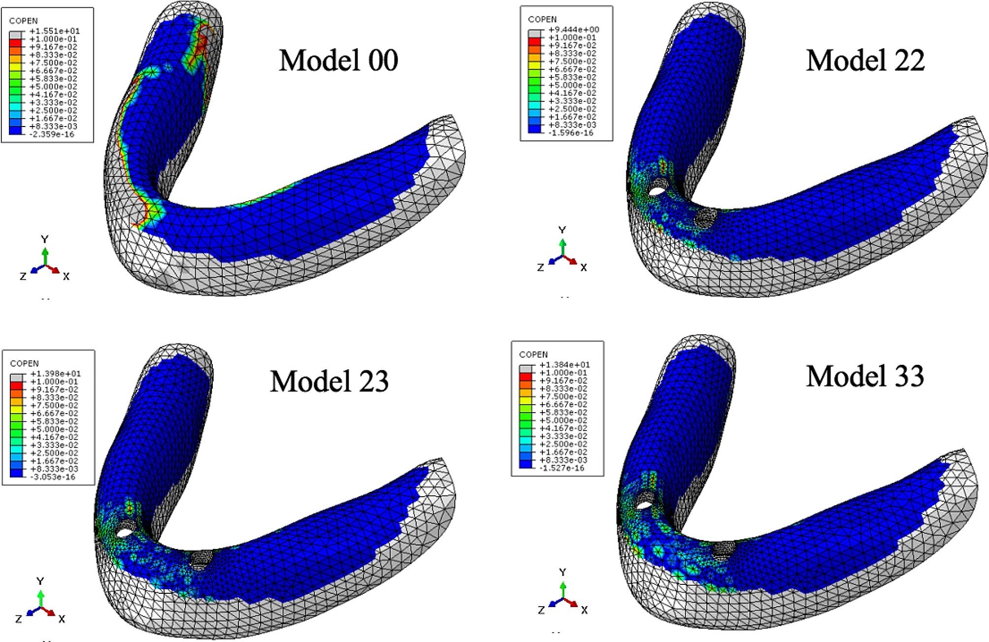
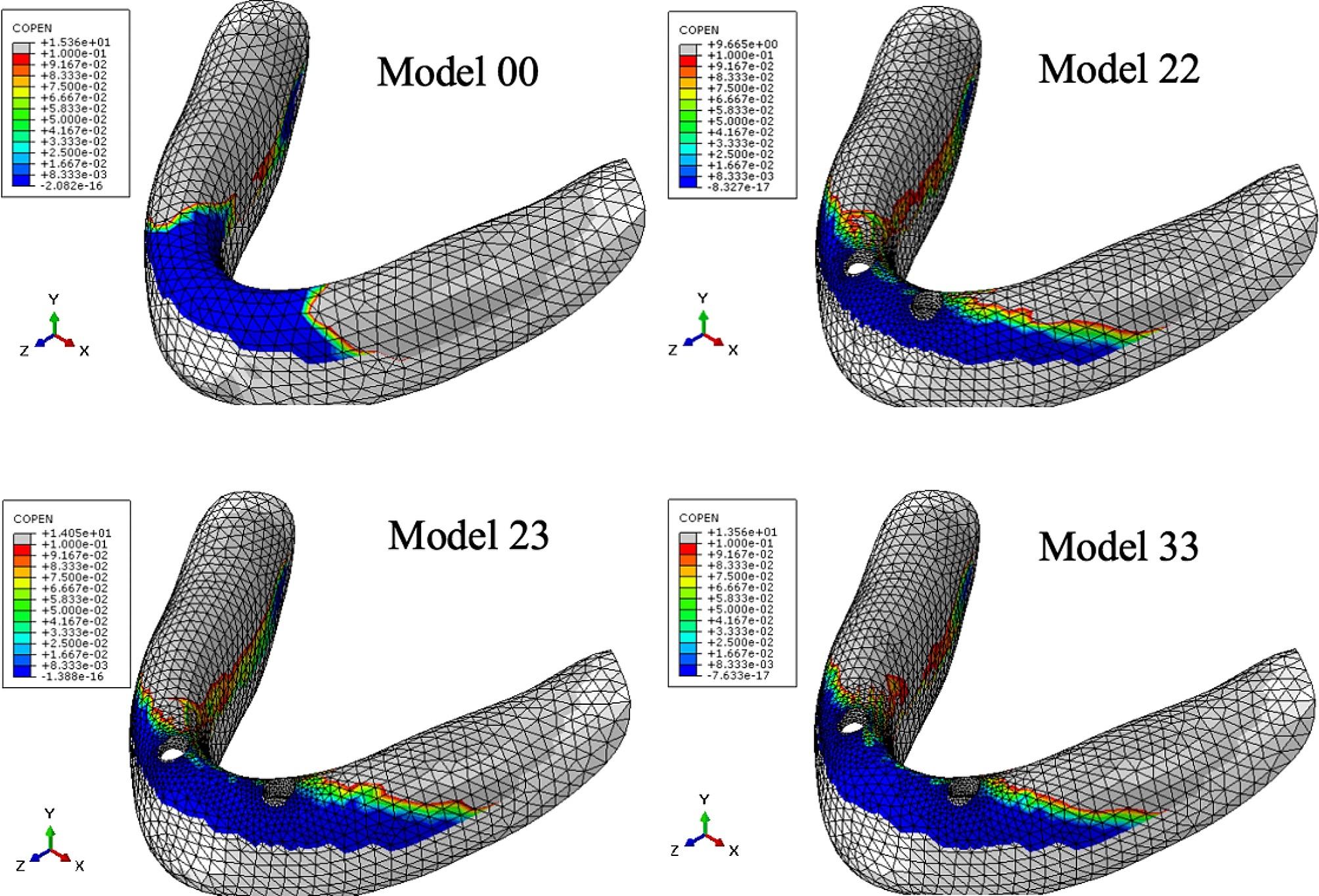
Results: Despite different distribution of implants, the maximum stress values in peri-implant bone remained within the physiological threshold for all models across three loading conditions. The dispersed implant distribution design (implant in the canine area) exhibited the highest maximum stress in peri-implant bone (822.8 µe) and the attachments (275 MPa) among the three IOD models. The CD model demonstrated highest peak pressure on mucosa under three loading conditions (0.8188 Mpa). The contact area between the denture and mucosa of the CD model was smaller than that in the IOD models under molar loading, yet it was larger in the CD model compared to the IOD model under anterior loading. However, the contact area between the denture and mucosa under anterior loading in all models was significantly smaller than those under molar loading. The IOD in all three models exhibited significantly less rotational movement than the complete denture. Different implant positions had minimal impact on the rotational movement of the IOD.
Conclusion: IOD with implants in canine area exhibited the highest maximum stress in the peri-implant bone and attachments, and demonstrated increased rotational movement. The maximum principal stress was concentrated around the neck of the small diameter one-piece implant, rather than in the abutment. An overdenture retained by two implants showed better stability than a complete denture.
Keywords: Distribution of implants; Implant overdenture; Three dimensional finite element analysis.
© 2024. The Author(s).
Conflict of interest statement
The authors declare no conflict of interest.
Figures
References
-
- Matthys C, Vervaeke S, Besseler J, De Bruyn H. Five-year study of mandibular overdentures on stud abutments: clinical outcome, patient satisfaction and prosthetic maintenance-influence of bone resorption and implant position. Clin Oral Implants Res. 2019;30(9):940–51. doi: 10.1111/clr.13501. - DOI - PubMed
-
- Schuster AJ, Possebon APDR, Schinestsck AR, Chagas-Júnior OL, Faot F. Effect of mandibular bone atrophy on maxillary and mandibular bone remodeling and quality of life with an implant-retained mandibular overdenture after 3 years. J Prosthet Dent. 2023;130(2):220–8. doi: 10.1016/j.prosdent.2021.08.019. - DOI - PubMed
-
- Stacchi C, Troiano G, Montaruli G, et al. Changes in implant stability using different site preparation techniques: osseodensification drills versus piezoelectric surgery. A multi-center prospective randomized controlled clinical trial. Clin Implant Dent Relat Res. 2023;25(1):133–40. doi: 10.1111/cid.13140. - DOI - PMC - PubMed
MeSH terms
Substances
Grants and funding
- No.TJWJ2021QN069/Tianjin Health Research Project
- No.KF2020120104/Open Funding of Guangdong Provincial Key Laboratory of Stomatology
- No.KF2020120104/Open Funding of Guangdong Provincial Key Laboratory of Stomatology
- No. 7222228/Beijing Natural Science Foundation
- Grant number: PKUSS-2023CRF501/Clinical Research Foundation of Peking University School and Hospital of Stomatology
LinkOut - more resources
Full Text Sources
