. 2024:156:51-119.
doi: 10.1016/bs.ctdb.2024.01.007. Epub 2024 Mar 15.

RNA binding proteins in cardiovascular development and disease

Affiliations

RNA binding proteins in cardiovascular development and disease

Sunil K Verma et al. Curr Top Dev Biol. 2024.

Abstract

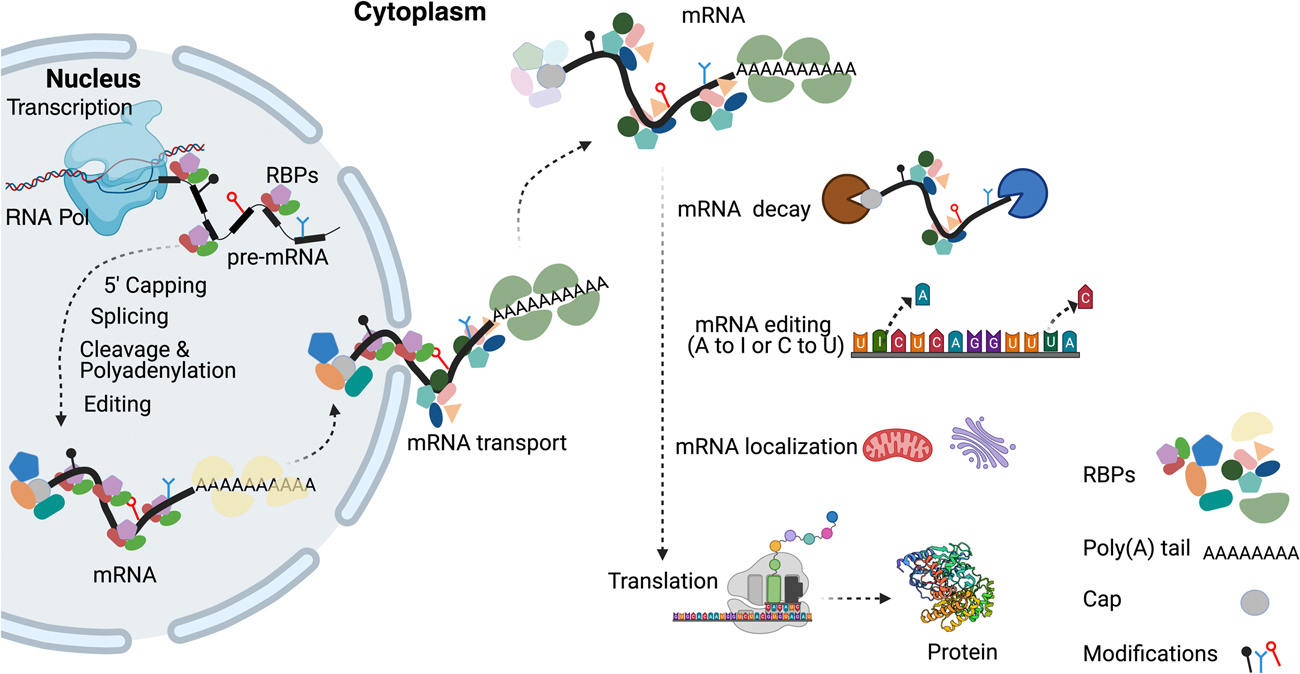
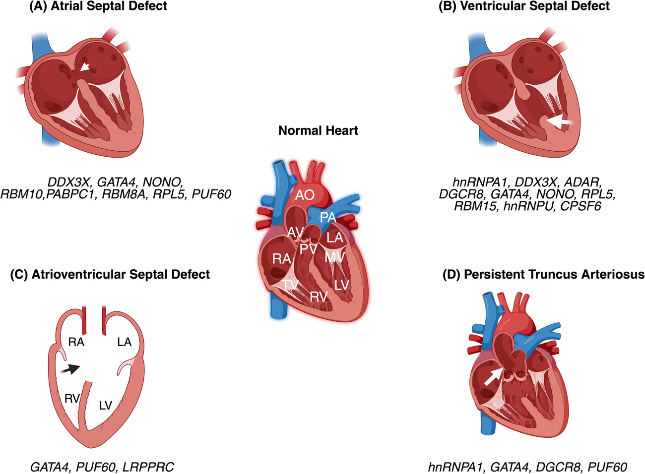
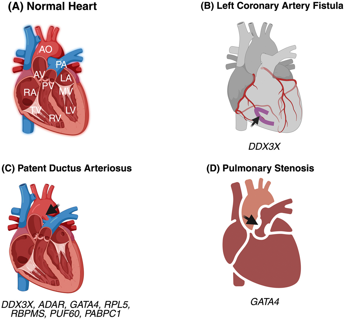
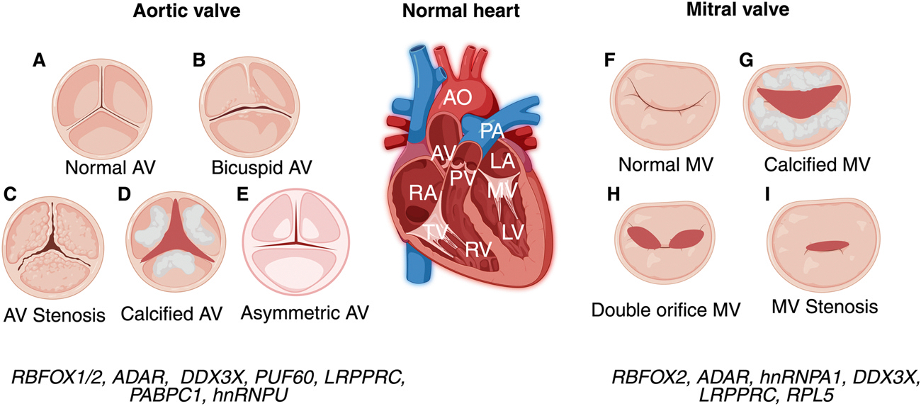
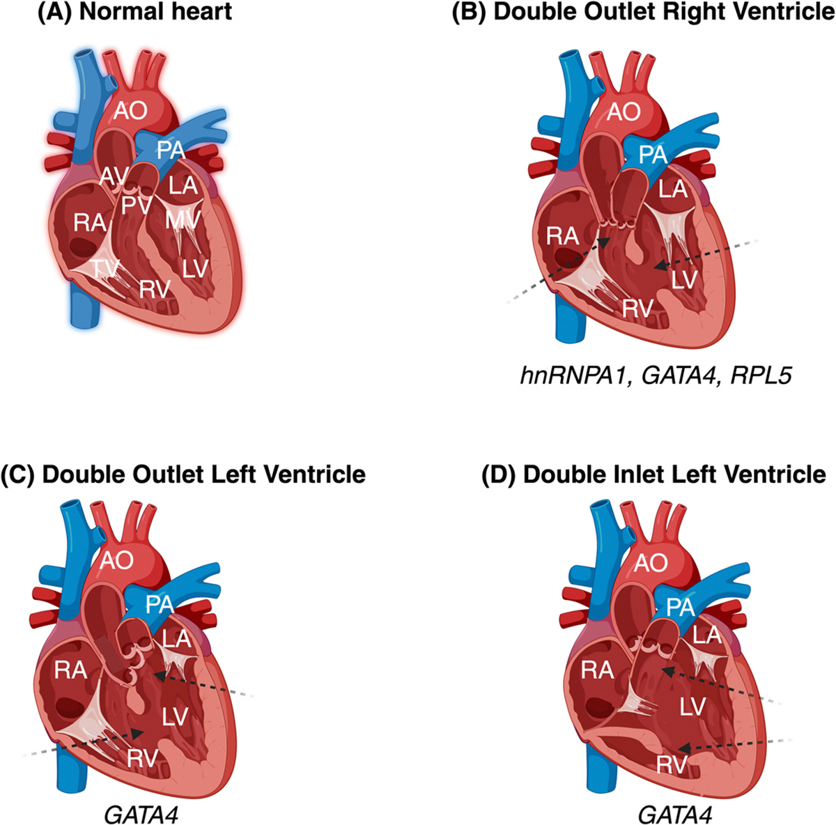
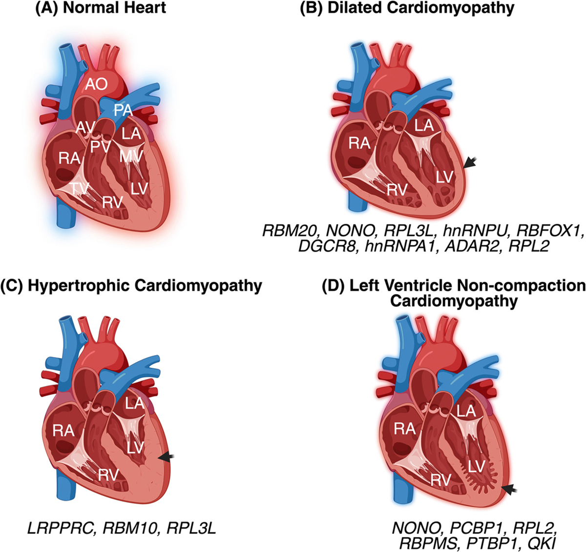
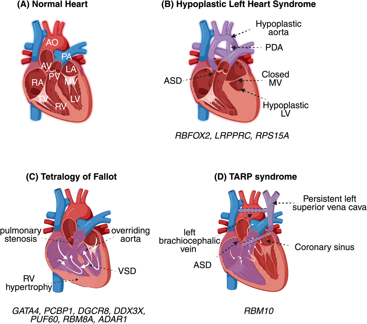
Congenital heart disease (CHD) is the most common birth defect affecting>1.35 million newborn babies worldwide. CHD can lead to prenatal, neonatal, postnatal lethality or life-long cardiac complications. RNA binding protein (RBP) mutations or variants are emerging as contributors to CHDs. RBPs are wizards of gene regulation and are major contributors to mRNA and protein landscape. However, not much is known about RBPs in the developing heart and their contributions to CHD. In this chapter, we will discuss our current knowledge about specific RBPs implicated in CHDs. We are in an exciting era to study RBPs using the currently available and highly successful RNA-based therapies and methodologies. Understanding how RBPs shape the developing heart will unveil their contributions to CHD. Identifying their target RNAs in the embryonic heart will ultimately lead to RNA-based treatments for congenital heart disease.

Keywords: Alternative polyadenylation; Alternative splicing; Congenital heart defects; Heart development; RNA binding protein; mRNA.


Conflict of interest statement

Declaration of interests

Authors declare no conflict of interest.

Figures

Fig. 1
Fig. 1. Schematic representation of mRNA life cycle mediated by RNA binding proteins.
RNA binding proteins (RBPs) bind pre-mRNAs in the nucleus and regulate editing, capping at the 5′ end, splicing and cleavage and polyadenylation of pre-mRNAs at the 3′ end. mRNA is then exported into the cytoplasm by specific RBPs. In the cytoplasm, a different set of cytoplasmic RBPs bind mRNAs to control mRNA decay, localization, editing and translation. Figures are generated using Biorender.
Fig. 2
Fig. 2. Septation defects with RBP involvement:
(A) Cross-sectional view of a normal adult heart representing all four chambers and valves. (B) Atrial septal defect (ASD) marked by white arrow indicating incomplete septation between the left and right atrium. (C) Ventricular septal defect (VSD) marked by white arrow indicating incomplete septation between the left and right ventricle. (D) Graphical representation of atrioventricular septal defect (AVSD) with incomplete septation between atrium and ventricle with a single valve. (E) Persistent truncus arteriosus (PTA) marked by white arrow indicating incomplete septation between the aorta and pulmonary artery. RBPs implicated in these defects are labeled below each picture.
Fig. 3
Fig. 3. Congenital heart defects affecting blood flow to/from the heart with RBP involvement.
(A) Cross- sectional view of a normal adult heart representing all four chambers, valves, and major blood vessels in the heart. (B) Left coronary artery fistula (in purple) marked by a black arrow indicates an abnormal connection of the left coronary artery. (C) Patent ductus arteriosus (PDA) is marked by a black arrow showing residual ductus arteriosus. (D) Pulmonary stenosis marked with a black arrow depicting narrowing of the pulmonary artery affecting blood flow. RBPs implicated in these defects are labeled below each picture.
Fig. 4
Fig. 4. Graphical representation of valve defects with RBP involvement:
Representative view of a normal aortic heart valve with three flaps (A) and mitral valve with two flaps (F). (B) Aortic valve with only two flaps. View of a constricted aortic valve (C) and mitral valve (I) displaying incomplete opening as seen in patients with valve stenosis. View of an aortic valve (D) and mitral valve (G) with calcified flap and partial opening. (E) Aortic valve view with asymmetric flaps. (H) View of a mitral valve with double opening. RBPs implicated in these CHDs are labeled below each picture. RA, right atrium; LA, left atrium; RV, right ventricle; LV, left ventricle; AV, aortic valve, MV, mitral valve, TV, tricuspid valve, PA, pulmonary artery, PV, pulmonary valve, Ao, aorta.
Fig. 5
Fig. 5. Other CHDs with RBP involvement:
(A) Cross-sectional view of a normal adult heart representing cardiac chambers, valves, and major blood vessels. (B) Cross-sectional view of double outlet right ventricle (DORV) adult heart showing opening of both aorta and pulmonary artery into the right ventricle. (C) Cross- sectional view of double outlet left ventricle (DOLV) adult heart showing opening of both aorta and pulmonary artery into the left ventricle. (D) Cross-sectional view of double inlet left ventricle (DILV) adult heart representing opening of both tricuspid and mitral valve into left ventricle with a severely hypertrophic right ventricle. RBPs implicated in these CHDs are labeled below each picture. RA: right atrium, LA: left atrium, RV: right ventricle, LV: left ventricle, AV: aortic valve, MV: mitral valve, TV: tricuspid valve, PA: pulmonary artery, PV: pulmonary valve, Ao: aorta.
Fig. 6
Fig. 6. Cardiomyopathies with RBP involvement.
(A) Cross-sectional view of a normal adult heart. (B) Graphical illustration of dilated cardiomyopathy with enlarged heart and thin ventricular wall (black arrow). (C) Hypertrophic cardiomyopathy represented as thickening of left ventricular wall and hypertrophic left ventricle. (D) Non-compaction cardiomyopathy (left ventricle or right ventricle). Left ventricle non compaction cardiomyopathy is represented as increased endocardial trabeculation and thin left ventricular wall (black arrow). RBPs implicated in these CHDs are labeled below each picture. RA, right atrium; LA, left atrium; RV, right ventricle; LV, left ventricle, AV, aortic valve, MV, mitral valve, TV, tricuspid valve, PA, pulmonary artery, PV, pulmonary valve, Ao, aorta.
Fig. 7
Fig. 7. Complex congenital heart diseases and syndromes with RBP involvement.
(A) Cross-sectional view of a normal adult heart representing all four chambers, valves, and major blood vessels. (B) A graphical representation of HLHS heart portraying the cardiac defects seen in human patients that includes atrial septal defect (ASD), hypoplastic aorta, patent ductus arteriosus (PDA), closed mitral valve (MV) and severely hypoplastic left ventricle (LV). (C) Graphical representation of heart cross-section displaying overriding aorta, stenosis of pulmonary artery, incomplete septation between right and left ventricle with right ventricle hypertrophy represented in Tetralogy of Fallot (TOF). (D) Representative illustration of TARP syndrome heart with persistent left superior vena cava, left brachiocephalic vain, coronary sinus and incomplete septation between left and right atrium. RBPs implicated in these CHDs are labeled below each picture. RA, right atrium, LA, left atrium, RV, right ventricle, LV, left ventricle, AV, aortic valve, MV, mitral valve, TV, tricuspid valve, PA, pulmonary artery, PV, pulmonary valve, Ao, aorta.

Similar articles

Cited by

References

    1. Abdin D, Rump A, Tzschach A, Sarnow K, Schrock E, Hackmann K, & Di Donato N (2019). European Journal of Medical Genetics, 62, 103587. - PubMed
    1. Akerberg AA, Burns CE, & Burns CG (2019). Pediatric Cardiology, 40, 1410–1418. - PMC - PubMed
    1. Akerberg AA, Trembley M, Butty V, Schwertner A, Zhao L, Beerens M, … Burns CG (2022). Circulation Research, 131, 980–1000. - PMC - PubMed
    1. Albers CA, Paul DS, Schulze H, Freson K, Stephens JC, Smethurst PA, … Ghevaert C (2012). Nature Genetics, 44, 435–439. S1–2. - PMC - PubMed
    1. Altaf F, Vesely C, Sheikh AM, Munir R, Shah STA, & Tariq A (2019). PLoS One, 14, e0200968. - PMC - PubMed

Publication types

LinkOut - more resources