. 2024 Mar 31;14(1):7615.
doi: 10.1038/s41598-024-57551-8.

Gene targeting in adult organs using in vivo cleavable donor plasmids for CRISPR-Cas9 and CRISPR-Cas12a

Affiliations

Gene targeting in adult organs using in vivo cleavable donor plasmids for CRISPR-Cas9 and CRISPR-Cas12a

Riki Ishibashi et al. Sci Rep. .

Abstract

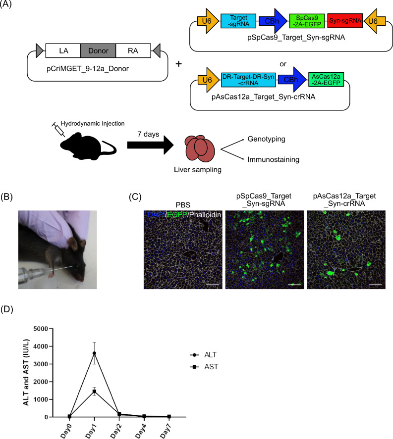
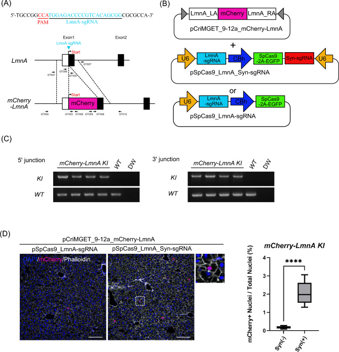
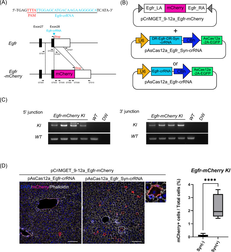
The CRISPR-Cas system for in vivo genome editing is a powerful tool for gene therapy against several diseases. We have previously developed the pCriMGET_9-12a system, an in vivo cleavable donor plasmid for precise targeted knock-in of exogenous DNA by both Cas9 and Cas12a. Here, we show that the pCriMGET_9-12a system can be applied for in vivo in-frame knock-in of exogenous DNA in adult mouse liver by hydrodynamic delivery of the targeting plasmids. The in vivo cleavable pCriMGET_9-12a donor plasmids significantly increased the knock-in efficiency of both CRISPR-Cas9 and CRISPR-Cas12a in the adult mouse liver compared to uncleavable donor plasmids. This strategy also achieved in-frame reporter gene knock-in without indel mutations. Therefore, in vivo gene targeting using the pCriMGET_9-12a system may contribute to the establishment of safer, more precise, versatile and efficient gene therapy methods in adult organs.


Conflict of interest statement

The authors declare no competing interests.

Figures

Figure 1
Figure 1
Hydrodynamic based retro-orbital sinus injection with the pCriMGET_9-12a system. (A) Schematic of the combination with hydrodynamic retro-obital sinus injection and the pCriMGET_9-12a system. pCriMGET_9-12a_donor and pSpCas9_Target_Syn-sgRNA, expressed as SpCas9-2A-EGFP and target-sgRNA and Syn-crRNA-TS-sgRNA, or pAsCas12a_Target_Syn-crRNA, expressed as AsCas12a-2A-EGFP and target-crRNA and Syn-crRNA-TS-crRNA, are injected from the retro-orbital sinus. The liver was corrected for genotyping and immunostaining on day 7 after injection. (B) Hydrodynamic retro-orbital sinus injection technique. (C) Representative immunofluorescence images of mouse liver sections injected with PBS, pSpCas9_Target_syn-sgRNA or pAsCas12a_Target_Syn-crRNA on one day after injection. EGFP (green), phalloidin (white) and DAPI (blue). Scale bar, 20 μm. The experiment was repeated in 4 individual mice. (D) Serum levels of ALT and AST after retro-orbital sinus injection of plasmid DNA. Day 0 as uninjected control. The experiment was repeated in 3 individual mice at each time point.
Figure 2
Figure 2
Precise in-frame knock-in of mCherry-LmnA using the pCriMGET_9-12a/CRISPR-Cas9 systems. (A) Targeting map of mCherry-LmnA. The LmnA sgRNA target site (blue arrow head) and sequence (blue letters), and the CRISPR-Cas9 PAM sequence (red letters) are shown. (B) Use of plasmids for mCherry-LmnA gene targeting. (C) Genotyping PCR for mCherry-LmnA knock-in liver. PCR for WT band detection was performed using LmnA-mCherry_GT001 and _GT002 primers for 5′ junction loci and LmnA-mCherry_GT006 and _GT007 primers for 3′ junction loci. PCR for KI band detection was performed using LmnA-mCherry_GT004 and _GT005 primers for 1st PCR and _GT001 and _GT003 primers for nested PCR at 5′ junction loci, and LmnA-mCherry_GT009 and _GT0010 primers for 1st PCR and _GT007 and _GT008 primers for nested PCR at 3′ junction loci (see (A)). (D) Representative immunofluorescence images of liver tissue injected with pCriMGET_9-12a_mCherry-LmnA and pSpCas9_LmnA-sgRNA or pSpCas9_LmnA_Syn-sgRNA plasmids. DAPI (blue), mCherry (magenta) and phalloidin (white). Scale bar, 20 μm. Enlarged image is shown as dashed box area. Knock-in ratio was calculated as the percentage of mCherry + nuclei among total nuclei (n > 13,000 nuclei from four individual slides). Mean ± s.d. from four individual slides. ****P < 0.001, by two-tailed Student's t-test.
Figure 3
Figure 3
Bicistronic H2B-mCherry reporter cassette targeting the albumin 3′ UTR locus by the pCriMGET_9-12a/CRISPR-Cas12a system. (A) Targeting map of Albumin-H2BmCherry. Alb-crRNA target site (blue arrow head) and sequence (blue letters), and CRISPR-Cas12a PAM sequence (red letters) are shown. (B) Use of plasmids for Albumin-H2BmCherry gene targeting. (C) Genotyping PCR for Alb-H2BmCherry knock-in liver. PCR for WT band detection was performed using Alb-H2BmCherry_GT001 and _GT002 primers for 5′ junction loci and Alb-H2BmCherry _GT006 and _GT007 primers for 3′ junction loci. PCR for KI band detection was performed using Alb-H2BmCherry_GT004 and _GT005 primers for 1st PCR and _GT001 and _GT003 primers for nested PCR at 5′ junction loci, and Alb-H2BmCherry _GT009 and _GT0010 primers for 1st PCR and _GT007 and _GT008 primers for nested PCR at 3′ junction loci (see (A)). (D) Representative immunofluorescence images of liver tissue injected with pCriMGET_9-12a_ Alb-H2BmCherry and pAsCas12a_Alb-crRNA or pAsCas12a_Alb_Syn-crRNA plasmids. DAPI (blue), mCherry (magenta) and phalloidin (white). Scale bar, 20 μm. Enlarged image is shown as dashed box area. Knock-in ratio was calculated as the percentage of mCherry + nuclei among total nuclei (n > 12,000 nuclei from four individual slides). Mean ± s.d. from four individual slides. ****P < 0.001, by two-tailed Student's t-test.
Figure 4
Figure 4
Precise in-frame knock-in of Egfr-mCherry by the pCriMGET_9-12a/CRISPR-Cas12a systems. (A) Targeting map of Egfr-mCherry. Egfr-crRNA target site (arrowhead) and sequence (blue letters), and CRISPR-Cas12a PAM sequence (red letters) are shown. (B) Use of plasmids for Egfr-mCherry gene targeting. (C) Genotyping PCR for Egfr-mCherry knock-in liver. PCR for WT band detection was performed using Egfr-mCherry _GT001 and _GT002 primers for 5′ junction loci and Egfr-mCherry _GT006 and _GT007 primers for 3′ junction loci. PCR for KI band detection was performed using Egfr-mCherry _GT004 and _GT005 primers for 1st PCR and _GT001 and _GT003 primers for nested PCR at 5′ junction loci, and Egfr-mCherry _GT009 and _GT0010 primers for 1st PCR and _GT007 and _GT008 primers for nested PCR at 3′ junction loci (see (A)) Asterisk indicates non-specific amplicon. (D) Representative immunofluorescence images of liver tissue injected with pCriMGET_9-12a_Egfr-mCherry and pAsCas12a_Egfr-crRNA or pAsCas12a_Egfr_Syn-crRNA plasmids. DAPI (blue), mCherry (magenta) and phalloidin (white). Scale bar, 20 μm. Enlarged image is shown as dashed box area. Knock-in ratio was calculated as the percentage of mCherry + cells among total cells (n > 13,000 cells from four individual slides). Mean ± s.d. from four individual slides. ****P < 0.001, by two-tailed Student's t-test.

References

    1. Raguram A, et al. Therapeutic in vivo delivery of gene editing agents. Cell. 2022;185(15):2806–2827. doi: 10.1016/j.cell.2022.03.045. - DOI - PMC - PubMed
    1. Taha EA, et al. Delivery of CRISPR-Cas tools for in vivo genome editing therapy: Trends and challenges. J. Control. Release. 2022;342:345–361. doi: 10.1016/j.jconrel.2022.01.013. - DOI - PubMed
    1. Li T, Yang Y, Qi H, et al. CRISPR/Cas9 therapeutics: Progress and prospects. Sig. Transduct Target Ther. 2023;8:36. doi: 10.1038/s41392-023-01309-7. - DOI - PMC - PubMed
    1. Bhavesh DK, et al. Dlivery of gene editing thrapeutics. Nanomedicine. 2023;54:102711. doi: 10.1016/j.nano.2023.102711. - DOI - PMC - PubMed
    1. Adli M. The CRISPR tool kit for genome editing and beyond. Nat. Commun. 2018;9:1911. doi: 10.1038/s41467-018-04252-2. - DOI - PMC - PubMed