. 2024 Apr;56(4):946-958.
doi: 10.1038/s12276-024-01203-4. Epub 2024 Apr 1.

Ceramide kinase-mediated C1P metabolism attenuates acute liver injury by inhibiting the interaction between KEAP1 and NRF2

Affiliations

Ceramide kinase-mediated C1P metabolism attenuates acute liver injury by inhibiting the interaction between KEAP1 and NRF2

Wei Dong et al. Exp Mol Med. 2024 Apr.

Abstract

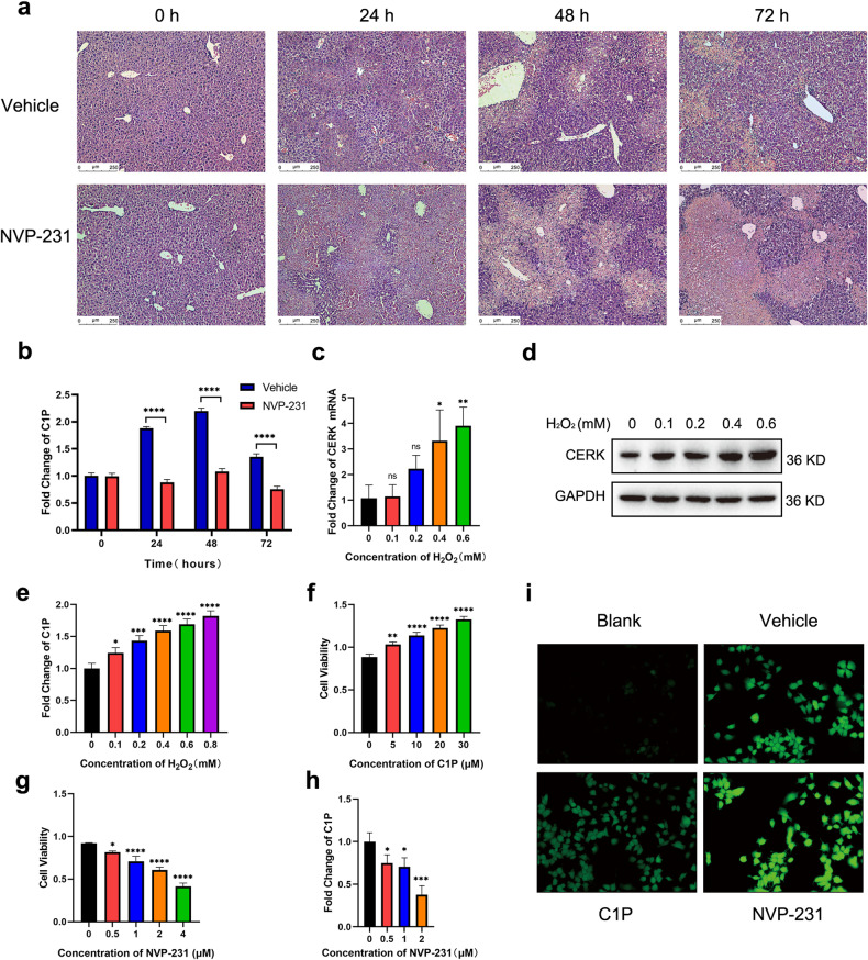
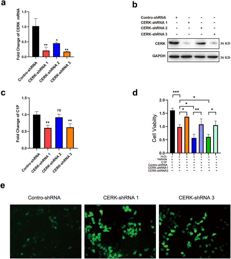
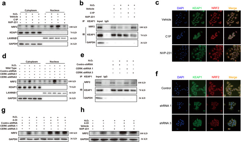
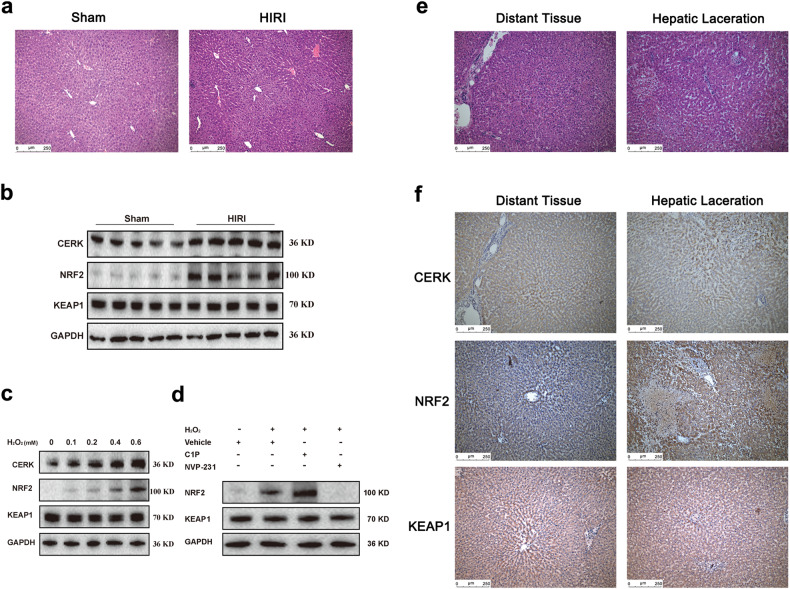
Acute liver injury is the basis of the pathogenesis of diverse liver diseases. However, the mechanism underlying liver injury is complex and not completely understood. In our study, we revealed that CERK, which phosphorylates ceramide to produce ceramide-1-phosphate (C1P), was the sphingolipid pathway-related protein that had the most significantly upregulated expression during acute liver injury. A functional study confirmed that CERK and C1P attenuate hepatic injury both in vitro and in vivo through antioxidant effects. Mechanistic studies have shown that CERK and C1P positively regulate the protein expression of NRF2, which is a crucial protein that helps maintain redox homeostasis. Furthermore, our results indicated that C1P disrupted the interaction between NRF2 and KEAP1 by competitively binding to KEAP1, which allowed for the nuclear translocation of NRF2. In addition, pull-down assays and molecular docking analyses revealed that C1P binds to the DGR domain of KEAP1, which allows it to maintain its interaction with NRF2. Importantly, these findings were verified in human primary hepatocytes and a mouse model of hepatic ischemia‒reperfusion injury. Taken together, our findings demonstrated that CERK-mediated C1P metabolism attenuates acute liver injury via the binding of C1P to the DGR domain of KEAP1 and subsequently the release and nuclear translocation of NRF2, which activates the transcription of cytoprotective and antioxidant genes. Our study suggested that the upregulation of CERK and C1P expression may serve as a potential antioxidant strategy to alleviate acute liver injury.


Conflict of interest statement

The authors declare no competing interests.

Figures

Fig. 1
Fig. 1. High expression of CERK is involved in CCl4-induced acute liver injury.
a Representative images of liver injury sites stained with hematoxylin and eosin (H&E) are shown. Scale bar: 250 μm. b qRT‒PCR analysis of the seven top genes involved in sphingolipid metabolism (fold change>6) in the validation groups was performed. (n = 5/group). c qRT‒PCR analysis of CERK expression was performed in acute liver injury time course experiments. (n = 5/group). d The protein expression of CERK in acute liver injury time course experiments was determined by western blotting. e Representative images of IHC staining for CERK in tissues at different time points after acute liver injury are shown. The arrow indicates severely injured hepatocytes, and the asterisk indicates mildly injured hepatocytes. Scale bar: 500 μm (top), 100 μm (bottom). One-way ANOVA was used to analyze the significant differences. **P< 0.01, ***P< 0.001, and ****P < 0.0001.
Fig. 2
Fig. 2. CERK inhibition exacerbates liver injury in vivo and in vitro.
a Representative images of liver injury sites stained with hematoxylin and eosin (H&E) are shown. Scale bar: 250 μm. b The relative concentration of total C1P in acute liver injury time course experiments induced by CERK inhibition was measured via ELISA (n = 5/group). c The mRNA expression of CERK in AML12 cells with H2O2-induced oxidative damage was measured by qRT‒PCR (n = 3/group). d The protein expression of CERK in AML12 cells with H2O2-induced oxidative damage was measured via WB. e The relative concentration of total C1P in AML12 cells with H2O2-induced oxidative damage was measured via ELISA (n = 3/group). f The viability of AML12 cells treated with C1P for 24 h and exposed to H2O2 (0.4 mM) was determined (n = 3/group). g The viability of AML12 cells treated with NVP-231 for 24 h and exposed to H2O2 (0.4 mM) was determined (n = 3/group). h The relative concentrations of total C1P in AML12 cells treated with NVP-231 for 24 h were measured via ELISA following exposure to H2O2 (n = 3/group). i The intracellular ROS level was measured by an ROS assay kit. H2O2 was added to 20 μM C1P and 2 μM NVP-231 for 24 h, while the same volume of vehicle was added to the vehicle group. b Two-way ANOVA; c, eh one-way ANOVA. *P < 0.05, **P < 0.01, ***P < 0.001, ****P < 0.0001, and ns not significant.
Fig. 3
Fig. 3. CERK knockdown exacerbated acute liver injury in vitro.
a The mRNA expression of CERK in AML12 cells with stable CERK knockdown was measured by qRT‒PCR. b The protein expression of CERK in AML12 cells with stable CERK knockdown was measured by western blotting. c The relative concentrations of total C1P in AML12 cells with stable CERK knockdown were measured via ELISA. d The viability of stable CERK-knockdown AML12 cells treated with C1P. e The level of intracellular ROS in AML12 cells with stable CERK knockdown was measured via an ROS assay kit. a, c Student’s t test; d one-way ANOVA. *P < 0.05, **P < 0.01, ***P < 0.001, and ns not significant.
Fig. 4
Fig. 4. CERK/C1P protects against oxidative damage via the upregulation of NRF2 protein expression.
a The protein expression of CERK in mice with acute liver injury was measured via WB at different time points. b The mRNA expression of CERK in mice with acute liver injury was measured by qRT‒PCR at different time points. c The protein expression of CERK in AML12 cells with oxidative damage induced by different concentrations of H2O2 was measured via western blotting. d The mRNA expression of CERK in AML12 cells with oxidative damage induced by different concentrations of H2O2 was measured via qRT‒PCR. e The protein expression of CERK in AML12 cells with stable CERK knockdown was measured by western blotting. f The mRNA expression of CERK in AML12 cells with stable CERK knockdown was measured by qRT‒PCR. g The protein expression of CERK in AML12 cells treated with NVP-231 (upper panel) or C1P (lower panel) was measured by western blotting. h The mRNA expression of CERK in AML12 cells treated with NVP-231 or C1P was measured by qRT‒PCR. One-way ANOVA was used to analyze the significant differences. ns not significant.
Fig. 5
Fig. 5. CERK and C1P do not lead to changes in KEAP1 expression, which is not involved in the upregulation of NRF2 protein expression induced by CERK/C1P.
a The protein expression of KEAP1 in mice with acute liver injury was measured via WB at different time points. b The mRNA expression of KEAP1 in mice with acute liver injury was measured by qRT‒PCR at different time points. c The protein expression of KEAP1 in AML12 cells with oxidative damage induced by different concentrations of H2O2 was measured via western blotting. d The mRNA expression of KEAP1 in AML12 cells with oxidative damage induced by different concentrations of H2O2 was measured via qRT‒PCR. e The protein expression of KEAP1 in AML12 cells with stable CERK knockdown was measured by western blotting. f The mRNA expression of KEAP1 in AML12 cells with stable CERK knockdown was measured by qRT‒PCR. g The protein expression of KEAP1 in AML12 cells treated with NVP-231 (upper panel) or C1P (lower panel) was measured via western blotting. h The mRNA expression of KEAP1 in AML12 cells treated with NVP-231 or C1P was measured by qRT‒PCR. One-way ANOVA was used to analyze the significant differences. ns not significant.
Fig. 6
Fig. 6. CERK and C1P facilitate the activation of NRF2 by promoting its dissociation from KEAP1.
a The protein expression of NRF2 and KEAP1 in the cytoplasm and nucleus of AML12 cells treated with C1P or NVP-231 was measured via WB. The negative control groups were not exposed to H2O2, while the vehicle, C1P, and NVP-231 groups were exposed to H2O2. b The interaction between KEAP1 and NRF2 in AML12 cells treated with C1P or NVP-231 was assessed by co-IP. The negative control groups (lane 2) were incubated with the same quality rabbit IgG as the co-IP baits. c Fluorescence images of the colocalization of KEAP1 and NRF2 in AML12 cells treated with C1P or NVP-231 were obtained via IF. d The protein expression of NRF2 and KEAP1 in the cytoplasm and nucleus of AML12 cells with stable CERK knockdown was measured via western blotting. e The interaction between KEAP1 and NRF2 in stable CERK-knockdown AML12 cells was assessed by co-IP. The negative control groups (lane 2) were incubated with the same quality rabbit IgG as the co-IP baits. f IF images of the colocalization of KEAP1 and NRF2 in AML12 cells with stable CERK knockdown were obtained. g The protein expression of NRF2 in AML12 cells cotreated with 2 μM NVP-231 and 4-OI was measured by western blotting. h The protein expression of NRF2 in stable CERK-knockdown AML12 cells treated with 4-OI was measured by western blotting. One-way ANOVA was used to analyze the significant differences. *P < 0.05, **P < 0.01, ***P < 0.001, ****P < 0.0001, and ns, not significant.
Fig. 7
Fig. 7. C1P binds to the DGR domain of KEAP1 to promote the dissociation of NRF2.
a C1P-coated beads were used to pull down the proteins that directly bind to C1P in AML12 cells with H2O2-induced oxidative damage. The KEAP1 and NRF2 levels in the pulled-down proteins were measured via western blotting. b Ceramide-coated beads were used to pull down the proteins that directly bind to ceramide in AML12 cells with H2O2-induced oxidative damage. The KEAP1 and NRF2 levels in the pulled-down proteins were measured via western blotting. c The binding mode and binding site of the C1P and KEAP1 proteins (PDBID: 7C60) were analyzed. AutoDock Vina was used for docking. d The domain structure of the mouse KEAP1 protein is shown. Only the deletion of the DGR domain inhibited the binding of C1P to KEAP1. e FLAG-tagged full-length KEAP1 and domain deletion mutants of KEAP1 were overexpressed in HEK-293T cells and pulled down with C1P-coated beads. In the pull-down assay of domain deletion mutants of KEAP1 with C1P-coated beads, the input proteins were analyzed. f The domain deletion mutants of KEAP1 that can bind to C1P were pulled down and analyzed by western blotting.
Fig. 8
Fig. 8. CERK is associated with the severity of liver injury in humans.
a Representative images of mice subjected to HIRI and stained with hematoxylin and eosin (H&E) are shown. Scale bar: 250 μm. b The protein expression levels of CERK, NRF2, and KEAP1 in mice with HIRI were determined (n = 5/group). c The protein expression levels of CERK, NRF2, and KEAP1 in human primary hepatocytes treated with different concentrations of H2O2 for 24 h were determined (n = 3/group). d The protein expressions of NRF2 and KEAP1 were determined in human primary hepatocytes treated with 20 μM C1P or 2 μM NVP-231 for 24 h and exposed to H2O2 (0.4 mM) (n = 3/group). e Representative images of H&E staining from patients with hepatic lacerations are shown. Scale bar: 250 μm. f Representative images of IHC staining for CERK, NRF2, and KEAP1 in patients with hepatic lacerations are shown. Scale bar: 250 μm.
Fig. 9
Fig. 9. The schematic depiction shows the mechanism underlying the regulatory effects of CERK-mediated C1P on NRF2 through binding to KEAP1.
Oxidative stress in the liver induces upregulated CERK expression, and CERK mediates the accumulation of C1P, which binds to the DGR region of KEAP1 and disrupts the binding of KEAP1 to NRF2. Then, NRF2 escapes KEAP1-mediated degradation and translocates to the nucleus, where it promotes the transcription of antioxidant genes.

References

    1. Diseases GBD, Injuries C. Global burden of 369 diseases and injuries in 204 countries and territories, 1990-2019: a systematic analysis for the Global Burden of Disease Study 2019. Lancet. 2020;396:1204–1222. doi: 10.1016/S0140-6736(20)30925-9. - DOI - PMC - PubMed
    1. Steinhorn B, et al. Chemogenetic generation of hydrogen peroxide in the heart induces severe cardiac dysfunction. Nat. Commun. 2018;9:4044. doi: 10.1038/s41467-018-06533-2. - DOI - PMC - PubMed
    1. Kattoor AJ, Pothineni NVK, Palagiri D, Mehta JL. Oxidative stress in atherosclerosis. Curr. Atheroscler. Rep. 2017;19:42. doi: 10.1007/s11883-017-0678-6. - DOI - PubMed
    1. Kietzmann T. Liver zonation in health and disease: hypoxia and hypoxia-inducible transcription factors as concert masters. Int. J. Mol. Sci. 2019;20:2347. doi: 10.3390/ijms20092347. - DOI - PMC - PubMed
    1. Li Y, Shi X, Zhang J, Zhang X, Martin RC. Hepatic protection and anticancer activity of curcuma: a potential chemopreventive strategy against hepatocellular carcinoma. Int. J. Oncol. 2014;44:505–513. doi: 10.3892/ijo.2013.2184. - DOI - PMC - PubMed

Publication types