. 2024 Jul;23(7):e14161.
doi: 10.1111/acel.14161. Epub 2024 Mar 31.

Selective targeting of dipeptidyl-peptidase 4 (DPP-4) positive senescent chondrocyte ameliorates osteoarthritis progression

Affiliations

Selective targeting of dipeptidyl-peptidase 4 (DPP-4) positive senescent chondrocyte ameliorates osteoarthritis progression

Du Hyun Ro et al. Aging Cell. 2024 Jul.

Abstract

Senescent cells increase in many tissues with age and induce age-related pathologies, including osteoarthritis (OA). Senescent chondrocytes (SnCs) are found in OA cartilage, and the clearance of those chondrocytes prevents OA progression. However, targeting SnCs is challenging due to the absence of a senescent chondrocyte-specific marker. Therefore, we used flow cytometry to screen and select senescent chondrocyte surface markers and cross-validated with published transcriptomic data. Chondrocytes expressing dipeptidyl peptidase-4 (DPP-4), the selected senescent chondrocyte-specific marker, had multiple senescence phenotypes, such as increased senescence-associated-galactosidase, p16, p21, and senescence-associated secretory phenotype expression, and showed OA chondrocyte phenotypes. To examine the effects of DPP-4 inhibition on DPP-4+ SnCs, sitagliptin, a DPP-4 inhibitor, was treated in vitro. As a result, DPP-4 inhibition selectively eliminates DPP-4+ SnCs without affecting DPP-4- chondrocytes. To assess in vivo therapeutic efficacy of targeting DPP-4+ SnCs, three known senolytics (ABT263, 17DMAG, and metformin) and sitagliptin were comparatively verified in a DMM-induced rat OA model. Sitagliptin treatment specifically and effectively eliminated DPP-4+ SnCs, compared to the other three senolytics. Furthermore, Intra-articular sitagliptin injection to the rat OA model increased collagen type II and proteoglycan expression and physical functions and decreased cartilage destruction, subchondral bone plate thickness and MMP13 expression, leading to the amelioration of OA phenotypes. Collectively, OARSI score was lowest in the sitagliptin treatment group. Taken together, we verified DPP-4 as a surface marker for SnCs and suggested that the selective targeting of DPP-4+ chondrocytes could be a promising strategy to prevent OA progression.

Keywords: dipeptidyl peptidase 4; osteoarthritis; senescence; senolytics; sitagliptin.


Conflict of interest statement

The authors have no relevant financial or non‐financial interests to disclose.

Figures

FIGURE 1
FIGURE 1
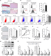
Increases in senescent chondrocytes in the cartilage of OA patients are closely related to cartilage damage. (a) (Left) Safranin‐O and IHC staining of p16 for Non‐OA and OA cartilage (severe‐ and mild‐damaged lesion), showing representative two donors. (Right) Quantification of p16 positive cells from Non‐OA and OA patients (n = 10). (b) (Left) SA β‐gal staining for Non‐OA and OA chondrocytes and (right) quantification of SA β‐gal positive cells (n = 5). (c) (Left) Representative image of western blot of p16 and p21 for chondrocytes from non‐OA and OA patients. (Right) Quantification of western blot analysis of p16 and p21 expression in non‐OA and OA chondrocytes (n = 5). (d) Cell counts of non‐OA and OA chondrocyte for 7 days to evaluate cell proliferation (n = 5). *p < 0.05.
FIGURE 2
FIGURE 2
Screening for a surface factor selectively expressed on senescent and OA chondrocytes. (a) The expression heatmap of surface marker genes in non‐OA chondrocytes, OA chondrocytes and oxidative stress‐induced senescent chondrocytes (n = 3, data showing the average of the positive cell percentage from three donors). (b) Analysis of surface markers (DPP‐4, CD221, CD36, CD24, CD28 and CD61) for Non‐OA, OA and oxidative stress‐induced senescent chondrocytes (SnCs) (n = 3). (c) The expression heatmap of 35 surface marker genes in non‐OA (n = 18) and OA patients (n = 20) was obtained from the NCBI Gene Expression Omnibus (GEO) under the accession code GSE114007. (d) volcano plot of 35 surface markers in osteoarthritis patients, using fold‐differences and p‐values of the expression level, based on the student's t‐test between non‐OA patients and OA patients (the names of significantly regulated genes (log2 fold‐change >1 or < −1 and p < 0.05) are indicated) and (e) log2 expression of significantly upregulated genes (CD28 and DPP‐4) between patients with either non‐OA or OA. * p < 0.05.
FIGURE 3
FIGURE 3
DPP‐4+ chondrocytes showed multiple senescent phenotypes. (a) Representative Safranin‐O and IHC staining of DPP‐4 for Non‐OA and OA cartilage. (b) (Left) Representative flow cytometry analysis of DPP‐4 expression from non‐OA and OA chondrocytes. (Right) Quantification of DPP‐4 positive cells determined by flow cytometry (n = 5 and n = 10, respectively). (c) (Left) Representative flow cytometry DPP‐4 (x‐axis) vs forward scatter (FSC: y‐axis) plot from IgG control, non‐OA, replicative (passage 15; P15), H2O2‐ and RAS‐induced senescent chondrocytes. (Right) Bar graph showing the percentage of DPP‐4 positive cells determined by flow cytometry (n = 5). (d) (Left) Representative SA β‐gal staining for non‐OA, DPP‐4‐ and DPP‐4+ chondrocytes. (Right) Quantification of SA β‐gal positive cells in non‐OA, DPP‐4‐ and DPP‐4+ chondrocytes (n = 5). (e) Cell counts of non‐OA, DPP‐4‐ and DPP‐4+ chondrocyte for 7 days to evaluate cell proliferation (n = 5). (f) The mRNA expression of p16 and p21 of non‐OA, DPP‐4‐ and DPP‐4+ chondrocytes using RT‐qPCR (n = 5). (g) (Top) Representative image of western blot analysis of p16 and p21 of DPP‐4‐ and DPP‐4+ chondrocytes. (Bottom) Quantification of western blot analysis of p16 and p21 expression in non‐OA, DPP‐4‐ and DPP‐4+ chondrocytes (n = 5). (h) The mRNA expression of SASPs (IL‐6, IL‐8, CCL2, BMP2, IGFBP7, TGF‐β, GDF15 and MMP13) in non‐OA, DPP‐4‐ and DPP‐4+ chondrocytes using RT‐qPCR. * p < 0.05.
FIGURE 4
FIGURE 4
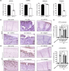
DPP‐4+ chondrocytes had OA phenotypes. (a) The mRNA expression of chondrocyte markers (SOX9, COL2A1, ACAN), hypertrophic chondrocyte markers (RUNX2, COL10A1) and catabolic markers (MMP13) in non‐OA, DPP‐4‐ and DPP‐4+ chondrocytes (n = 5). (b) Representative H&E and safranin‐o staining for pellets generated from non‐OA, DPP‐4‐ and DPP‐4+ chondrocytes and (c) Quantification of safranin‐O positive area of non‐OA, DPP‐4‐ and DPP‐4+ pellets (n = 5). * p < 0.05.
FIGURE 5
FIGURE 5
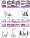
Sitagliptin could specifically and effectively target DPP‐4 positive chondrocytes. (a) Evaluation of cell viability of DPP‐4‐ and DPP‐4+ chondrocytes upon treatment of 20 μM ABT263, 100 nM 17DMAG, 20 μM metformin or 5 μM sitagliptin (n = 5). (b, d) Representative histological analysis using IHC for DPP‐4 and p16 staining to evaluate in vivo senolytic effects of ABT263, 17DMAG, metformin and sitagliptin and (c, e) Quantification of DPP‐4 and p16 positive cells (n = 7 per group). * p < 0.05.
FIGURE 6
FIGURE 6
Intra‐articular injection of the DPP‐4‐inhibitor sitagliptin prevented OA development in the rat DMM model. (a) Representative histological analysis using safranin‐O to evaluate OA progression in sham control and DMM rats after PBS, ABT263, 17DMAG, metformin or sitagliptin injection, (b) quantification of subchondral bone plate thickness of sham control and DMM rats after PBS, ABT263, 17DMAG, metformin or sitagliptin injection and (c) scoring of OA using OARSI grading system in sham control and DMM‐induced OA rat that had undergone intra‐articular injection of PBS, ABT263, 17DMAG, metformin or sitagliptin (n = 7). Representative collagen type II (d) and MMP13 (e) IHC staining images of sham control and DMM rats injected with PBS, ABT263, 17DMAG, metformin or sitagliptin and (f, g) quantification collagen type II and MMP13, respectively, using scoring system (n = 7). (h) Treadmill running distance and time (n = 7), and # and * indicate p < 0.05 when compared to PBS groups, respectively. *p < 0.05.

References

    1. Acosta, J. C. , Banito, A. , Wuestefeld, T. , Georgilis, A. , Janich, P. , Morton, J. P. , Athineos, D. , Kang, T.‐W. , Lasitschka, F. , Andrulis, M. , Pascual, G. , Morris, K. J. , Khan, S. , Jin, H. , Dharmalingam, G. , Snijders, A. P. , Carroll, T. , Capper, D. , Pritchard, C. , … Gil, J. (2013). A complex secretory program orchestrated by the inflammasome controls paracrine senescence. Nature Cell Biology, 15(8), 978–990. 10.1038/ncb2784 - DOI - PMC - PubMed
    1. Ashraf, S. , Cha, B.‐H. , Kim, J.‐S. , Ahn, J. , Han, I. , Park, H. , & Lee, S.‐H. (2016). Regulation of senescence associated signaling mechanisms in chondrocytes for cartilage tissue regeneration. Osteoarthritis and Cartilage, 24(2), 196–205. 10.1016/j.joca.2015.07.008 - DOI - PubMed
    1. Baker, D. J. , Wijshake, T. , Tchkonia, T. , LeBrasseur, N. K. , Childs, B. G. , van de Sluis, B. , Kirkland, J. L. , & van Deursen, J. M. (2011). Clearance of p16Ink4a‐positive senescent cells delays ageing‐associated disorders. Nature, 479(7372), 232–236. 10.1038/nature10600 - DOI - PMC - PubMed
    1. Bi, J. , Cai, W. , Ma, T. , Deng, A. , Ma, P. , Han, Y. , Lou, C. , & Wu, L. (2019). Protective effect of vildagliptin on TNF‐α‐induced chondrocyte senescence. IUBMB Life, 71(7), 978–985. 10.1002/iub.2049 - DOI - PubMed
    1. Boehm, A. K. , Seth, M. , Mayr, K. G. , & Fortier, L. A. (2007). Hsp90 mediates insulin‐like growth factor 1 and interleukin‐1β signaling in an age‐dependent manner in equine articular chondrocytes. Arthritis & Rheumatism, 56(7), 2335–2343. 10.1002/art.22664 - DOI - PubMed

MeSH terms

Substances