. 2024 Mar 31;11(1):20.
doi: 10.1186/s40779-024-00523-w.

FOXO1 reshapes neutrophils to aggravate acute brain damage and promote late depression after traumatic brain injury

Affiliations

FOXO1 reshapes neutrophils to aggravate acute brain damage and promote late depression after traumatic brain injury

Mi Zhou et al. Mil Med Res. .

Abstract

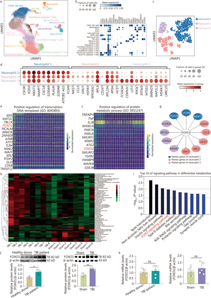
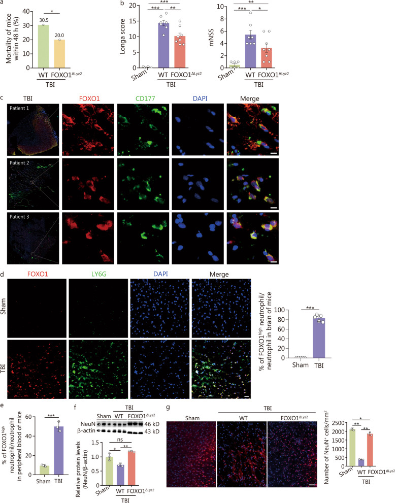
Background: Neutrophils are traditionally viewed as first responders but have a short onset of action in response to traumatic brain injury (TBI). However, the heterogeneity, multifunctionality, and time-dependent modulation of brain damage and outcome mediated by neutrophils after TBI remain poorly understood.

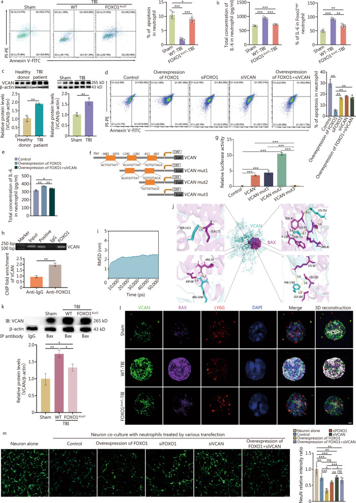
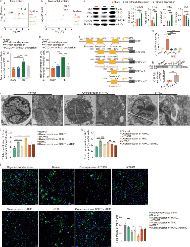
Methods: Using the combined single-cell transcriptomics, metabolomics, and proteomics analysis from TBI patients and the TBI mouse model, we investigate a novel neutrophil phenotype and its associated effects on TBI outcome by neurological deficit scoring and behavioral tests. We also characterized the underlying mechanisms both in vitro and in vivo through molecular simulations, signaling detections, gene expression regulation assessments [including dual-luciferase reporter and chromatin immunoprecipitation (ChIP) assays], primary cultures or co-cultures of neutrophils and oligodendrocytes, intracellular iron, and lipid hydroperoxide concentration measurements, as well as forkhead box protein O1 (FOXO1) conditional knockout mice.

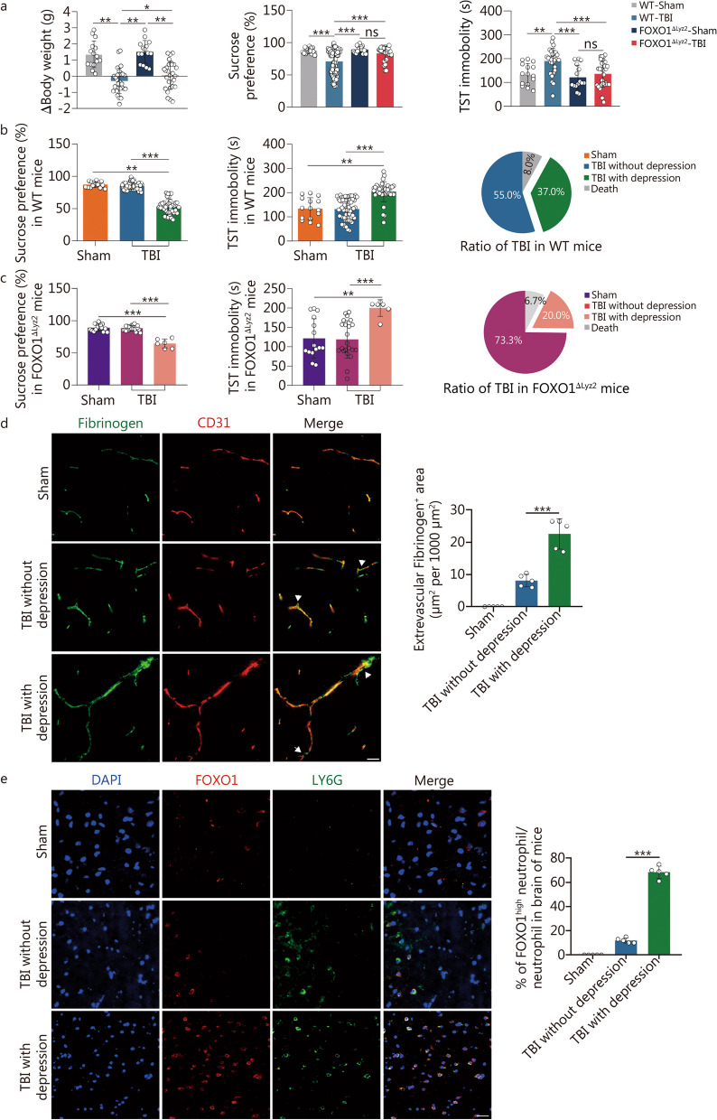
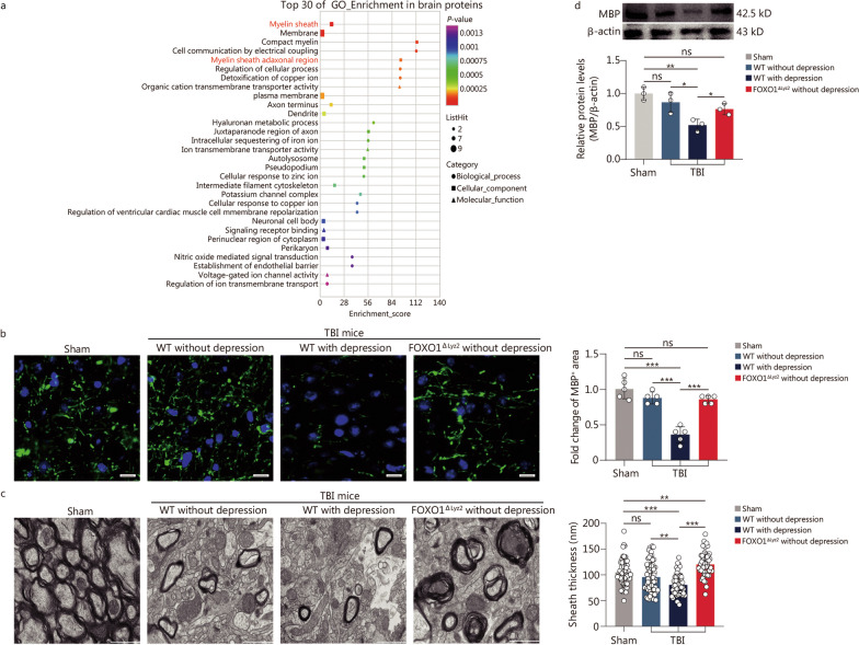
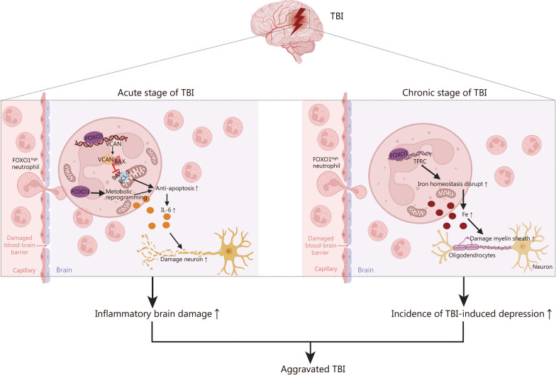
Results: We identified that high expression of the FOXO1 protein was induced in neutrophils after TBI both in TBI patients and the TBI mouse model. Infiltration of these FOXO1high neutrophils in the brain was detected not only in the acute phase but also in the chronic phase post-TBI, aggravating acute brain inflammatory damage and promoting late TBI-induced depression. In the acute stage, FOXO1 upregulated cytoplasmic Versican (VCAN) to interact with the apoptosis regulator B-cell lymphoma-2 (BCL-2)-associated X protein (BAX), suppressing the mitochondrial translocation of BAX, which mediated the antiapoptotic effect companied with enhancing interleukin-6 (IL-6) production of FOXO1high neutrophils. In the chronic stage, the "FOXO1-transferrin receptor (TFRC)" mechanism contributes to FOXO1high neutrophil ferroptosis, disturbing the iron homeostasis of oligodendrocytes and inducing a reduction in myelin basic protein, which contributes to the progression of late depression after TBI.

Conclusions: FOXO1high neutrophils represent a novel neutrophil phenotype that emerges in response to acute and chronic TBI, which provides insight into the heterogeneity, reprogramming activity, and versatility of neutrophils in TBI.

Keywords: Acute stage; Chronic stage; Forkhead box protein O1 (FOXO1); Neutrophil; Traumatic brain injury (TBI).


Conflict of interest statement

The authors declare that they have no competing interests.

Figures

Fig. 1
Fig. 1
Multilevel omics integration reveals high expression of FOXO1 in the acute stage following TBI. a UMAP embedding of the entire dataset colored by orthogonally generated clusters labeled by manual cell type annotation, which were the scRNA-seq data analysis for peripheral blood samples from healthy donors and TBI patients (n = 3 in each group). b Dot plot showing the top 10 changed genes in every cell cluster of the human scRNA-seq. c UMAP plot depicting the subpopulation of human neutrophils by scRNA-seq, with each cell color-coded for clusters. d Dot plot showing the expression of the top 10 changed genes in each human neutrophil cluster. e GO analysis depicting the detailed gene of positive regulation of transcription, DNA-templated. f GO analysis depicting the detailed gene of positive regulation of protein metabolic process. g Representative regulatory network of target genes by FOXO1 in each neutrophil cluster. The same color represents the gene from the same cluster. h Heatmap showing the differential metabolites in neutrophils between the sham and TBI groups in mice. Higher expression levels are indicated by red and lower by green. i Bubble chart showing the pathway activities through the differential metabolites of neutrophils from mice (sham group vs. TBI group). Each group had 7 repeated samples of neutrophils per group. j Western blotting showing the expression of FOXO1 in neutrophils from humans or mice. Human: TBI patients vs. healthy donors; mice: TBI group vs. sham group, n = 3 for each group. k The relative mRNA expressions of FOXO1 in neutrophils from human or mice detected by qRT-PCR. Human: TBI patients vs. healthy donors; mice: TBI group vs. sham group, n = 5 for each group. Data are represented as the mean ± SEM. *P < 0.05, **P < 0.01, ns non-significant. FOXO1 forkhead box protein O1, TBI traumatic brain injury, UMAP uniform manifold approximation and projection, GO Gene Ontology, qRT‑PCR quantitative real‑time PCR, MAIT mucosal-associated invariant T, NK natural killer cell, DC dendritic cellsDC, ILC innate lymphoid cell, CSF3R granulocyte colony-stimulating factor receptor, SOD2 superoxide dismutase [Mn], mitochondrial, MEAT1 Terminal uridylyltransferase 7, NAMPT Nicotinamide Phosphoribosyltransferase, CXCL8 C-X-C Motif Chemokine Ligand 8, S100A9 S100 calcium binding protein A9, PLAUR plasminogen activator, urokinase receptor, S100A8 S100 calcium binding protein A8, ATP2B1-AS1 ATP2B1 antisense RNA 1, NKG7 natural killer cell granule protein 7, CCL5 C–C motif chemokine ligand 5, GNLY granulysin, HSPA5 heat shock protein family A (Hsp70) member 5, PPP2R5C protein phosphatase 2 regulatory subunit B'gamma, RPL3 ribosomal protein L3, CD247 CD247 molecule, RPL23A, ribosomal protein L23a, PSA ribosomal protein sa, HCST hematopoietic cell signal transducer, FCGR3A A fc gamma receptor iiia, CDKN1C cyclin dependent kinase inhibitor 1c, LST1 leukocyte specific transcript 1, RHOC ras homolog family member, IFITM3 interferon induced transmembrane protein 3, MTSS1 MTSS I-BAR domain containing 1, SMIM25 plaque enriched lncRNA in atherosclerotic and inflammatory bowel macrophage regulation, NAP1L1 nucleosome assembly protein 1 like 1, MS4A7 membrane spanning 4-domains a7, LRRC25 LRRC25 leucine rich repeat containing 25, IER5 immediate early response 5, KMT2C lysine methyltransferase 2c, TBL1X transducin beta like 1 x-linked, PICALM phosphatidylinositol binding clathrin assembly protein, WWOX ww domain containing oxidoreductase, ZNF609 zinc finger protein 609, JUNB JunB proto-oncogene, AP-1 transcription factor subunit, ATF3 activating transcription factor 3, TLR4 toll like receptor 4, NME2 nme/nm23 nucleoside diphosphate kinase 2, MAFF maf bzip transcription factor f, IER2 immediate early response 2, TFDP2 transcription factor dp-2, GLI3 gli family zinc finger 3, CDK8 cyclin dependent kinase 8, TNFSF11 TNF superfamily member 11, TNFAIP3 TNF α induced protein 3, TNF tumor necrosis factor, IL1B interleukin 1 β, NFKB1A nuclear factor kappa b subunit 1, SMURF1 smad specific e3 ubiquitin protein ligase 1, WWP2 WW domain containing E3 ubiquitin protein ligase 2, ATG2 autophagy related 2, SMURF2 smad specific e3 ubiquitin protein ligase 2, TGFB1 transforming growth factor β 1, PANBP9, MAPK8 mitogen-activated protein kinase 8, GSK3B glycogen synthase kinase 3 β. RAB7A member RAS oncogene family, RAB1A member RAS oncogene family, RAN binding protein 9
Fig. 2
Fig. 2
High expression of FOXO1 in neutrophils aggravates neuronal damage in the acute stage of TBI. a Mortality of mice within 48 h after from TBI (n = 50). b Behavioural recovery of mice was assessed using the Longa score and mNSS at 48 h post-TBI (sham group, n = 6; WT TBI mice, n = 7; FOXO1△Lyz2 TBI mice, n = 8). c Immunostaining of the neutrophil marker CD177 (green), FOXO1 (red), and nuclei (DAPI, blue) in brain tissue from 3 TBI patients. Scale bar = 5 μm. d Immunostaining of the neutrophil markers LY6G (green), FOXO1 (red), and nuclei (DAPI, blue) in brain tissue from the TBI mouse model (n = 5). Scale bar = 10 μm. e Flow cytometry analysis of the percentage of FOXO1high neutrophils among leukocytes in peripheral blood of sham group and TBI mice. The experiment was repeated three times and n = 8 mice per group each time. f Western blotting showing the expression of NeuN in brain injury areas of mice. g Immunostaining of the neuron marker NeuN (red) and nuclei (DAPI, blue) in brain tissue from mice. Scale bar = 50 μm. In f and g, n = 6 per group for assay while here showed 3 biologically independent samples to represent the average status. Data are represented as the mean ± SEM. *P < 0.05, **P < 0.01, ***P < 0.001, ns non-significant. FOXO1 forkhead box protein O1, TBI traumatic brain injury, WT wild-type, mNSS modified neurological severity score, DAPI 4,6-diamidino-2-phenylindole dihydrochloride, NeuN neuronal nuclear antigen
Fig. 3
Fig. 3
FOXO1 enhances the anti-apoptosis and IL-6 release abilities of neutrophils. a Flow cytometry showing the percentage of apoptosis in neutrophil infiltrate to brain injury in TBI mice (n = 5). b ELISA analysis (left) and flow cytometry (right) of IL-6 levels in neutrophils of mice collected after TBI (n = 5). c Western blotting showing the expression of VCAN in neutrophils in both TBI patients and TBI mouse models. Three biologically independent samples of each group represented the average status here. d Flow cytometry showing the percentage of apoptosis in neutrophils transfected with different over/si-expression plasmids. Three biologically independent samples to represent the average status. e ELISA analysis of IL-6 in neutrophils of mice. The experiment was repeated three times. f Putative FOXO1 binding sequence of the mouse VCAN promoter gene. VCAN mut 1: deletion of one FOXO1 binding site at − 1874 to − 1885 bp; VCAN mut 2: deletion of two FOXO1 binding sites at − 1874 to − 1885 bp and − 1281 to − 1292 bp; VCAN mut 3: deletion of three FOXO1 binding sites at − 1874 to − 1885 bp, − 1281 to − 1292 bp and − 802 to − 813 bp. g Luciferase activity of FOXO1 co-transfected with mutated reporters under specific conditions. All transfected cells were treated under the indicated conditions for 6 h and lysed for dual-luciferase measurements. h ChIP of the FOXO1 binding sequence from the murine VCAN promoter gene. After treating neutrophils with the indicated conditions for 6 h, the total chromatin was collected and amplified as input (positive control). Antibodies against FOXO1 were used to pull down the binding segments, of which IgG was introduced as a negative control. i The RMSD showed by the backbone atoms of the VCAN-BAX system during the molecular docking. j Pymol MOE software was used to predict the binding between VCAN and BAX. k Co-IP of VCAN and BAX in neutrophils from bone marrow in a mouse model. β-actin was detected in the supernatant after IP. l Immunostaining showing the expression, location, and binding between BAX and VCAN in neutrophils from bone marrow in the mouse model of TBI. Scale bar = 1 μm. m Immunostaining showing the expression of the neuron marker NeuN (green) and nuclei (DAPI, blue) in neurons co-cultured with neutrophils treated as described above. Scale bar = 50 μm. The experiment was repeated 5 times. Data are represented as the mean ± SEM. *P < 0.05, **P < 0.01, ***P < 0.001, ns non-significant. FOXO1 forkhead box protein O1, TBI traumatic brain injury, ELISA enzyme‐linked immunosorbent assay, ORF open reading frame, mut mutant, ChIP chromatin immunoprecipitation, VCAN Versican, RMSD root mean square deviation, BAX BCL-2-associated X protein, DAPI 4,6-diamidino-2-phenylindole dihydrochloride, Co-IP co-immunoprecipitation
Fig. 4
Fig. 4
High expression of FOXO1 in neutrophils reduces the incidence of depression after TBI. FOXO1△Lyz2 mice and their WT littermate were randomly divided into a sham group and a TBI group. a Body weight and behavioral tests, including the sucrose preference and TST in mice, n = 30 in the sham group, n = 100 in WT TBI mice, n = 30 in FOXO1△Lyz2 sham mice, and n = 30 in FOXO1△Lyz2 TBI mice. b The behavioral tests of sucrose preference and TST were assayed in WT TBI mice for the identification of depression after TBI. Sham group, n = 15; TBI with depression group, n = 37; TBI without depression group, n = 55. c The behavioral tests of sucrose preference and TST were assayed in FOXO1△Lyz2 TBI mice for the identification of depression after TBI. Sham group, n = 15; TBI with depression group, n = 6; TBI without depression group, n = 22. d Immunostaining of fibrinogen (green) and CD31 (red) in the brain tissue of TBI mice (scale bar = 20 μm). e Immunostaining of FOXO1 (red), LY6G (green), and nuclei (DAPI, blue) in brain injury tissue of the TBI mouse model (scale bar = 20 μm). In d and e, in each group, n = 5 for data collection. Data are represented as the mean ± SEM. *P < 0.05, **P < 0.01, ***P < 0.001, ns non-significant. FOXO1 forkhead box protein O1, TBI traumatic brain injury, WT wild-type, TST tail suspension test, DAPI 4,6-diamidino-2-phenylindole dihydrochloride
Fig. 5
Fig. 5
High expression of FOXO1 in neutrophils increases myelin damage. a Proteomic analysis of brain tissues from mice. The top 30 proteins are enriched terms by GO analysis (group of TBI with depression vs. group of TBI without depression in WT mice). b Immunostaining of MBP (green) and nuclei (DAPI, blue) in the brain tissue of different groups (n = 5). Scale bar = 50 μm. c Electron microscopy image of corpus callosum sections and quantification of myelinated axon number and g-ratio (n = 6). Scale bar = 1 μm. d Western blotting showing the expression of MBP in neutrophils in the TBI mouse model (n = 3). In each group, three biologically independent samples were represented. Data are represented as the mean ± SEM. *P < 0.05, **P < 0.01, ***P < 0.001, ns non-significant. FOXO1 forkhead box protein O1, GO Gene Ontology, TBI traumatic brain injury, MBP myelin basic protein, DAPI 4,6-diamidino-2-phenylindole dihydrochloride
Fig. 6
Fig. 6
FOXO1 disrupts iron homeostasis in neutrophils and oligodendrocytes. a Volcano plot showing variation in protein expression in brain-insured tissues between the group of TBI with depression and the group of TBI without depression in WT mice. The fold change (FC) log (base 2) is on the X-axis, and the negative false log discovery rate (P-value) (base 10) is on the Y-axis. Higher expression levels are indicated by red and lower by green. b Volcano plot showing variation in protein expression of neutrophils between the TBI-induced depression group and the insusceptible group. The FC log (base 2) is on the X-axis, and the negative false log discovery rate (P-value) (base 10) is on the Y-axis. Higher expression levels are indicated by red and lower by green. c Western blotting showing the expression of FOXO1, FTL11, TFRC, and xCT in neutrophils from the TBI mice with and without depression (n = 6). The sham group served as the control. In each group, three biologically independent samples were represented here to show the average status. d Intracellular iron colorimetric assay showing the concentration of total Fe in neutrophils from bone marrow of the TBI mice with and without depression (n = 5). The sham group served as the control. e LPO assay showing the concentration of LPO in neutrophils from the bone marrow of the TBI mice with and without depression (n = 5). The sham group served as the control. f Putative FOXO1 binding sequence of the mouse TFRC promoter gene. TFRC wt1: the reporter plasmid containing the two FOXO1 binding sites at − 1070 to − 1080 bp and − 1085 to − 1095 bp; TFRC mut1: the reporter plasmid containing the two mutated FOXO1 binding sites at − 1070 to − 1080 bp and − 1085 to − 1095 bp; TFRC wt2: the reporter plasmid containing the FOXO1 binding site at − 526 to − 536 bp; TFRC mut2: the reporter plasmid containing the mutated FOXO1 binding site at − 526 to − 536 bp. g Luciferase activity of FOXO1 co-transfected with mutated reporters under specific conditions. All transfected cells were treated under the indicated conditions for 6 h and lysed for dual-luciferase measurements. h ChIP of the FOXO1 binding sequence from the murine TFRC promoter gene. After treating neutrophils with the indicated conditions for 6 h, the total chromatin was collected and amplified as input (positive control). Antibodies against FOXO1 were used to pull down the binding segments, of which IgG was introduced as a negative control. i Electron microscopy image of the form of mitochondria in neutrophils. j Intracellular iron colorimetric assay showing the concentration of total Fe in neutrophils. k LPO assay showing the concentration of LPO in neutrophils from bone marrow. l Immunostaining of MBP (green) and nuclei (DAPI, blue) in oligodendrocytes (scale bar = 50 μm). Each experiment was repeated three times. Data are represented as the mean ± SEM. *P < 0.05, **P < 0.01, ***P < 0.001. FOXO1 forkhead box protein O1, TBI traumatic brain injury, wt wild-type, FTH1 ferritin heavy chain 1, FTL1 ferritin heavy chain 1, TFRC transferrin receptor, xCT system xc(−) cystine/glutamate antiporter, mut mutant, LPO lipid hydroperoxide, ChIP chromatin immunoprecipitation, MBP myelin basic protein, DAPI 4,6-diamidino-2-phenylindole dihydrochloride
Fig. 7
Fig. 7
The effects and mechanism of FOXO1 high neutrophils in the acute and chronic stages of TBI. FOXO1 forkhead box protein O1, TBI traumatic brain injury, VCAN Versican, TFRC transferrin receptor, MBP myelin basic protein, BAX B-cell lymphoma-2 (BCL-2)-associated X protein, BCL-2 B-cell lymphoma-2, IL-6 interleukin-6

Comment in

References

    1. Corps KN, Roth TL, McGavern DB. Inflammation and neuroprotection in traumatic brain injury. JAMA Neurol. 2015;72(3):355–62. - PMC - PubMed
    1. Kong LZ, Zhang RL, Hu SH, Lai JB. Military traumatic brain injury: a challenge straddling neurology and psychiatry. Mil Med Res. 2022;9(1):2. - PMC - PubMed
    1. Jamjoom AAB, Rhodes J, Andrews PJD, Grant SGN. The synapse in traumatic brain injury. Brain. 2021;144(1):18–31. - PMC - PubMed
    1. Wilson L, Stewart W, Dams-O’Connor K, Diaz-Arrastia R, Horton L, Menon DK, et al. The chronic and evolving neurological consequences of traumatic brain injury. Lancet Neurol. 2017;16(10):813–25. - PMC - PubMed
    1. Jassam YN, Izzy S, Whalen M, McGavern DB, El Khoury J. Neuroimmunology of traumatic brain injury: time for a paradigm shift. Neuron. 2017;95(6):1246–65. - PMC - PubMed