Establishment of replication-competent vesicular stomatitis virus recapitulating SADS-CoV entry
- PMID: 38557247
- PMCID: PMC11092325
- DOI: 10.1128/jvi.01957-23
Establishment of replication-competent vesicular stomatitis virus recapitulating SADS-CoV entry
Abstract
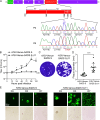
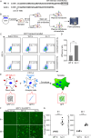
Zoonotic coronaviruses pose a continuous threat to human health, with newly identified bat-borne viruses like swine acute diarrhea syndrome coronavirus (SADS-CoV) causing high mortality in piglets. In vitro studies indicate that SADS-CoV can infect cell lines from diverse species, including humans, highlighting its potential risk to human health. However, the lack of tools to study viral entry, along with the absence of vaccines or antiviral therapies, perpetuates this threat. To address this, we engineered an infectious molecular clone of Vesicular Stomatitis Virus (VSV), replacing its native glycoprotein (G) with SADS-CoV spike (S) and inserting a Venus reporter at the 3' leader region to generate a replication-competent rVSV-Venus-SADS S virus. Serial passages of rVSV-Venus-SADS S led to the identification of an 11-amino-acid truncation in the cytoplasmic tail of the S protein, which allowed more efficient viral propagation due to increased cell membrane anchoring of the S protein. The S protein was integrated into rVSV-Venus-SADS SΔ11 particles, susceptible to neutralization by sera from SADS-CoV S1 protein-immunized rabbits. Additionally, we found that TMPRSS2 promotes SADS-CoV spike-mediated cell entry. Furthermore, we assessed the serum-neutralizing ability of mice vaccinated with rVSV-Venus-SADS SΔ11 using a prime-boost immunization strategy, revealing effective neutralizing antibodies against SADS-CoV infection. In conclusion, we have developed a safe and practical tool for studying SADS-CoV entry and exploring the potential of a recombinant VSV-vectored SADS-CoV vaccine.IMPORTANCEZoonotic coronaviruses, like swine acute diarrhea syndrome coronavirus (SADS-CoV), pose a continual threat to human and animal health. To combat this, we engineered a safe and efficient tool by modifying the Vesicular Stomatitis Virus (VSV), creating a replication-competent rVSV-Venus-SADS S virus. Through serial passages, we optimized the virus for enhanced membrane anchoring, a key factor in viral propagation. This modified virus, rVSV-Venus-SADS SΔ11, proved susceptible to neutralization, opening avenues for potential vaccines. Additionally, our study revealed the role of TMPRSS2 in SADS-CoV entry. Mice vaccinated with rVSV-Venus-SADS SΔ11 developed potent neutralizing antibodies against SADS-CoV. In conclusion, our work presents a secure and practical tool for studying SADS-CoV entry and explores the promise of a recombinant VSV-vectored SADS-CoV vaccine.
Keywords: SADS-CoV; VSV; VSV-vectored vaccine; replication-competent; viral entry.
Conflict of interest statement
Q.D. and Z.Z. have submitted a patent application (202311684558.1) for the utilization of the rVSV-SADS S system in the development of a SADS-CoV vaccine.
Figures


Similar articles
-
Swine Acute Diarrhea Syndrome Coronavirus: An Overview of Virus Structure and Virus-Host Interactions.Animals (Basel). 2025 Jan 9;15(2):149. doi: 10.3390/ani15020149. Animals (Basel). 2025. PMID: 39858149 Free PMC article. Review.
-
Interplay of swine acute diarrhoea syndrome coronavirus and the host intrinsic and innate immunity.Vet Res. 2025 Jan 9;56(1):5. doi: 10.1186/s13567-024-01436-1. Vet Res. 2025. PMID: 39789633 Free PMC article. Review.
-
A vesicular stomatitis virus-based prime-boost vaccination strategy induces potent and protective neutralizing antibodies against SARS-CoV-2.PLoS Pathog. 2021 Dec 16;17(12):e1010092. doi: 10.1371/journal.ppat.1010092. eCollection 2021 Dec. PLoS Pathog. 2021. PMID: 34914812 Free PMC article.
-
A Replication-Competent Vesicular Stomatitis Virus for Studies of SARS-CoV-2 Spike-Mediated Cell Entry and Its Inhibition.Cell Host Microbe. 2020 Sep 9;28(3):486-496.e6. doi: 10.1016/j.chom.2020.06.020. Epub 2020 Jul 3. Cell Host Microbe. 2020. PMID: 32738193 Free PMC article.
-
Optimized Pseudotyping Conditions for the SARS-COV-2 Spike Glycoprotein.J Virol. 2020 Oct 14;94(21):e01062-20. doi: 10.1128/JVI.01062-20. Print 2020 Oct 14. J Virol. 2020. PMID: 32788194 Free PMC article.
Cited by
-
Adaptive truncation of the S gene in IBV during chicken embryo passaging plays a crucial role in its attenuation.PLoS Pathog. 2024 Jul 30;20(7):e1012415. doi: 10.1371/journal.ppat.1012415. eCollection 2024 Jul. PLoS Pathog. 2024. PMID: 39078847 Free PMC article.
-
Development and Immunogenic Evaluation of a Recombinant Vesicular Stomatitis Virus Expressing Nipah Virus F and G Glycoproteins.Viruses. 2025 Jul 31;17(8):1070. doi: 10.3390/v17081070. Viruses. 2025. PMID: 40872782 Free PMC article.
-
Swine Acute Diarrhea Syndrome Coronavirus: An Overview of Virus Structure and Virus-Host Interactions.Animals (Basel). 2025 Jan 9;15(2):149. doi: 10.3390/ani15020149. Animals (Basel). 2025. PMID: 39858149 Free PMC article. Review.
-
Interplay of swine acute diarrhoea syndrome coronavirus and the host intrinsic and innate immunity.Vet Res. 2025 Jan 9;56(1):5. doi: 10.1186/s13567-024-01436-1. Vet Res. 2025. PMID: 39789633 Free PMC article. Review.
References
-
- Yang YL, Qin P, Wang B, Liu Y, Xu GH, Peng L, Zhou J, Zhu SJ, Huang YW. 2019. Broad cross-species infection of cultured cells by bat HKU2-related swine acute diarrhea syndrome coronavirus and identification of its replication in murine dendritic cells in vivo highlight its potential for diverse interspecies transmission. J Virol 93:e01448-19. doi:10.1128/JVI.01448-19 - DOI - PMC - PubMed
MeSH terms
Substances
Supplementary concepts
Grants and funding
- 82341084, 82272302, 82241077, 32070153/MOST | National Natural Science Foundation of China (NSFC)
- 2021YFC2300200-04, 2023YFC2305900/MOST | National Key Research and Development Program of China (NKPs)
- Z220018/| Beijing Municipal Natural Science Foundation ()
- 2022Z82WKJ013/Tsinghua University Vanke Special Fund for Public Health and Health Discipline Development
- SXMU-Tsinghua Collaborative Innovation Center for Frontier Medicine
LinkOut - more resources
Full Text Sources

