. 2024 Oct 1;80(4):859-871.
doi: 10.1097/HEP.0000000000000865. Epub 2024 Apr 1.

Variants in autophagy genes MTMR12 and FAM134A are putative modifiers of the hepatic phenotype in α1-antitrypsin deficiency

Affiliations

Variants in autophagy genes MTMR12 and FAM134A are putative modifiers of the hepatic phenotype in α1-antitrypsin deficiency

Edgar N Tafaleng et al. Hepatology. .

Abstract

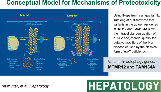
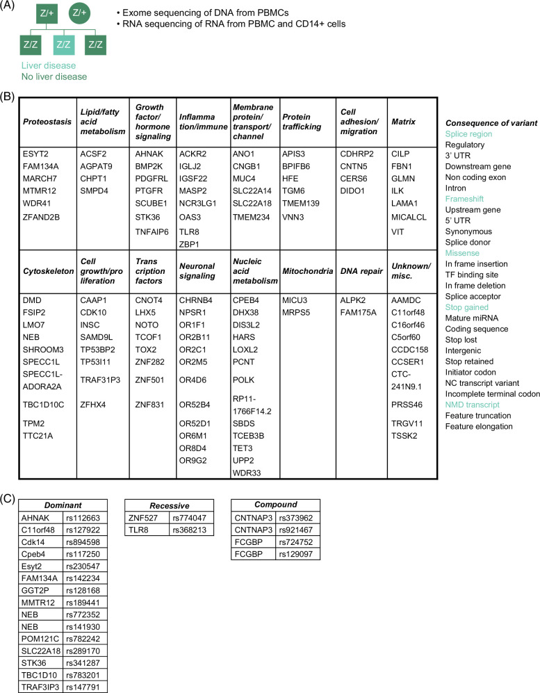
Background and aims: In the classical form of α1-antitrypsin deficiency, a misfolded variant α1-antitrypsin Z accumulates in the endoplasmic reticulum of liver cells and causes liver cell injury by gain-of-function proteotoxicity in a sub-group of affected homozygotes but relatively little is known about putative modifiers. Here, we carried out genomic sequencing in a uniquely affected family with an index case of liver failure and 2 homozygous siblings with minimal or no liver disease. Their sequences were compared to sequences in well-characterized cohorts of homozygotes with or without liver disease, and then candidate sequence variants were tested for changes in the kinetics of α1-antitrypsin variant Z degradation in iPS-derived hepatocyte-like cells derived from the affected siblings themselves.

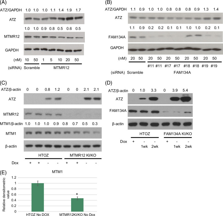
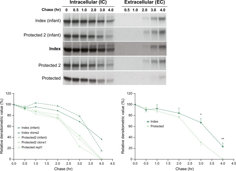
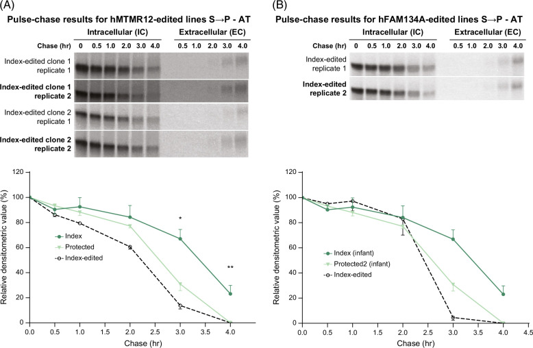
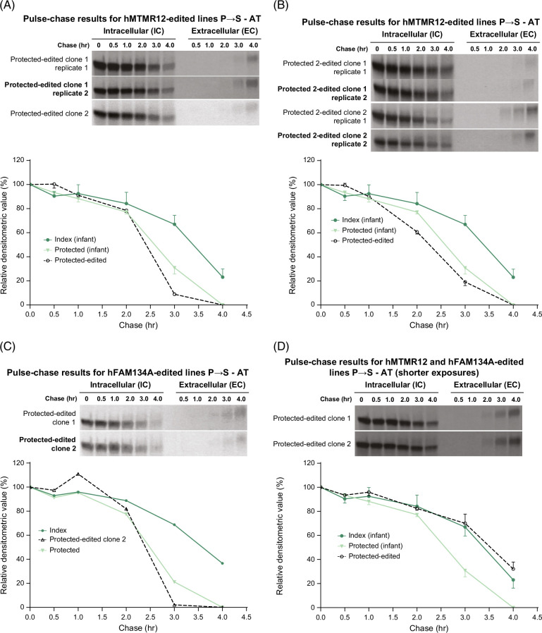
Approach and results: Specific variants in autophagy genes MTMR12 and FAM134A could each accelerate the degradation of α1-antitrypsin variant Z in cells from the index patient, but both MTMR12 and FAM134A variants were needed to slow the degradation of α1-antitrypsin variant Z in cells from a protected sib, indicating that inheritance of both variants is needed to mediate the pathogenic effects of hepatic proteotoxicity at the cellular level. Analysis of homozygote cohorts showed that multiple patient-specific variants in proteostasis genes are likely to explain liver disease susceptibility at the population level.

Conclusions: These results validate the concept that genetic variation in autophagy function can determine susceptibility to liver disease in α1-antitrypsin deficiency and provide evidence that polygenic mechanisms and multiple patient-specific variants are likely needed for proteotoxic pathology.


Conflict of interest statement

Alex Soto-Gutierrez owns stock in Pittsburgh ReLiver and Von Baer Wolff. Adam E. Locke is employed by and owns stock in Regeneron. Michael H. Cho received grants from Bayer. Edwin K Silverman received grants from Bayer and Northpond Laboratories. Ira J. Fox consults for Miromatrix. He owns stock in Pittsburgh ReLiver and Von Baer Wolff. The remaining authors have no conflicts to report.

Figures

None
Graphical abstract
FIGURE 1
FIGURE 1
Unique family (A), potentially damaging variants (B), and those prioritized for further analysis (C) are described in the text. The variants in fibrillin-1 (FBN) and homeostatic iron regulator (HFE) are described as “ conflicting interpretation of pathogenicity” by the ClinVar database. Abbreviaton: PBMC, peripheral blood mononuclear cells.
FIGURE 2
FIGURE 2
Immunoblot analysis of the effect of knocking down and/or knocking in the MTMR12 and FAM134A variants in the HTO/Z cell line with inducible expression of ATZ. (A) Knocking down MTMR12 with siRNA (relative densitometric values for ATZ levels compared to GAPDH levels are shown at the top and relative densitometric values for MTMR12 levels are shown above the MTMR12 blot); (B) Knocking down FAM134A with siRNA (relative densitometric values for ATZ levels compared to GAPDH levels are shown at the top and relative densitometric values for FAM134A levels are shown above the FAM134A blot); (C) ATZ, MTMR12, MTM1 and β-actin levels in HTO/Z cell line with MTMR12 variant knock-in/knockout background (relative densitometric values for ATZ levels compared to β-actin levels are shown at the top and relative densitometric values for MTM1 levels compared to β-actin levels are shown above the MTM1 blot); (D) ATZ, FAM134A and β-actin levels in HTO/Z cell line with FAM134A variant knock-in/knockout background (relative densitometric values for ATZ levels compared to β-actin are shown at the top); (E) MTM1 levels in HTO/Z cell line after removal of dox for 3 weeks, parent on the left and with MTMR12 variant knock-in/knockout background on the right (relative densitometric values on the vertical axis; n = 4 separate experiments for each bar; *p < 0.001 for students t-test). Abbreviations: ATZ, α1-antitrypsin variant Z; FAM134A, family with sequence similarity 134 member A; GAPDH, glyceraldehyde-3-phosphate dehydrogenase; HTO, HeLa tet-off cell line; MTMR12, myotubularin-related protein 12; siRNA, small interfering RNA.
FIGURE 3
FIGURE 3
Kinetics of ATZ fate in index versus protected subjects. Analysis of pulse-chase radiolabeling in iHeps from index subjects and protected siblings. Fluorograms are shown at the top and densitometric analysis at the bottom. The densitometric results for each line are shown at the left bottom, and the composite for index versus protected at the right bottom. The lines were derived from skin fibroblasts collected during infancy for index and protected 2 subjects and separate PBMCs collected from the index and both protected subjects as adults. The younger sibling is referred to as “protected” and the older sib as “protected 2”. Multiple replicates are shown in the graph at the lower left. The composite of replicates (n = 3 for index; n = 4 for protected) is shown in the graph at the lower right. Bars represent SEM. The difference between the index and protected subjects for the composite of replicates is significant at 3 hours (*p = 0.0092) and 4 hours (**p = 0.0096). Abbreviations: ATZ, α1-antitrypsin variant Z; EC, extracellular; iHeps, induced hepatocyte-like cells; IC, intracellular; PBMC, peripheral blood mononuclear cells.
FIGURE 4
FIGURE 4
Effect of correcting MTMR12 (A) and FAM134A (B) variants in the index subject on the kinetics of ATZ fate. Fluorograms for the edited clones are shown at the top, and densitometric analysis compared to unedited iHeps from the index and protected subjects is shown in the graphs at the bottom. Bars represent SEM. In (A), the difference between the index control (n = 3) and index-edited cells (n = 4) was significant at 3 hours (*p = 0.0006) and at 4 hours (**p = 0.0096). In (B), the difference between index control (n = 3) and the total (n = 6) of index-edited together with protected control was significant at 3 hours (p < 0.01) and 4 hours (p < 0.01). Abbreviations: AT, α1-antitrypsin; ATZ, α1-antitrypsin variant Z; EC, extracellular; FAM134A, family with sequence similarity 134 member A; iHeps, induced hepatocyte-like cells; IC, intracellular; MTMR12, myotubularin-related protein 12.
FIGURE 5
FIGURE 5
Effect of introducing MTMR12 and FAM134A variants into iHeps from protected subjects on the kinetics of ATZ fate. Fluorograms from the edited clones are shown at the top and densitometric analysis compared to unedited index and protected subjects is shown in the graphs below in each case. The MTMR12 variant was introduced into iHeps from the younger sibling (“protected”) in (A) and from the older sibling (“protected 2”) in (B) and the FAM134A variant was introduced into iHeps from ‘protected’ sib in (C). In (D) both of the variants have been introduced together in the “protected” subject. Bars represent SEM. In (A), (B), and (C), the difference between index control (n = 3) and the total (n = 6) of protected-edited and protected control was significant at 3 and 4 hours (p < 0.01). In (D), the difference between protected control (n = 4) and the total (n = 5) of protected-edited and index control was significant at 3 and 4 hours (p < 0.01). Abbreviations: AT, α1-antitrypsin; ATZ, α1-antitrypsin variant Z; EC, extracellular; FAM134A, family with sequence similarity 134 member A; iHeps, induced hepatocyte-like cells; IC, intracellular; MTMR12, myotubularin-related protein 12.

References

    1. Perlmutter DH. α1-antitrypsin deficiency: A misfolded protein variant with unique effects on the endoplasmic reticulum. Endoplasmic Reticulum Stress Dis. 2016;3:63–72. - PMC - PubMed
    1. Strnad P, McElvaney NG, Lomas DA. Alpha1-antitrypsin deficiency. N Engl J Med. 2020;382:1443–1455. - PubMed
    1. Chu AS, Chopra KB, Perlmutter DH. Is severe progressive liver disease caused by alpha-1-antitrypsin deficiency more common in children or adults? Liver Transpl. 2016;22:886–894. - PubMed
    1. Sveger T. Liver disease in alpha1-antitrypsin deficiency detected by screening of 200,000 infants. N Engl J Med. 1976;294:1316–1321. - PubMed
    1. Mostafavi B, Diaz S, Tanash HA, Piitulainen E. Liver function in alpha-1-antitrypsin deficient individuals at 37 to 40 years of age. Medicine. 2017;96:e6180. - PMC - PubMed

Publication types

Substances