

Primary Serous Cystadenocarcinoma of the Spleen
- PMID: 38558745
- PMCID: PMC10980539
- DOI: 10.7759/cureus.55165
Primary Serous Cystadenocarcinoma of the Spleen
Abstract
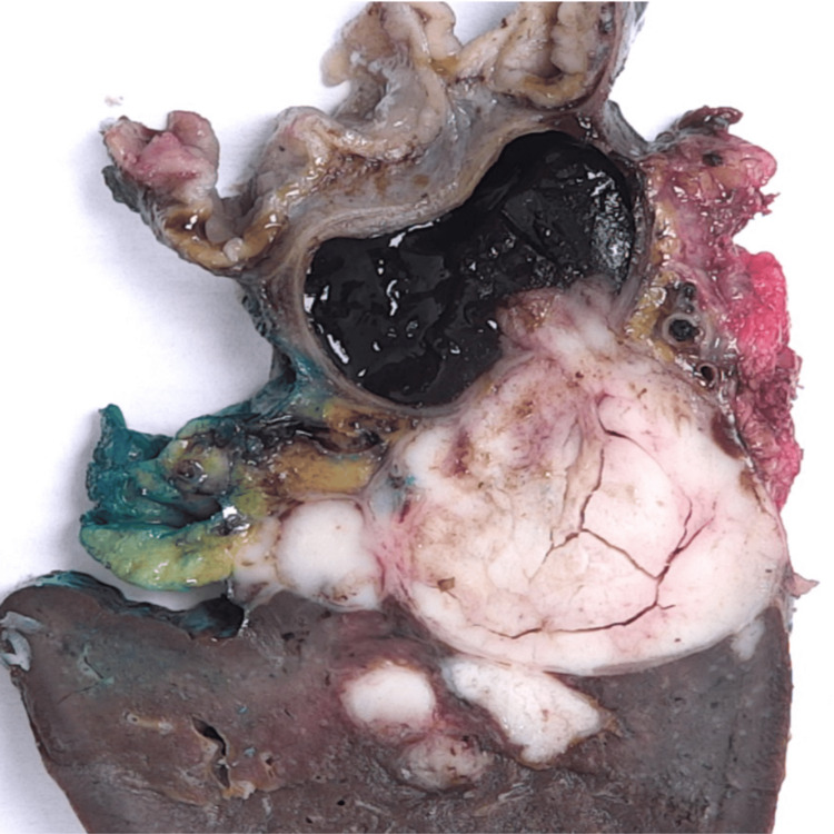
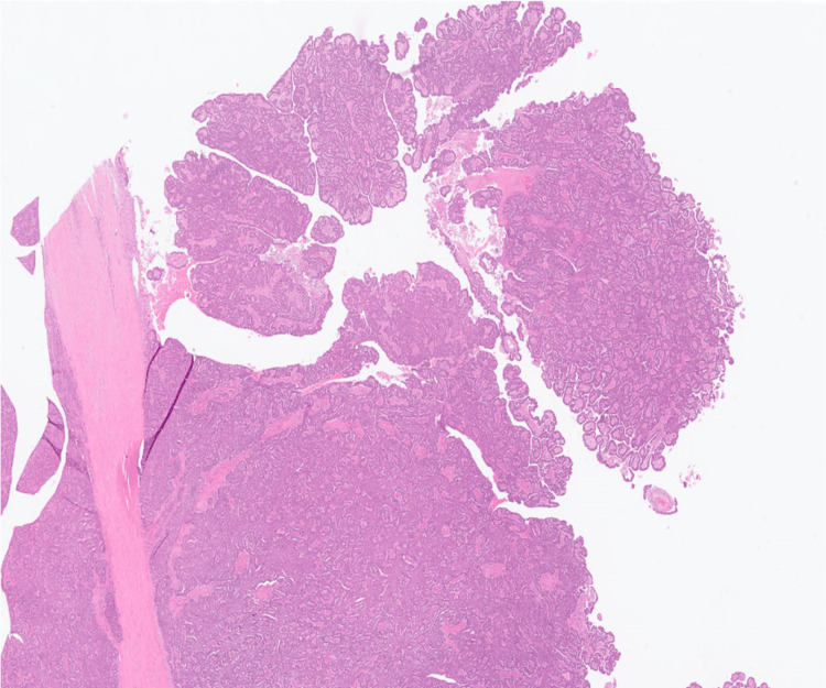
Tumours of the spleen are uncommon, and most are metastases from primaries in other organs. Primary splenic malignancies are subdivided into two main groups: lymphoid and non-lymphoid. Primary splenic cystadenocarcinomas are extremely rare, and only reports of the mucinous variant exist. We present the case of a female in her eighth decade of life who was found to have an incidental complex splenic mass with a cystic component, which showed an interval increase in size on serial imaging. After further investigation, including positron emission tomography (PET), endoscopic ultrasound (EUS), and laparoscopy, she successfully underwent distal pancreatectomy, splenectomy, and partial gastrectomy for a suspected locally invasive pancreatic malignancy. Histology and immunohistochemical analyses were consistent with the first recorded case of primary serous cystadenocarcinoma of the spleen in the literature.
Keywords: cancer; hepatopancreatobiliary; serous cystadenocarcinoma; spleen; surgery.
Copyright © 2024, Sciberras et al.
Conflict of interest statement
The authors have declared that no competing interests exist.
Figures


References
-
- Mucinous cystadenocarcinoma of the spleen - a very rare case of a primary splenic MCN. Wlaźlak M, Grząsiak O, Wierzchniewska-Ławska A, Hogendorf P, Durczyński A, Strzelczyk J. Pol Przegl Chir. 2020;93:1–5. - PubMed
-
- Pancreatic, hepatic, splenic, and mesenteric mucinous cystic neoplasms (MCN) are lumped together as extra ovarian MCN. Shiono S, Suda K, Nobukawa B, et al. Pathol Int. 2006;56:71–77. - PubMed
-
- Patterns of metastatic cancer in the spleen. MA JH Jr, GR S. Am J Clin Pathol. 1963;40:58–66. - PubMed
Publication types
LinkOut - more resources
Full Text Sources