

Eosinophilic Pneumonia Induced by Daptomycin
- PMID: 38558746
- PMCID: PMC10978459
- DOI: 10.7759/cureus.55095
Eosinophilic Pneumonia Induced by Daptomycin
Abstract
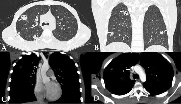
Daptomycin-induced eosinophilic pneumonia (DIEP) is a rare but serious complication associated with the use of this broad-spectrum antibiotic. We present the case of a teenager with a history of nasopharyngeal cancer who developed DIEP while receiving daptomycin to treat an infection associated with an implanted chamber catheter. Symptoms included recurrent dyspnea and peripheral eosinophilia, with radiological findings consistent with DIEP. The pathophysiology involves an immune response triggered by daptomycin, resulting in eosinophilic pulmonary inflammation. Diagnosis requires a thorough evaluation of medical history, clinical laboratory tests, and radiological findings. The main treatment involves discontinuation of daptomycin and, in severe cases, the use of steroids. It is essential to consider DIEP in patients with respiratory failure and bilateral pulmonary opacities who have used daptomycin and to suspect it in those with blood eosinophilia or in bronchoalveolar lavage.
Keywords: daptomycin; drug-related side effects and adverse reactions; eosinofilia pulmonar; pulmonary eosinophilia; radiology.
Copyright © 2024, Ayala Torres et al.
Conflict of interest statement
The authors have declared that no competing interests exist.
Figures


Similar articles
-
A Case of Daptomycin-Induced Eosinophilic Pneumonia and Its Management Insights.Cureus. 2025 Apr 29;17(4):e83195. doi: 10.7759/cureus.83195. eCollection 2025 Apr. Cureus. 2025. PMID: 40443582 Free PMC article.
-
Eosinophilic pneumonia caused by daptomycin: Six cases from two institutions and a review of the literature.J Infect Chemother. 2017 Apr;23(4):245-249. doi: 10.1016/j.jiac.2016.09.001. Epub 2016 Dec 18. J Infect Chemother. 2017. PMID: 28003110
-
Seventeen Cases of Daptomycin-Induced Eosinophilic Pneumonia in a Cohort of Patients Treated for Bone and Joint Infections: Proposal for a New Algorithm.Open Forum Infect Dis. 2022 Nov 3;9(11):ofac577. doi: 10.1093/ofid/ofac577. eCollection 2022 Nov. Open Forum Infect Dis. 2022. PMID: 36447615 Free PMC article.
-
Daptomycin-induced eosinophilic pneumonia - a systematic review.Antimicrob Resist Infect Control. 2016 Dec 12;5:55. doi: 10.1186/s13756-016-0158-8. eCollection 2016. Antimicrob Resist Infect Control. 2016. PMID: 27999664 Free PMC article. Review.
-
Acute Eosinophilic Pneumonia Secondary to Menthol Cigarette Use: A Rare Phenomenon With a Review of Literature.J Investig Med High Impact Case Rep. 2020 Jan-Dec;8:2324709620925978. doi: 10.1177/2324709620925978. J Investig Med High Impact Case Rep. 2020. PMID: 32462944 Free PMC article. Review.
References
-
- Drug-induced eosinophilic pneumonia: high-resolution CT findings in 14 patients. Souza CA, Müller NL, Johkoh T, Akira M. AJR Am J Roentgenol. 2006;186:368–373. - PubMed
-
- Eosinophilic pneumonia in patients treated with daptomycin: review of the literature and US FDA adverse event reporting system reports. Kim PW, Sorbello AF, Wassel RT, Pham TM, Tonning JM, Nambiar S. Drug Saf. 2012;35:447–457. - PubMed
Publication types
LinkOut - more resources
Full Text Sources