Editorial
. 2024 Mar 1;6(1):obae005.
doi: 10.1093/iob/obae005. eCollection 2024.

The Metameric Echinoderm

Affiliations
Editorial

The Metameric Echinoderm

R L Turner. Integr Org Biol. .

Abstract

Animal phyla are distinguished by their body plans, the ways in which their bodies are organized. A distinction is made, for example, among phyla with bodies of many segments (metameric; e.g., annelids, arthropods, and chordates), others with completely unsegmented bodies (americ; e.g., flatworms and mollusks), and a few phyla with bodies of 2 or 3 regions (oligomeric; e.g., echinoderms and hemichordates). The conventional view of echinoderms as oligomeric coelomates adequately considers early development, but it fails to recognize the metameric body plan that develops in the juvenile rudiment and progresses during indeterminate adult growth. As in the 3 phyla traditionally viewed to be metameric (annelids, arthropods, and chordates), metamery, or metamerism, in echinoderms occurs by (1) subterminal budding of (2) serially repeated components of (3) mesodermal origin. A major difference in most echinoderms is that metamery is expressed along multiple body axes, usually 5. The view of a metameric echinoderm might invite new discussions of metazoan body plans and new approaches to the study of morphogenesis, particularly in comparative treatments with annelids, arthropods, and chordates.

Tierstämme zeichnen sich durch ihren Körperbauplan, das heist, die Art und Weise aus, wie ihre Körper organisiert sind. Man unterscheidet beispielsweise Stämme mit Körpern aus vielen Segmenten (metamerer; z. B. Ringelwürmer, Arthropoden, Akkordaten), andere mit völlig unsegmentierten Körpern (amerer; z. B. Plattwürmer, Mollusken) und einige wenige Phyla mit Körpern aus zwei oder drei Regionen (oligomerer; z. B. Stachelhäuter, Hemichordaten). Die herkömmliche Sichtweise von Stachelhäutern als oligomere Coelomate berücksichtigt die frühe Entwicklung angemessen, berücksichtigt jedoch nicht den metameren Körperplan, der sich im jugendlichen Rudiment entwickelt und während des unbestimmten Erwachsenenwachstums fortschreitet. Wie bei den drei Phyla, die traditionell als metamer angesehen werden (Anneliden, Arthropoden, Chordaten), erfolgt Metamerie bei Stachelhäutern durch (1) subterminale Knospung von (2) seriell wiederholten Komponenten (3) mesodermalen Ursprungs. Ein wesentlicher Unterschied bei den meisten Stachelhäutern besteht darin, dass die Metamerie entlang mehrerer Körperachsen, normalerweise fünf, ausgedrückt wird. Die Sicht auf einen metameren Stachelhäuter könnte zu neuen Diskussionen über Metazoen-Körperpläne und neue Ansätze zur Untersuchung der Morphogenese führen, insbesondere bei vergleichenden Behandlungen mit Ringelwürmern, Arthropoden und Chordaten.


Conflict of interest statement

The author declares no competing interests

Figures
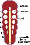
Fig. 1
Fig. 1
Schematic of a representative metameric metazoon. The anterior (acron) and posterior (pygidium) regions of the animal become increasingly separated by the insertion of segments (nos. 1–6) between them in a growth zone immediately anterior to the pygidium. The oldest segment (no. 1) lies behind the acron, the youngest (no. 6) anterior to the pygidium. This pattern of growth is called subterminal, penultimate, or teloblastic budding. Segments have parts of the coelom within tissue of mesodermal origin. The coelomic lining is not colored because metazoan coeloms can derive from embryonic ectoderm (blue), endoderm (yellow), or mesoderm (red).
Fig. 2
Fig. 2
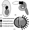
Organization of the oligomeric body plan. (A) In the embryo or larva, the coelom divides into 3 paired or unpaired cavities: protocoel, mesocoel, and metacoel. (B) In an acorn worm, the 3 body regions that contain the three coelomic cavities are easily recognized as the proboscis (prosoma with contained protocoel), collar (mesosoma with mesocoel), and trunk (metasoma with metacoel). (C) Oligomeric organization in a sea urchin, as a representative echinoderm, is obscured by expansion of the mesosoma around the other 2 regions. The prosoma includes the perforated madreporite at the surface and the axial organ descending from it. The perivisceral coelom, mostly comprised of the left metacoel, occupies much of the internal space along with viscera (not shown). The mesosoma has an internal component (stone canal) associated with the axial organ. The stone canal connects by a ring canal to 5 (one shown here) radial canals, which supply the externally visible parts of the water-vascular system, seen as podia extending from the body surface. Five paired rows (ambulacra) of podia alternate with 5 interambulacra; these 10 sections of the body form the body wall (external mesosoma). Much of the body wall is calcified as ossicles, pieces of the articulated skeleton of mesodermal origin.
Fig. 3
Fig. 3
Serial repetition along the arm of a brittlestar. (A) External expression of serial repetition is seen in the arm of Ophiactis savignyi. Repeated parts include arms spines (s), the plates of each segment (dorsal arm plates [dap], lateral arm plates [lap], ventral arm plates [not in view]), and podia (p). Dashed line indicates the plane of the cross-section in frame (B). Scale bar 200 µm. (Scanning electron micrograph by Y. Demirci.) (B) Schematic of repeated elements of one segment. Triplet sets of arm spines (s) articulate on lateral arm plates (lap) of the skeleton. The bulk of each segment consists of a centrally located skeletal vertebra (v). Adjacent vertebrae are joined and operated by paired ligaments (not shown) and by dorsal and ventral intervertebral muscles (im). The radial canal of the water-vascular system (wvs) runs beneath the vertebra and sends a lateral water vessel to each podium (p), which protrudes externally between a lateral arm plate and the ventral arm plate (vap). The nervous system, within the connective tissue space (cts), includes segmental ganglia below the radial canal and gives rise to nerve tracts that connect to the podial ring ganglia and spine ganglia. For simplicity, the ectoneural and hyponeural nervous systems are not distinguished, and the hemal system is not shown.
Fig. 4
Fig. 4
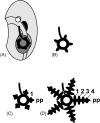
Early development of the water-vascular system in the juvenile rudiment (cf. Fig. 2A). (A) The left mesocoel encircles the early gut. (B) Five radial canals grow from the ring canal. (C) The growing tip of each radial canal is a primary podium (pp), in front of which is budded a succession of paired segmental secondary podia, the first pair (no. 1) shown here. (D) As the axis grows, additional podial pairs are added between the primary podium and the youngest pair (here, no. 4), the oldest pair (no. 1) lying nearest the mouth.
Fig. 5
Fig. 5
Subterminal addition of ossicles and podia in the sea urchin Tripneustes ventricosus. Near the top of the globe-like test, an ocular plate, bearing a single pore for the primary podium (pp), produces 2 columns of ambulacral plates (e.g., a[n], a[n-1], a[n-2] in one column) and shares production of the flanking interambulacral columns of plates (e.g., i[n], i[n-1], i[n-2]) with adjoining genital plates. As more ambulacral plates are added, they group together as triads in this species as compound plates (cp). Members of a triad eventually fuse together, as in the compound plate composed of a(n-10–n-12) and the next older compound plate outlined here for emphasis. The youngest (nth) externally visible ambulacral plate of one column has a simple podial pore and lies between the ocular plate and the next older ambulacral plate (a[n-1]). With growth of an ambulacral plate, the podial pore appears doubled on the surface. Ambulacral plates and compound plates of 2 ambulacral columns are staggered, forming a zigzag suture between the columns. Interambulacral plates in this species are larger than ambulacral and compound plates and are not in one-to-one correspondence; in this case, interambulacral plate i(n-2) shares sutures with ambulacral plates a(n-4–n-13). Soft tissue such as podia has been removed by treatment with bleach. Scanning electron micrograph.

References

    1. Allievi A, Carnavesi M, Ferrario C, Sugni M, Bonasoro F. 2022. An evo-devo perspective on the regeneration patterns of continuous arm structures in stellate echinoderms. Eur Zool J 2022:241–62.
    1. Arthur W. 1997. The origin of animal body plans: a study in evolutionary developmental biology. New York, NY: Cambridge University Press.
    1. Ausich WI, Brett CE, Hess H, Simms MJ. 1999. Crinoid form and function. In: Hess H, Ausich WI, Brett CE, Simms MJ, editors. Fossil crinoids. Cambridge: Cambridge University Press. p. 3–30.
    1. Balavoine G, Adoutte A. 2003. The segmented Urbilateria: a testable scenario. Integr Comp Biol 43:137–47. - PubMed
    1. Barnes RD. 1963. Invertebrate zoology. Philadelphia, PA: Saunders.

Publication types

LinkOut - more resources