[Preprint]. 2024 Mar 19:2024.03.13.584759.
doi: 10.1101/2024.03.13.584759.

A Foxf1-Wnt-Nr2f1 cascade promotes atrial cardiomyocyte differentiation in zebrafish

Affiliations

A Foxf1-Wnt-Nr2f1 cascade promotes atrial cardiomyocyte differentiation in zebrafish

Ugo Coppola et al. bioRxiv. .

Update in

Abstract

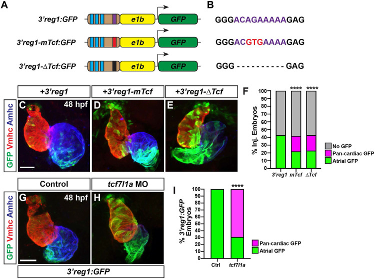
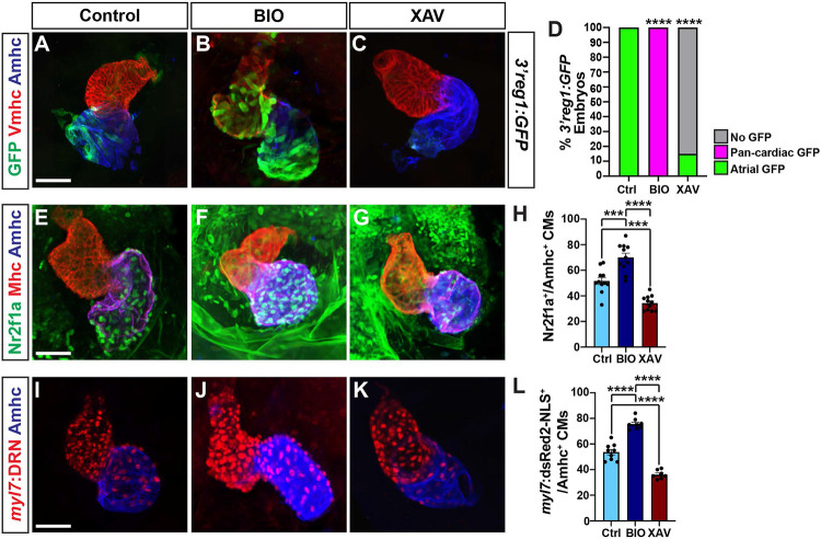
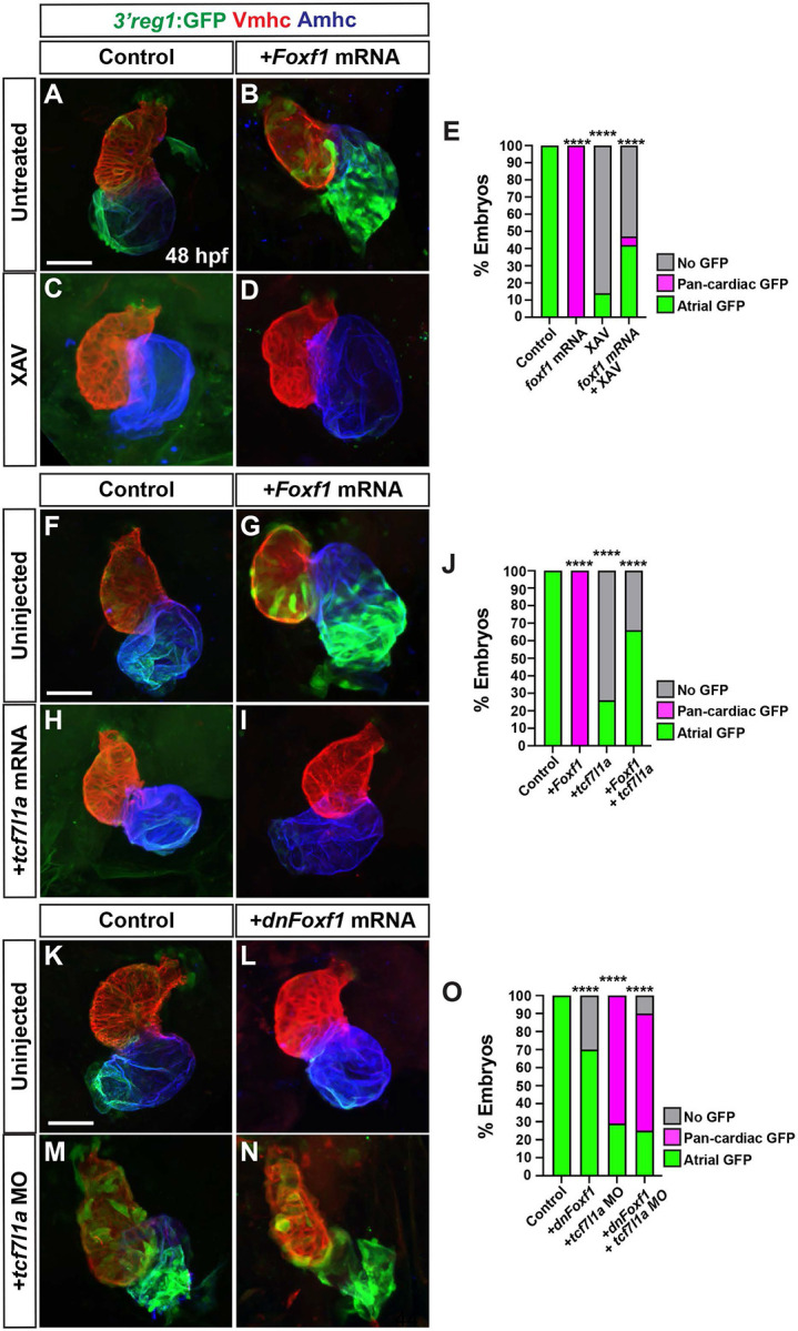
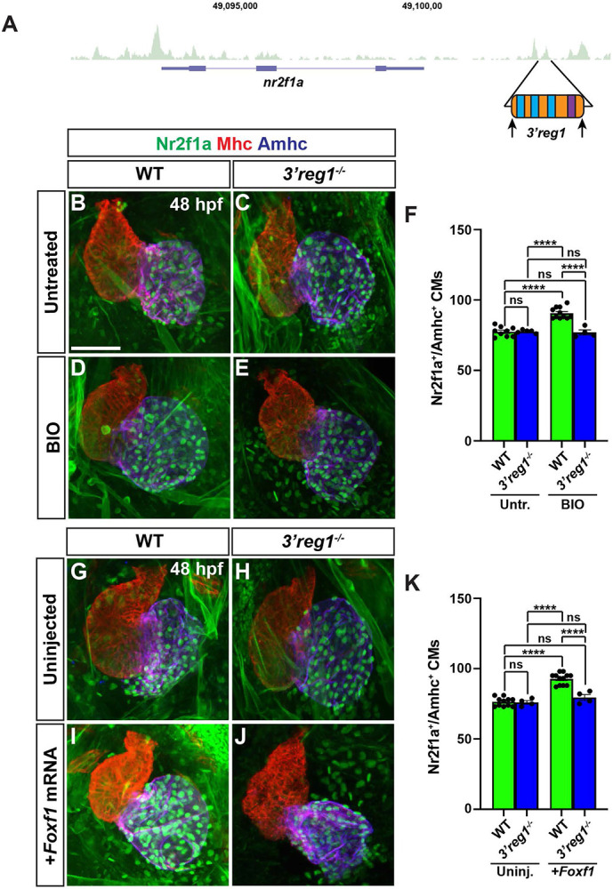
Nr2f transcription factors (TFs) are conserved regulators of vertebrate atrial cardiomyocyte (AC) differentiation. However, little is known about the mechanisms directing Nr2f expression in ACs. Here, we identified a conserved enhancer 3' to the nr2f1a locus, which we call 3'reg1-nr2f1a (3'reg1), that can promote Nr2f1a expression in ACs. Sequence analysis of the enhancer identified putative Lef/Tcf and Foxf TF binding sites. Mutation of the Lef/Tcf sites within the 3'reg1 reporter, knockdown of Tcf7l1a, and manipulation of canonical Wnt signaling support that Tcf7l1a is derepressed via Wnt signaling to activate the transgenic enhancer and promote AC differentiation. Similarly, mutation of the Foxf binding sites in the 3'reg1 reporter, coupled with gain- and loss-of-function analysis supported that Foxf1 promotes expression of the enhancer and AC differentiation. Functionally, we find that Wnt signaling acts downstream of Foxf1 to promote expression of the 3'reg1 reporter within ACs and, importantly, both Foxf1 and Wnt signaling require Nr2f1a to promote a surplus of differentiated ACs. CRISPR-mediated deletion of the endogenous 3'reg1 abrogates the ability of Foxf1 and Wnt signaling to produce surplus ACs in zebrafish embryos. Together, our data support that downstream members of a conserved regulatory network involving Wnt signaling and Foxf1 function on a nr2f1a enhancer to promote AC differentiation in the zebrafish heart.


Figures

Figure 1.
Figure 1.. The 3’reg1-nr2f1a enhancer is expressed in the atrium.
A) ATAC-seq profiling from ACs showing regions of open chromatin near the nr2f1a locus (purple). Black bars indicate called regions of open chromatin. Black box indicates 3’reg1. B) VISTA plot showing conservation of zebrafish 3’reg1 enhancer in Callorhincus milli (Australian ghostshark), Xenopus tropicalis (Tropical clawed frog), Anolis carolinensis (Green Anole), Mus musculus (House mouse), and Homo sapiens (human). Pink indicates >50% conservation of regulatory regions with zebrafish 3’reg1. Median lines in individual VISTA plots indicate 75% conservation. C) Clustal alignment of 3’reg1 from the VISTA plot in indicated species. Black asterisks indicate conserved nucleotides. Red asterisks indicate partially conserved nucleotides. Conserved putative binding sites for Foxf (blue shade) and Lef/Tcf (purple shade) TFs. D) Schematic of the 3’reg1:GFP reporter vector. E) In situ hybridization (ISH) for gfp in transgenic 3’reg1:GFP embryo at 24 hpf (n=24). Venous pole of the heart tube (black arrowhead). View is lateral with anterior left and dorsal up. Scale bar: 200μm. F-H) Confocal images of hearts from transgenic 3’reg1:GFP embryos stained for 3’reg1:GFP (green), Vmhc (VCs – red), and Amhc (ACs – blue). 36 hpf (n=5), 48 hpf (n=7), 72 hpf (n=5). 3’reg1:GFP in the atria of the hearts (white arrowhead). Images are frontal views with the arterial pole up. n indicates the number of embryos examined for a representative experiment. Scale bar: 50 μm. **** indicate P < 0.0001.
Figure 2.
Figure 2.. Tcf7l1a restricts 3’reg1 reporter expression within the heart.
A) Schematics of 3’reg1:GFP reporter constructs. Foxf sites (blue), Lef/Tcf site (purple), mutated Lef/Tcf site (red), and deleted Lef/Tcf site (black). B) Sequences of the WT (purple), mutated (red), and deleted Left/Tcf sites within the 3’reg1 enhancer. C-E) Confocal images of hearts from embryos injected with the 3’reg1:GFP, 3’reg1-mTcf:GFP, 3’reg1-ΔTcf:GFP constructs. F) The percentage of transient transgenic embryos with reporter atrial, pan-cardiac, and lacking expression in their hearts. 3’reg1:GFP (n=200); 3’reg1-mTcf:GFP (n=101); 3’reg1-ΔTcf:GFP (n=102). G,H) Confocal images of hearts from control and tcf7l1a MO-injected transgenic 3’reg1:GFP embryos. I) The percentage of stable 3’reg1:GFP embryos with reporter atrial and pan-cardiac expression in their hearts. Control (n=152); tcf7l1a-MO (n=92). Images are frontal views with the arterial pole up. Hearts are stained for 3’reg1:GFP (green), Vmhc (red), Amhc (blue). Scale bars: 50 μm. **** indicate P < 0.0001.
Figure 3.
Figure 3.. Wnt signaling promotes 3’reg1 reporter expression and AC differentiation.
A-C) Confocal images of hearts from control, BIO-, and XAV-treated 3’reg1:GFP embryos stained for 3’reg1:GFP (green), Vmhc (red), Amhc (blue). D) The percentage of control and treated 3’reg1:GFP embryos with atrial, pan-cardiac, and lacking expression in their hearts. Control (n=35); BIO (n=32); XAV (n=49). E-G) Confocal images of hearts from control, BIO-, and XAV-treated embryos stained for Nr2f1a (green), Mhc (pan-cardiac, red), Amhc (blue). H) The number of Nr2f1a+/Amhc+ cardiomyocytes with the hearts of control, BIO-, and XAV-treated embryos. Control (n=12); BIO (n=11); XAV (n=11). I-K) Confocal images of hearts from control, BIO-, and XAV-treated myl7:DsRed2-NLS (myl7:DRN) embryos stained for DsRed-NLS (pan-cardiac nuclei - red) and Amhc (blue). L) The number of myl7:DsRed2-NLS+/Amhc+ cardiomyocytes (ACs) within the hearts of control, BIO-, and XAV-treated myl7:DsRed2-NLS embryos. Control (n=9); BIO (n=10); XAV (n=8). Scale bars: 50 μm. Error bars in graphs indicate s.e.m.. *** indicate P < 0.001. **** indicate P < 0.0001.
Figure 4.
Figure 4.. Foxf1 promotes 3’reg1 reporter expression within the heart.
A) Schematics of 3’reg1:GFP reporter constructs with WT, deleted, and mutated Foxf sites. Foxf sites (blue), mutated Foxf sites (red), deleted Foxf sites (black), and Lef/Tcf site (purple). B) Sequences of the WT (blue), mutated (red), and deleted (dashes) Foxf sites within the 3’reg1 enhancer. C) The percentage of transient transgenic embryos with reporter expression in the atria. Control (3’reg1) (n=150); mFoxf-A (n=60); ΔFoxf-A (n=101); mFoxf-B (n=49); ΔFoxf-B (n=200); mFoxf-C (n=98); ΔFoxf-C (n=195). D-F) Confocal images of hearts from control uninjected, Foxf1 mRNA and dnFoxf1 mRNA injected 3’reg1:GFP embryos stained for 3’reg1:GFP (green), Vmhc (red), Amhc (blue). G) The percentage of injected 3’reg1:GFP embryos with atrial expression, pan-cardiac expression, and lacking expression. Control (n=35); Foxf1 (n=29); dnFoxf1 (n=59). H-J) Confocal images of hearts from control, Foxf1 mRNA, and dnFoxf1 mRNA injected embryos stained for Nr2f1a (green), Mhc (red), Amhc (blue). K) The number of Nr2f1a+/Amhc+ cardiomyocytes (ACs) within the hearts of control, Foxf1 mRNA, and dnFoxf1 injected embryos. Control (n=8); Foxf1 (n=11); dnFoxf1 (n=8). L-N) Confocal images of hearts from control, Foxf1 mRNA, and dnFoxf1 injected myl7:DsRed2-NLS (myl7:DRN) embryos stained for DsRed-NLS (red) and Amhc (blue). O) Quantification of the number of myl7:DsRed2-NLS+/Amhc+ cardiomyocytes (ACs) within the hearts of control, Foxf1 mRNA, and dnFoxf1 injected myl7:DsRed2-NLS embryos. Control (n=10); Foxf1 (n=13); dnFoxf1 (n=11). Scale bars: 50 μm. Error bars in graphs indicate s.e.m.. *** indicate P < 0.001. **** indicate P < 0.0001.
Figure 5.
Figure 5.. Tcf7l1a limits the ability of Foxf1 to promote 3’reg1 reporter expression.
A-D) Confocal images of hearts from control, Foxf1 mRNA injected, XAV-treated, and Foxf1 mRNA injected plus XAV-treated 3’reg1:GFP embryos . E) The percentage of control, Foxf1 mRNA injected, XAV-treated, and Foxf1 mRNA injected plus XAV-treated 3’reg1:GFP embryos with atrial expression, pan-cardiac expression, and lacking expression. Control (n=26); Foxf1 (n=24); XAV n=98; Foxf1-XAV (n=103). F-I) Confocal images of hearts from control, Foxf1 mRNA injected, tcf7l1a mRNA injected, and Foxf1 mRNA and tcf7l1a mRNA co-injected 3’reg1:GFP embryos. E) The percentage of control, Foxf1 mRNA injected, tcf7l1a mRNA injected, and Foxf1 mRNA and tcf7l1a mRNA co-injected 3’reg1:GFP embryos with atrial expression, pan-cardiac expression, and lacking expression. Control (n=39), Foxf1 (n=34); tcf7l1a (n=95); Foxf1-tcf7l1a (n=51). K-N) Confocal images of hearts from control, dnFoxf1 mRNA injected, tcf7l1a MO injected, and Foxf1 mRNA and tcf7l1a MO co-injected 3’reg1:GFP embryos. O) The percentage of control, dnFoxf1 mRNA injected, tcf7l1a MO injected, and Foxf1 mRNA and tcf7l1a MO 3’reg1:GFP embryos with atrial expression, pan-cardiac expression, and lacking expression. Control (n=27), dnFoxf1 (n=102); Tcf7l1a-MO (n=96); dnFoxf1-tcf7l1aMO (n=100). Hearts are stained for 3’reg1:GFP (green), Vmhc (red), Amhc (blue)Scale bars: 50 μm. **** indicate P < 0.0001.
Figure 6.
Figure 6.. Wnt signaling and Foxf1 require Nr2f1a to promote a surplus of cardiomyocytes.
A-D) Confocal images of hearts from untreated and BIO-treated WT and nr2f1a−/− myl7:DsRed2-NLS embryos. E) The number of myl7:DsRed2-NLS+/Amhc+ cardiomyocytes in hearts of untreated and BIO-treated WT and nr2f1a−/− myl7:DsRed2-NLS embryos. Untreated WT (n=12); untreated nr2f1a−/− (n=7); treated WT (n=11); treated nr2f1a−/− (n= 10). F-I) Confocal images of hearts from uninjected and Foxf1 mRNA-injected WT and nr2f1a−/− myl7:DsRed2-NLS embryos. J) The number of myl7:DsRed2-NLS+/Amhc+ cardiomyocytes in uninjected and Foxf1 mRNA-injected WT and nr2f1a−/−myl7:DsRed2-NLS embryos. Uninjected WT (n=7); Uninjected nr2f1a−/− (n=5); Inj. WT (n=10); Injected nr2f1a−/− (n=5). Hearts are stained for DsRed-NLS (red) and Amhc (blue). Scale bars: 50 μm. Error bars in graphs indicate s.e.m.. ** indicate P < 0.01. **** indicate P < 0.001. **** indicate P < 0.0001. ns indicates not a statistically significant difference.
Figure 7.
Figure 7.. Wnt signaling and Foxf1 require 3’reg1 to promote a surplus ACs.
A) Schematic of 3’reg1 enhancer deletion generated using CRISPR/Cas12a. Arrows indicate location of the guides. B-E) Confocal images of hearts from untreated and BIO-treated WT and 3’reg1Δ/Δ embryos stained for Nr2f1a (green), Mhc (red), and Amhc (blue). BIO was unable to promote an increase in ACs in 3’reg1−/− embryos. E) The number of Nr2f1a+/Amhc+ cardiomyocytes in untreated and BIO-treated WT and 3’reg1−/− embryos. Untreated WT (n=9); untreated 3’reg1−/− (n=5); treated WT (n=11); treated 3’reg1−/− (n=4). F-I) Confocal images of hearts from uninjected and Foxf1 mRNA-injected WT and 3’reg1−/− embryos stained for Nr2f1a (green), Mhc (red), and Amhc (blue). J) The number of Nr2f1a+/Amhc+ cardiomyocytes in uninjected and Foxf1 mRNA-injected WT and Nr2f1a+ 3’reg1−/− embryos. Uninjected WT (n=11); Uninjected 3’reg1−/− (n=4); Injected WT (n=12); Injected 3’reg1−/− (n= 4). Scale bar: 50 μm. Error bars in graphs indicate s.e.m.. **** indicate P < 0.0001. ns indicates not a statistically significant difference.
Figure 8.
Figure 8.. A Wnt-Foxf1-Nr2f1a cascade directs atrial cardiomyocyte differentiation.
Model depicting the Wnt signaling and Foxf1-dependent regulatory network that controls Nr2f1a expression and the role of the 3’reg1 enhancer in the atrial cardiomyocyte differentiation during early heart tube development.

Similar articles

References

    1. Targoff KL, Colombo S, George V, Schell T, Kim S-H, Solnica-Krezel L, et al. Nkx genes are essential for maintenance of ventricular identity. Development. 2013;140: 4203–4213. doi:10.1242/dev.095562 - DOI - PMC - PubMed
    1. van Weerd JH, Christoffels VM. The formation and function of the cardiac conduction system. Development. 2016;143: 197–210. doi:10.1242/dev.124883 - DOI - PubMed
    1. Bruneau BG, Riley P. Heart Development and Disease. Cold Spring Harbor Laboratory Press; 2020. Available: https://play.google.com/store/books/details?id=pwE6yAEACAAJ
    1. Pereira FA, Qiu Y, Zhou G, Tsai M-J, Tsai SY. The orphan nuclear receptor COUP-TFII is required for angiogenesis and heart development. Genes & Development. 1999. pp. 1037–1049. doi:10.1101/gad.13.8.1037 - DOI - PMC - PubMed
    1. Devalla HD, Schwach V, Ford JW, Milnes JT, El-Haou S, Jackson C, et al. Atrial-like cardiomyocytes from human pluripotent stem cells are a robust preclinical model for assessing atrial-selective pharmacology. EMBO Mol Med. 2015;7: 394–410. doi:10.15252/emmm.201404757 - DOI - PMC - PubMed

Publication types