[Preprint]. 2024 Mar 12:rs.3.rs-3929981.
doi: 10.21203/rs.3.rs-3929981/v1.

State-dependent central synaptic regulation by GLP-1 is essential for energy homeostasis

Affiliations

State-dependent central synaptic regulation by GLP-1 is essential for energy homeostasis

Le Wang et al. Res Sq. .

Update in

Abstract

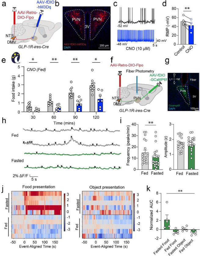
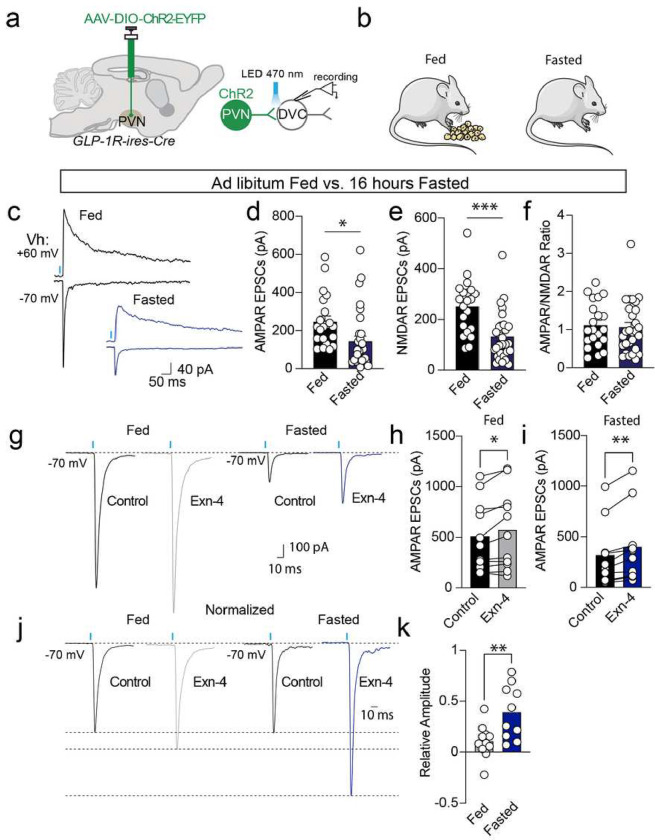
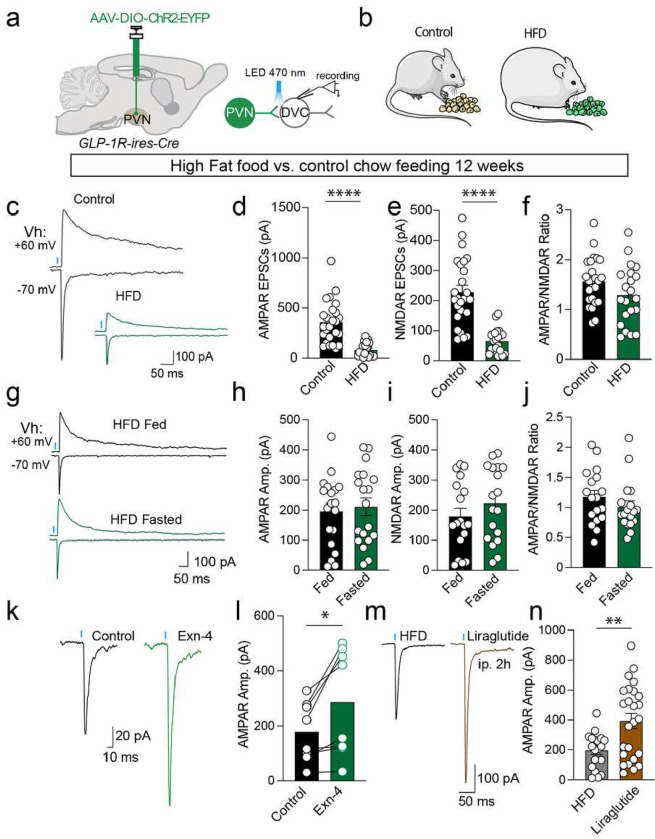
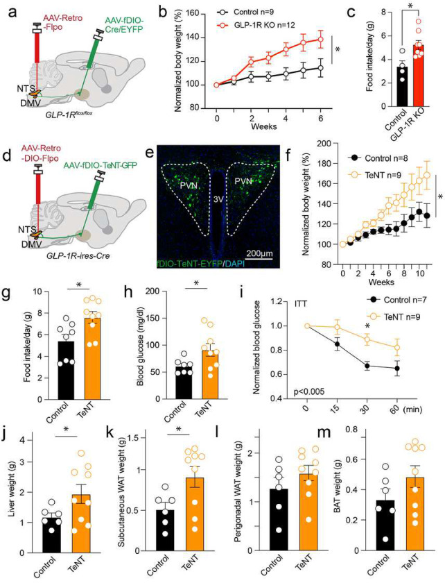
Central nervous system (CNS) control of metabolism plays a pivotal role in maintaining energy homeostasis. Glucagon-like peptide-1 (GLP-1, encoded by Gcg), secreted by a distinct population of neurons located within the nucleus tractus solitarius (NTS), suppresses feeding through projections to multiple brain targets1-3. Although GLP-1 analogs are proven clinically effective in treating type 2 diabetes and obesity4, the mechanisms of GLP-1 action within the brain remain unclear. Here, we investigate the involvement of GLP-1 receptor (GLP-1R) mediated signaling in a descending circuit formed by GLP-1R neurons in the paraventricular hypothalamic nucleus (PVNGLP-1R) that project to dorsal vagal complex (DVC) neurons of the brain stem in mice. PVNGLP- 1R→DVC synapses release glutamate that is augmented by GLP-1 via a presynaptic mechanism. Chemogenetic activation of PVNGLP-1R→DVC neurons suppresses feeding. The PVNGLP-1R→DVC synaptic transmission is dynamically regulated by energy states. In a state of energy deficit, synaptic strength is weaker but is more profoundly augmented by GLP-1R signaling compared to an energy-replete state. In an obese state, the dynamic synaptic strength changes in the PVNGLP-1R→DVC descending circuit are disrupted. Blocking PVNGLP-1R→DVC synaptic release or ablation of GLP-1R in the presynaptic compartment increases food intake and causes obesity, elevated blood glucose, and impaired insulin sensitivity. These findings suggest that the state-dependent synaptic plasticity in this PVNGLP-1R→DVC descending circuit mediated by GLP-1R signaling is an essential regulator of energy homeostasis.


Conflict of interest statement

Conflict of Interests The authors declare no conflict of interest.

Figures

Figure 1:
Figure 1:. PVNGLP-1R→DVC descending circuit is regulated by GLP-1R-mediated signaling.
a. Experimental paradigm for AAV-DIO-ChR2-EYFP injection to label PVN GLP-1R neurons. b. Representative image of the PVN with AAV-mediated expression of ChR2-EYFP. c-d. Representative image of DVC with expression of ChR2-EYFP. e. DVC cells were patched and evaluated for synaptic connectivity with optogenetically evoked EPSCs (oEPSCs) that are blocked by CNQX (n=20 cells/12 mice). f. Percentage of neurons showing synaptic connections (numbers indicate the numbers of responsive cells/total cells). g-h. Representative traces and quantification of AMPAR mediated oEPSCs before and after application of Exn-4 (n=29 cells/12 mice). i-j. Representative traces and quantification of light-evoked PPR before and after application of Exn-4 (n=24 cells/12 mice). k. Representative traces of oEPSCs with or without Exn-4. AMPAR- and NMDAR-mediated oEPSCs were recorded at holding potentials of −70 mV and +60 mV, respectively. NMDAR- oEPSCs were measured at 50 ms after stimulations. l. Pooled data of AMPAR/NMDAR-EPSCs ratio before and after application of Exn-4 (n=15 cells/12 mice). m-n. Representative traces and quantification of AMPAR-mediated oEPSCs before and after application of H89 and Exn-4 (n=10 cells/3 mice). Data are presented as mean ± SEM and sample sizes are indicated in each plot; paired student’s t-tests are applied to (e) and (g-n). One-way ANOVA is applied to (f); ∗p< 0.05; ∗∗p< 0.01; ∗∗∗p< 0.001; ∗∗∗∗p< 0.0001.
Figure 2:
Figure 2:. State-dependent PVNGLP-1R→DVC neuronal activity suppresses feeding.
a. Experimental paradigm for virus delivery and chemogenetic stimulation of PVNGLP-1RDVC neurons. b. Representative image of AAV-fDIO-hM3Dq-mCherry expression in the PVNGLP-1R→DVC neurons. c. Representative traces of CNO application. d. Quantification of CNO application in PVN hM3Dq- or control-virus expressing cells (n=6 cells/2 mice). e. Food intake consumption upon activation of PVNGLP-1RDVC neurons in the dark cycle (control n=9 mice, hM3Dq n=7 mice). f. Experimental paradigm for virus delivery and fiber photometry imaging of PVNGLP-1RDVC neurons. G. Representative image of Gcamp6f expression in the PVNGLP-1RDVC neurons. h. Representative traces of PVNGLP-1RDVC neuron’s calcium activity in the fasted and fed state. i. Frequency and amplitude quantification of PVNGLP-1RDVC neurons’ calcium activity in the fast and fed state (n=20 trial/5 mice). j-k. Calcium signals of PVNGLP-1RDVC neurons during different food/object presentations and under different energy states (n=5 mice). Data are presented as mean ± SEM and sample sizes are indicated in each plot; paired student’s t-test is applied to (d) and (i); One-way ANOVA is applied to (k). Two-way ANOVA with Geisser-Greenhouse correction is applied to (e). *p< 0.05; **p<0.01.
Figure 3:
Figure 3:. State-dependent synaptic plasticity of PVNGLP-1R→DVC neurons.
a-b. Experimental paradigm for AAV-DIO-Chr2-EYFP injection and electrophysiology experiments. c-f. Representative traces and quantification of AMPAR-mediated oEPSCs (Fed n=21 cells/3 mice, Fasted n=30 cells/4 mice), NMDAR EPSCs (Fed n=21 cells/3 mice, Fasted n=29 cells/4 mice), and AMPAR/NMDAR EPSC ratio (Fed n=21 cells /3 mice, Fasted n=29 cells/4 mice) under different conditions. g-i. Representative traces and quantification of AMPAR oEPSCs witho or without Exn-4 under different energy states (Fed n=11 cells/4 mice, Fasted n=10 cells/3 mice). j-k. Representative traces and quantification of normalized AMPAR-mediated oEPSCs with or without Exn-4 under different energy states (Fed n=11 cells/4 mice, Fasted n=10 cells/3 mice). Data are presented as mean ± SEM and n numbers are indicated in each plot. Student’s t-tests are applied to (d-f) and (k), and paired student’s t-tests are applied to (h) and (i). *p< 0.05; **p<0.01, ***p<0.001.
Figure 4:
Figure 4:. High-fat diet (HFD) induced obesity blunts the state-dependent synaptic plasticity of PVNGLP-1R→DVC neurons.
a-b. Experimental paradigm for AAV-DIO-Chr2-EYFP injection and electrophysiology experiments. c-f. Representative traces and quantification of AMPAR-mediated oEPSCs (Control n=25 cells/3 mice, HFD n=23 cells/3 mice), NMDAR-oEPSCs (Control n=25 cells/3 mice, HFD n=21 cells/3 mice), and AMPAR/NMDAR EPSC ratio (Control n=25 cells/3 mice, HFD n=21 cells/3 mice) in control or HFD animal. g-i. Representative traces and quantification of AMPAR oEPSCs (Fed n=19 cells/3 mice, Fasted n=19 cells/3 mice), NMDAR oEPSCs (Fed n=18 cells/3 mice, Fasted n=18 cells/3 mice), and AMPAR/NMDAR EPSCs ratio (Fed n=18 cells/3 mice, Fasted n=18 cells/3 mice) under different energy states in HFD-induced obese animals. k-l. Representative traces and quantification of AMPAR-mediated oEPSCs before and after application of Exn-4 in HFD-induced obese animals (n=8 cells/7 mice). m-n. Representative traces and quantification of AMPAR-mediated oEPSCs after control or Liraglutide i.p. 2h injection (control n=19 cells/3 mice, Liraglutide n=25 cells/3 mice) in HFD obese mice. Data are presented as mean ± SEM and sample sizes are indicated in each plot; Student’s t-tests are applied to (d-j) and (n), and paired student’s t-tests are applied to (l). *p< 0.05; **p<0.01, ***p<0.001, ***p<0.0001.
Figure 5:
Figure 5:. Chronic perturbation of PVNGLP-1R→DVC neurons induces obesity.
a. Experimental paradigm for the depletion of PVNGLP-1RDVC neurons GLP-1R using AAV-Cre virus injection. b. Quantification of body weight gain after depletion of PVNGLP-1RDVC neurons GLP-1R (Control n=9 mice, Knockout n=12 mice). c. Quantification of daily food intake after GLP-1R ablation in PVNGLP-1RDVC neurons compared to control animals (Control n=4 mice, Knockout n=8 mice). d. Experimental paradigm for the chronic inactivation of PVNGLP-1RDVC neurons synaptic release. e. Representative image of the PVN showing the expression of TeNT-GFP in the PVNGLP-1RDVC neurons. f. Quantification of body weight gain after expressing TeNT/Control in PVNGLP-1RDVC neurons (Control n= 7–8 mice, TeNT n=9 mice). g. Quantification of daily food intake after expressing TeNT in PVNGLP-1RDVC neurons as compared to control animals (control n=8 mice, TeNT n=9 mice). h. Fasting glucose levels (Control n=7 mice, TeNT n=9 mice). i. Insulin tolerance test-induced glucose level changes (Control n=7 mice, TeNT n=9 mice). j-m Liver, subcutaneous WAT, perigonadal WAT, and BAT weight in TeNT-injected and control animals (Control n=6 mice, TeNT n=9 mice). Data are presented as mean ± SEM and sample sizes are indicated in each plot. Two-way ANOVA, with Geisser-Greenhouse correction, is applied to (b), (f), and (i); Student’s t-tests are applied to (c), (g-h), and (j-m). *p< 0.05; **p<0.01, ***p<0.001, ***p<0.0001.

References

    1. Sandoval D. CNS GLP-1 regulation of peripheral glucose homeostasis. Physiol Behav 94, 670–674 (2008). 10.1016/j.physbeh.2008.04.018 - DOI - PMC - PubMed
    1. Morton G. J., Cummings D. E., Baskin D. G., Barsh G. S. & Schwartz M. W. Central nervous system control of food intake and body weight. Nature 443, 289–295 (2006). 10.1038/nature05026 - DOI - PubMed
    1. Ly T. et al. Sequential appetite suppression by oral and visceral feedback to the brainstem. Nature 624, 130–137 (2023). 10.1038/s41586-023-06758-2 - DOI - PMC - PubMed
    1. Drucker D. J. GLP-1 physiology informs the pharmacotherapy of obesity. Mol Metab 57, 101351 (2022). 10.1016/j.molmet.2021.101351 - DOI - PMC - PubMed
    1. Zhang C. et al. Area Postrema Cell Types that Mediate Nausea-Associated Behaviors. Neuron 109, 461–472 e465 (2021). 10.1016/j.neuron.2020.11.010 - DOI - PMC - PubMed

Publication types