[Preprint]. 2024 Mar 15:rs.3.rs-3944211.
doi: 10.21203/rs.3.rs-3944211/v1.

TARGETING SOLUBLE AMYLOID-BETA OLIGOMERS WITH A NOVEL NANOBODY

Affiliations

TARGETING SOLUBLE AMYLOID-BETA OLIGOMERS WITH A NOVEL NANOBODY

Justin R Haynes et al. Res Sq. .

Update in

  • Targeting soluble amyloid-beta oligomers with a novel nanobody.
    Haynes JR, Whitmore CA, Behof WJ, Landman CA, Ong HH, Feld AP, Suero IC, Greer CB, Gore JC, Wijesinghe P, Matsubara JA, Wadzinski BE, Spiller BW, Pham W. Haynes JR, et al. Sci Rep. 2024 Jul 12;14(1):16086. doi: 10.1038/s41598-024-66970-6. Sci Rep. 2024. PMID: 38992064 Free PMC article.

Abstract

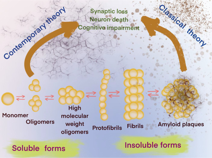
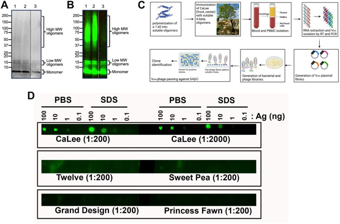
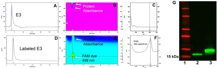
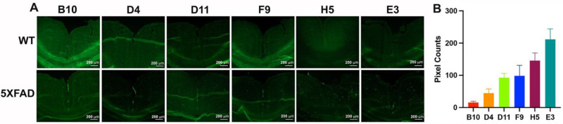
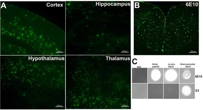
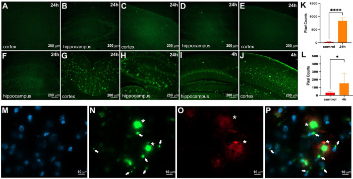
The classical amyloid cascade hypothesis postulates that the aggregation of amyloid plaques and the accumulation of intracellular hyperphosphorylated Tau tangles, together, lead to profound neuronal death. However, emerging research has demonstrated that soluble amyloid-β oligomers (SAβOs) accumulate early, prior to amyloid plaque formation. SAβOs induce memory impairment and disrupt cognitive function independent of amyloid-β plaques, and even in the absence of plaque formation. This work describes the development and characterization of a novel anti-SAβO (E3) nanobody generated from an alpaca immunized with SAβO. In-vitro assays and in-vivo studies using 5XFAD mice indicate that the fluorescein (FAM)-labeled E3 nanobody recognizes both SAβOs and amyloid-β plaques. The E3 nanobody traverses across the blood-brain barrier and binds to amyloid species in the brain of 5XFAD mice. Imaging of mouse brains reveals that SAβO and amyloid-β plaques are not only different in size, shape, and morphology, but also have a distinct spatial distribution in the brain. SAβOs are associated with neurons, while amyloid plaques reside in the extracellular matrix. The results of this study demonstrate that the SAβO nanobody can serve as a diagnostic agent with potential theragnostic applications in Alzheimer's disease.


Conflict of interest statement

BEW and BWS are co-founders of Turkey Creek Biotechnology LLC and have equity ownership. The authors declare no other competing conflicts of interest with the contents of this articles.

Figures

Figure 1.
Figure 1.. Contemporary and classical hypotheses of the amyloid-β cascade mechanisms.
Figure 2.
Figure 2.. Production of nanobody from SAβO-injected alpaca.
(A) Coomassie stained SDS-polyacrylamide gel, and (B) Western analysis to confirm the production of the low molecular weight SAβOs (16 kDa) and some higher molecular species. Lanes 1 and 2 are from two different SAβO preparations; lane 3 is from free Abeta peptide. (C) General procedures for generating antibodies/nanobodies after immunization of alpaca with SAβO antigen. (D) Dot-blot analysis showed that the plasma from the alpaca immunized with SAβO (CaLee) recognizes SAβO antigen, while no response was observed for other non-immunized alpacas (Twelve, Sweety Pea, Grand Design, and Princess Fawn).
Figure 3.
Figure 3.. Representative data for the characterization of a dye-labeled clone.
(A,B,C) Two-dimensional HPLC assay using a Cytiva HiTrap Sepharose column to characterize E3 nanobody and its labeled product. Unlabeled E3 nanobody. (D,E,F) FAM-labeled E3 nanobody. (G) Western blot analysis to show the purity of E3 (lane 2) and confirm FAM-labeled labeled E3 (lane 3). Molecular weight markers are shown in lane 1.
Figure 4.
Figure 4.. Screening for the specificity of the nanobody using 5XFAD mouse brain slides.
(A) Ex-vivo evaluation of SAβO-binding specificity of the nanobody clones using consecutive brain slides obtained from a WT (6-month-old) and a 5XFAD mouse (11-month-old). (B) Ranking of nanobody clones after pixel quantification of 5XFAD brain slides. The threshold of each image was adjusted until the background disappeared using ImageJ. This process was repeated 3 times, and the data were presented as a mean ± SD.
Figure 5.
Figure 5.. Specificity of E3 nanobody in different regions of the 5XFAD brains.
(A) Representative ex-vivo imaging of different regions of the brains of 5XFAD mice (n=3, 10-month-old) after staining with FAM-labeled E3 nanobody. (B) Ex-vivo staining of AF488-labeled 6E10 antibody on a 5XFAD brain slide. (C) Dot-blot analysis of E3 nanobody binding. Lanes 1–3: blots to test the binding specificity of E3 nanobody against different negative control protein and amyloid-β isoforms. Lane 4: immunoblot using the 6E10 antibody to confirm the presence of SAβO in the supernatant of ultracentrifuge 5XFAD brain sample. All ex-vivo staining of brain slides were incubated for 1 h at room temperature.
Figure 6.
Figure 6.. The E3 nanobody can cross the BBB and stain amyloid targets.
Representative data of hippocampus and cortex per cohort. All treatments were performed by intravenous injection via the tail veins. 5XFAD mice (n=3) were treated with E3 nanobody (A,B) resulting in no labeling. WT mice (n=3) were treated with free FAM fluorescent dye (C,D) resulting in no labeling. WT mice (n=3) were treated with FAM-labeled E3 nanobody (E,F) resulting in no labeling. 5XFAD mice (n=5) were treated with FAM-labeled E3 nanobody 24 h before brain collection (G,H) resulting in significant numbers of bright green fluorescent profiles in cortex and hippocampus. 5XFAD mice (n=3) were treated with FAM-labeled E3 Nb 4 h before brain collection (I,J) resulting in fewer bright green fluorescent profiles. The fluorescent pixels were quantified and compared between (A,B) (n=3) versus (G,H) (n=5) at ****p<0.0001 (K). And between (A,B) (n=3) versus (I,J) (n=3) at *p<0.005. Confocal imaging of ex-vivo 5XFAD hippocampus slides: DAPI staining of the nucleus (M). FAM-labeled E3 recognized SAβO (white arrows) and larger amyloid-β plaques (asterisks) (N). Amyloid-β plaques stained by Alexa 647-labeled 6E10 antibody (O). Merging of M, N and O (P) indicates that E3-positive staining around the nuclei (white arrows).

References

    1. Nguyen P. H. et al. Amyloid Oligomers: A Joint Experimental/Computational Perspective on Alzheimer’s Disease, Parkinson’s Disease, Type II Diabetes, and Amyotrophic Lateral Sclerosis. Chem Rev 121, 2545–2647 (2021). 10.1021/acs.chemrev.0c01122 - DOI - PMC - PubMed
    1. De-Paula V. J., Radanovic M., Diniz B. S. & Forlenza O. V. Alzheimer’s disease. Subcell Biochem 65, 329–352 (2012). 10.1007/978-94-007-5416-4_14 - DOI - PubMed
    1. Selkoe D. J. Alzheimer’s disease. Cold Spring Harb Perspect Biol 3, 1–16 (2011). - PMC - PubMed
    1. Cline E. N., Bicca M. A., Viola K. L. & Klein W. L. The Amyloid-beta Oligomer Hypothesis: Beginning of the Third Decade. J Alzheimers Dis 64, S567–S610 (2018). 10.3233/JAD-179941 - DOI - PMC - PubMed
    1. Chromy B. A. et al. Self-assembly of Abeta(1–42) into globular neurotoxins. Biochemistry 42, 12749–12760 (2003). 10.1021/bi030029q - DOI - PubMed

Publication types

LinkOut - more resources