[Preprint]. 2024 Mar 14:2024.03.14.585003.
doi: 10.1101/2024.03.14.585003.

Meiosis-specific functions of kinetochore protein SPC105R required for chromosome segregation in Drosophila oocytes

Meiosis-specific functions of kinetochore protein SPC105R required for chromosome segregation in Drosophila oocytes

Jay N Joshi et al. bioRxiv. .

Update in

Abstract

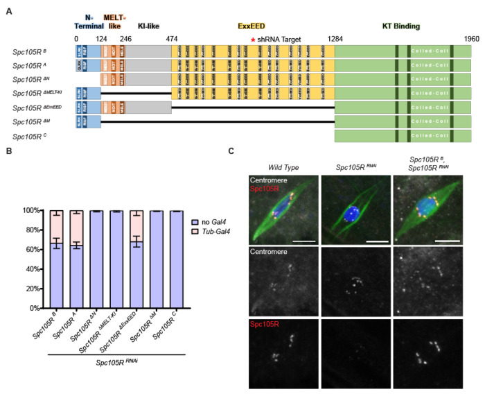
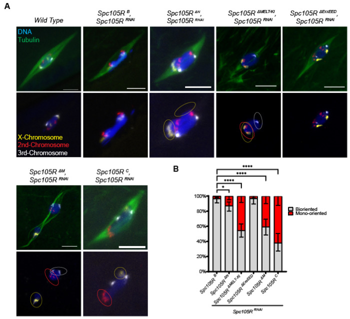
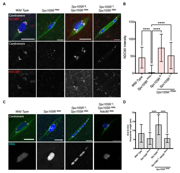
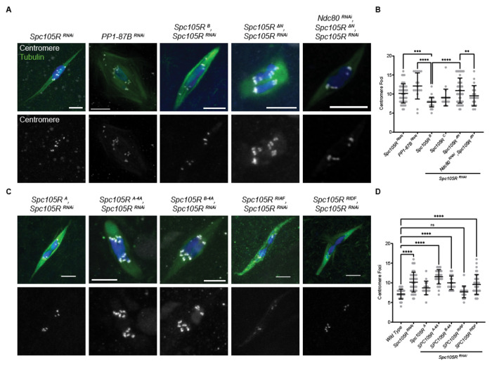
The reductional division of meiosis I requires the separation of chromosome pairs towards opposite poles. We have previously implicated the outer kinetochore protein SPC105R/KNL1 in driving meiosis I chromosome segregation through lateral attachments to microtubules and co-orientation of sister centromeres. To identify the domains of SPC105R that are critical for meiotic chromosome segregation, an RNAi-resistant gene expression system was developed. We found that SPC105R's C-terminal domain (aa 1284-1960) is necessary and sufficient for recruiting NDC80 to the kinetochore and building the outer kinetochore. Furthermore, the C-terminal domain recruits BUBR1, which in turn recruits the cohesion protection proteins MEI-S332 and PP2A. Of the remaining 1283 amino acids, we found the first 473 are most important for meiosis. The first 123 amino acids of the N-terminal half of SPC105R contain the conserved SLRK and RISF motifs that are targets of PP1 and Aurora B kinase and are most important for regulating the stability of microtubule attachments and maintaining metaphase I arrest. The region between amino acids 124 and 473 are required for two activities that are critical for accurate chromosome segregation in meiosis I, lateral microtubule attachments and bi-orientation of homologs.


Figures

Figure 1:
Figure 1:
Spc105RB can rescue most wild-type SPC105R functions. (A) A schematic of the two known Drosophila Spc105R isoforms and mutants used in this study. The first 9 amino acids in SPC105RB are changed from MNANKRRSS to MVDLLFLQ in SPC105R A. The coordinates on the schematic represent the first amino acid of each domain. The N-terminal includes the SLRK and RISF motifs and is where the two isoforms differ. Following this is a domain with three MELT-like motifs, a region that contains two KI-like repeats, a central domain containing repeats with the consensus ExxEED, and the C-terminal region containing coiled-coil motifs. All transgenes included missense mutations to make them resistant to the shRNA GL00392. (B) Viability of Spc105R mutants, all in a Spc105RRNAi background. Data shows the relative amounts of progeny expressing the Spc105RRNAi and mutants (Tub-Gal4) to siblings that did not (no Gal4) (n> 200). (C) Confocal images of wild-type, Spc105RRNAi, and Spc105RB, Spc105RRNAi oocytes. Merged images show DNA (blue), Tubulin (green), centromeres (white), and SPC105R (red). Centromeres and SPC105R are shown in separate channels. Scale bars are 5 μm.
Figure 2:
Figure 2:
SPC105R domains required for homologous chromosome bi-orientation. A) Confocal images of Spc105RRNAi oocytes expressing the indicated transgene. Merged images show DNA (blue), Tubulin (green), the X-Chromosome (Yellow), the second chromosome (Red), and the third chromosome (white). DNA and FISH probes are shown in a separate channel. Scale bars are 5 μm. B) Quantification for percent of chromosome mono-orientation: Spc105R B, Spc105RRNAi oocytes (n=93), Spc105R ΔN, Spc105RRNAi oocytes (n=117), Spc105R ΔExxEED, Spc105RRNAi oocytes (n=65), Spc105R ΔM, Spc105RRNAi oocytes (n=81), Spc105R ΔMELT-KI, Spc105RRNAi oocytes (n=123), Spc105R C, Spc105RRNAi oocytes (n=63). Significance in frequency of mono-orientation om oocytes determined by Fisher’s exact test, with * = p-values < 0.01, **** = p-value < 0.0001.
Figure 3:
Figure 3:
Spc105R’s C-Terminal Domain recruits NDC80. The N-terminal 1284 amino acids of SPC105R is deleted in Spc105R C, retaining only the last 676 amino acids (Figure 1). A) Confocal images of Spc105RRNAi oocytes expressing the indicated transgene, with DNA (blue), Tubulin (green), centromeres (white), and the kinetochore protein NDC80 (red). Centromeres and NDC80 are shown in separate channels. Scale bars are 5 μm. B) Quantification of NDC80 intensity at the centromeres in wild-type (n=68), Spc105RRNAi (n=77), Spc105RB, Spc105RRNAi (n=131), and Spc105RC, Spc105RRNAi (n=107) oocytes. Error bars indicate standard deviation and **** = p-value < 0.0001 by a one-sided unpaired t-test. C) Confocal images of Spc105RRNAi oocytes expressing the indicated transgene, with DNA (blue), Tubulin (green), and centromeres (white). DNA is shown in separate channels. Scale bars are 5 μm. D) Quantification of end-to-end chromosome length of wild-type (n=34), Spc105RRNAi (n=12), Spc105RC, Spc105RRNA (n=22), and Spc105R C, Spc105RRNAi, Ndc80RNAi (n=30) oocytes. Error bars indicate standard deviation, and *** = p-value < 0.001, **** = p-value < 0.0001, ns = not significant by a one-sided unpaired t-test.
Figure 4:
Figure 4:
Spc105R’s C-terminal domain is required for centromeric cohesion protection in meiosis I. A) Confocal images of Spc105RRNAi oocytes expressing the indicated transgene with DNA (blue), Tubulin (green), centromeres (white), and MEI-S332 (red) and shown as a separate channel. Scale bars are 5 μm. B) Quantification of MEI-S332 intensity at the centromeres for wild-type (n=71), Spc105RRNAi (n=202), Spc105RB, Spc105RRNAi (n=53) and Spc105RC, Spc105RRNAi (n=114) oocytes. Error bars indicate standard deviation, and **** = p-value < 0.0001by one-sided unpaired t-test. C) Confocal images of Spc105RRNAi oocytes expressing the indicated transgene, with DNA (blue), Tubulin (green), centromeres (white), and the PP2A subunit HA-WDB (red), also in a separate channel. D) Quantification of WDB intensity at the centromeres in Spc105RB, Spc105RRNAi (n=134), Spc105RRNAi (n=189) and Spc105RC Spc105RRNAi (n=155). Error bars indicate standard deviation and one-sided unpaired t-test show **** = p-value < 0.0001. E) Confocal images of Spc105RRNAi oocytes expressing the indicated transgene, with DNA (blue), Tubulin (green), centromeres (white), and GFP-BUBR1 (red) and in a separate channel. F) Quantification of BUBR1 intensity at the centromeres, in wild-type (n=247), Spc105RRNAi (n=153), Spc105RB, Spc105RRNA (n=67) Spc105RC, Spc105RRNAi (n =149) and Spc105RMELT-KI, Spc105RRNAi (n =89) oocytes. Error bars indicate standard deviation and significance is shown by a one-sided unpaired t-test **** = p-value < 0.0001.
Figure 5:
Figure 5:
SPC105R N-terminal domain interacts with PP1 to maintain sister centromere co-orientation. Mutants with a deletion of the N-terminal domain (Spc105RΔN) or mutations in the SLRK and RISF sequences are shown in Figure S 4A. A) Confocal images of PP1-87BRNAi or Spc105RΔN oocytes. Merged images show DNA (blue), Tubulin (green), and centromeres (white). Centromeres are shown in a separate channel. Scale bars are 5 μm. B) Quantification of centromere foci for images in A. One-sided unpaired t-test showed significance in number of foci. Sample sizes are: 46, 16, 37, 20, 48, 20, 16, in order of the graph. C) Confocal images of oocytes where the RISF site was changed to AAAA, RIAF or RIDF. D) Quantification of centromere foci for images in B. One-sided unpaired t-test showed significance in number of foci between all channels. Sample sizes are: 37, 46, 17, 29, 19, 23, 36, in order of the graph. Error bars indicate standard deviation, **** p < 0.0001, ** p = 0.001.
Figure 6:
Figure 6:
SPC105R regulates interactions with microtubules. A) Confocal images of oocytes in a mei-P22 mutant (mei-P22mut) background. The first image is only Ndc80 RNAi, while the rest express Ndc80RNAi, Spc105RRNAi, and a Spc105R transgene. Due to the absence of crossing over, mei-P22mut mutant oocytes fail to arrest in metaphase, and precociously enter anaphase. B) Oocytes in mei-P22mut, Spc105R RNAi background. The second image is also expressing a Spc105R-mis12 transgene. Merged images show DNA (blue), Tubulin (green), and centromeres (white). Chromosomes are shown in a separate channel. Scale bars are 5 μm. C) Quantification of the frequency of chromosome separation in oocytes. Sample sizes are: 25, 50, 23, 175, 115, 81, 83, 21, in order of the graph. Significance in frequency of oocytes entering precocious anaphase determined by Fisher’s exact test, with ** = p-value < 0.01, **** = p-value < 0.0001, and ns = not significant.
Figure 7:
Figure 7:
The N terminal domain of SPC105R fused to MIS12 A) Confocal images of stage 14 oocytes expressing Spc105RN-Mis12, either in the presence or absence of Spc105RRNAi. The fusion protein was detected using an antibody to the Myc tag (red), along with CENP-C (white), and tubulin (green). B) Spc105RN-MIS12, Spc105RRNAi oocyte, detected using both an antibody to the Myc tag (red) and SPC105R (yellow), along with CENP-C (white). C) Ndc80 (red) does not localize in Spc105RN-MIS12, Spc105RRNAi oocytes. D) Measurement of NDC80 intensity in wild-type (n=137) and Spc105R N-MIS12, Spc105RRNAi (n=160) oocytes. Error bars indicate standard deviation, and one-sided unpaired t-test showed significance **** = p < 0.0001. E) WDB (red) localization in Spc105R N-MIS12, Spc105RRNAi oocytes. F) Measurement of WDB intensity in wild-type (n=154), Spc105RRNAi (n=195), and Spc105RN-MIS12, Spc105RRNAi (n=183) oocytes. Error bars indicate standard deviation and one-sided unpaired t-test showed significance, *** = p < 0.001.

References

    1. Audett M.R., Johnson E.L., McGory J.M., Barcelos D.M., Szalai E.O., Przewloka M.R., and Maresca T.J.. 2022. The microtubule- and PP1-binding activities of Drosophila melanogaster Spc105 control the kinetics of SAC satisfaction. Mol Biol Cell. 33:ar1. - PMC - PubMed
    1. Bajaj R., Bollen M., Peti W., and Page R.. 2018. KNL1 Binding to PP1 and Microtubules Is Mutually Exclusive. Structure. 26:1327–1336 e1324. - PMC - PubMed
    1. Barbosa J., Sunkel C.E., and Conde C.. 2022. The Role of Mitotic Kinases and the RZZ Complex in Kinetochore-Microtubule Attachments: Doing the Right Link. Front Cell Dev Biol. 10:787294. - PMC - PubMed
    1. Bonner A.M., Hughes S.E., Chisholm J.A., Smith S.K., Slaughter B.D., Unruh J R., Collins K.A, Friederichs J.M., Florens L., Swanson S.K., Pelot M.C., Miller D.E., Washburn M.P., Jaspersen S.L., and Hawley R.S.. 2013. Binding of Drosophila Polo kinase to its regulator Matrimony is noncanonical and involves two separate functional domains. Proc Natl Acad Sci U S A. 110:E1222–1231. - PMC - PubMed
    1. Bonner A.M., Hughes S.E., and Hawley R.S.. 2020. Regulation of Polo Kinase by Matrimony Is Required for Cohesin Maintenance during Drosophila melanogaster Female Meiosis. Curr Biol. 30:715–722.e713. - PubMed

Publication types