[Preprint]. 2024 Mar 13:2024.03.12.584718.
doi: 10.1101/2024.03.12.584718.

Autophagosome turnover requires Arp2/3 complex-mediated maintenance of lysosomal integrity

Affiliations

Autophagosome turnover requires Arp2/3 complex-mediated maintenance of lysosomal integrity

Corey J Theodore et al. bioRxiv. .

Abstract

Autophagy is an intracellular degradation process that maintains homeostasis, responds to stress, and plays key roles in the prevention of aging and disease. Autophagosome biogenesis, vesicle rocketing, and autolysosome tubulation are controlled by multiple actin nucleation factors, but the impact of actin assembly on completion of the autophagic pathway is not well understood. Here we studied autophagosome and lysosome remodeling in fibroblasts harboring an inducible knockout (iKO) of the Arp2/3 complex, an essential actin nucleator. Arp2/3 complex ablation resulted in increased basal levels of autophagy receptors and lipidated membrane proteins from the LC3 and GABARAP families. Under both steady-state and starvation conditions, Arp2/3 iKO cells accumulated abnormally high numbers of autolysosomes, suggesting a defect in autophagic flux. The inability of Arp2/3 complex-deficient cells to complete autolysosome degradation and turnover is explained by the presence of damaged, leaky lysosomes. In cells treated with an acute lysosomal membrane-damaging agent, the Arp2/3-activating protein WHAMM is recruited to lysosomes, where Arp2/3 complex-dependent actin assembly is crucial for restoring intact lysosomal structure. These results establish the Arp2/3 complex as a central player late in the canonical autophagy pathway and reveal a new role for the actin nucleation machinery in maintaining lysosomal integrity.


Figures

Figure 1.
Figure 1.. Autophagy receptors and lipidated ATG8-family proteins accumulate in Arp2/3 complex induced knockout (iKO) cells.
(A) Mouse tail fibroblasts (MTFs) were treated with DMSO (Flox) or 4-OHT (iKO) for 6d and collected at 8d. Samples were then lysed, subjected to SDS-PAGE, and immunoblotted with antibodies to NBR1, TAX1BP1, Optineurin, SQSTM1/p62, NDP52, ArpC2, Tubulin, Actin, and GAPDH. (B) For quantification, the relative amounts of Optineurin, p62, and NDP52 were calculated by dividing their respective band intensities by the average loading control intensities. Each bar represents the mean ±SD from n = 3–5 experiments. AU, arbitrary units. (C) Flox and iKO cell extracts were collected at 8d and immunoblotted with antibodies to LC3, GABARAP, and the loading controls. (D) For quantification, the relative amounts of LC3-II and GABARAP-II were calculated by dividing their respective band intensities by the average loading control intensities. Each bar represents the mean ±SD from 3–5 experiments.
Figure 2.
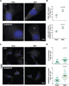
Figure 2.. Deletion of the Arp2/3 complex leads to an increase in mature autophagic rings in fed and starved cells.
(A) MTFs were treated with DMSO (Flox) or 4-OHT (iKO) for 6d and fixed at 8d. Cells were then stained with antibodies to detect LC3 or GABARAP (gray) and with DAPI to visualize DNA (blue). Arrowheads highlight mature autophagosomal rings. (B) The percentage of cells with mature rings was quantified. Large symbols represent the average %s from individual experiments in which 42–61 cells were examined. Lines denote the mean ±SD from n = 3 experiments. (C) Flox and iKO cells were treated with HBSS for 4h on day 8, fixed, and stained as in part (A). (D) Superplots depict the number of mature autophagosomal rings per cell. Small symbols represent the #s within individual cells. Large symbols represent the average #s from individual experiments in which 45–67 cells were examined. Lines denote the mean ±SD from n = 3 experiments.
Figure 3.
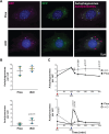
Figure 3.. The increase in autophagic membranes in iKO cells is not due to an increase in autophagosome biogenesis.
(A) Flox and iKO cells were treated with chloroquine (CQ) for 2h on day 8, fixed, and stained with antibodies to LC3 or GABARAP (gray) and DAPI (blue). Arrowheads highlight mature autophagosomal rings. (B) The number of mature rings per cell was quantified. Large symbols represent the average #s from individual experiments in which 30–42 cells were examined. Lines denote the mean ±SD from n = 3 experiments. (C) The fold change in the # of mature rings per cell following CQ treatment was calculated by dividing the value for CQ-treated cells by the value for control media-treated cells. Each large symbol represents the fold change in one experiment, and lines denote the mean ±SD from n = 3 experiments.
Figure 4.
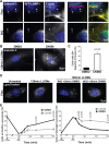
Figure 4.. Arp2/3 complex depletion causes a defect in autolysosome turnover.
(A) MTFs were treated with DMSO (Flox) or 4-OHT (iKO) for 2d, transfected with RFP-GFP-LC3, fixed at 6d, and stained with DAPI (blue). Autophagosomes exhibit both GFP (green) and RFP (magenta) fluorescence (white when merged), whereas autolysosomes display only RFP (magenta) fluorescence. Arrowheads highlight autolysosomes in an iKO cell. (B) The numbers of autophagosomes and autolysosomes per cell were quantified. Large symbols represent the average #s from individual experiments in which 10–23 cells were examined. Lines denote the mean ±SD from n = 5 experiments. (C) Flox and iKO cells were treated with chloroquine (CQ) for 2h on day 6, subjected to a media washout (W/O), fixed at the indicated timepoints, and imaged. Each symbol represents the mean # of autophagosomes or autolysosomes ±SD from n = 3 experiments.
Figure 5.
Figure 5.. Lysosomal damage is increased when the Arp2/3 complex is deleted.
(A) Flox and iKO cells were treated with LysoTracker for 30min and fixed at 8d. (B) Cells were stained with antibodies to IST1 or Galectin-3 (gray) and DAPI (blue). (C) The numbers of bright IST1 or Galectin-3 puncta per cell were quantified. Small symbols represent the #s within individual cells. Large symbols represent the average #s from individual experiments in which 48–85 cells were examined. Lines denote the mean ±SD from n = 3 experiments.
Figure 6.
Figure 6.. Arp2/3 complex-mediated actin assembly is important for lysosome repair.
(A) MTFs expressing RFP-LAMP1 (magenta) were treated with DMSO (Flox) or 4-OHT (iKO) for 6d, exposed to 1mM LLOMe for 15 min, fixed, and stained with antibodies to detect Galectin-3, with phalloidin to visualize filamentous-actin (F-actin), and with DAPI. (B) MTFs were exposed to LLOMe for 2h in media containing either DMSO or CK666, fixed, and stained with Galectin-3 antibodies and DAPI. (C) The percentage of cells with 20 or more Galectin-3 puncta was quantified. Each bar represents the mean ±SD from n = 3 experiments. (D) MTFs were exposed to LLOMe for 2h on day 6, subjected to a washout (W/O) with media containing either DMSO or CK666 for the indicated timepoints and LysoTracker for 30min, fixed, and imaged. (E) The % of cells with either a damaged/diffuse (<3 puncta) or healthy/punctate (>10 puncta) LysoTracker staining pattern was quantified. Each symbol represents the mean % of cells with the depicted LysoTracker phenotype ±SD from n = 3 experiments.
Figure 7.
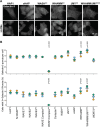
Figure 7.. WHAMM/JMY double knockout (DKO) cells experience increased lysosomal damage following LLOMe treatment.
(A) Parental (HAP1 or eHAP) or CRISPR-engineered knockout (KO) human fibroblasts lacking one or more Arp2/3-activating proteins were cultured in either normal growth media or media containing 0.5mM LLOMe for 3h before being fixed and stained with antibodies to Galectin-3. (B) The numbers of Galectin-3 puncta per cell were quantified. Symbols represent the average #s from individual experiments in which 57–109 cells were examined. Lines denote the mean ±SD from n = 3 experiments.
Figure 8.
Figure 8.. WHAMM is recruited to lysosomes following LLOMe-induced damage.
(A) HAP1 and eHAP cells were cultured in normal media or treated with 0.5mM LLOMe for 3h, fixed, and stained with antibodies to LAMP2 (magenta) and either WASH, JMY, or WHAMM (green). (B) Magnified images of boxes highlighted in (A). (C) The percentage of WASH or WHAMM puncta that co-stained for LAMP2 was quantified. Bars represent the average %s ±SD from independent experiments in which n = 53–56 cells were examined.
Figure 9.
Figure 9.. Model for Arp2/3 complex-mediated lysosomal maintenance in autophagy.
Upon lysosomal damage, WHAMM localizes to the lysosome in normal cells (Flox) and activates the Arp2/3 complex to nucleate actin filaments which mediate lysosomal repair, allowing degradation of autophagosomes during autophagy. In cells lacking the Arp2/3 complex (iKO), actin is not assembled at the lysosome, leading to an increase in recruitment of Galectin-3 and an accumulation of damaged lysosomes which are unable to degrade autophagosomes.

Similar articles

References

    1. Aits S., Kricker J., Liu B., Ellegaard A.-M., Hämälistö S., Tvingsholm S., Corcelle-Termeau E., Høgh S., Farkas T., Holm Jonassen A., et al. (2015). Sensitive detection of lysosomal membrane permeabilization by lysosomal galectin puncta assay. Autophagy 11, 1408–1424. - PMC - PubMed
    1. Alekhina O., Burstein E., and Billadeau D.D. (2017). Cellular functions of WASP family proteins at a glance. Journal of Cell Science 130, 2235–2241. - PMC - PubMed
    1. Aplin A., Jasionowski T., Tuttle D.L., Lenk S.E., and Dunn W.A. Jr. (1992). Cytoskeletal elements are required for the formation and maturation of autophagic vacuoles. Journal of Cellular Physiology 152, 458–466. - PubMed
    1. Bhattacharya A., Mukherjee R., Kuncha S.K., Brunstein M.E., Rathore R., Junek S., Münch C., and Dikic I. (2023). A lysosome membrane regeneration pathway depends on TBC1D15 and autophagic lysosomal reformation proteins. Nature Cell Biology 25, 685–698. - PubMed
    1. Boyle K.B., and Randow F. (2013). The role of ‘eat-me’ signals and autophagy cargo receptors in innate immunity. Current Opinion in Microbiology 16, 339–348. - PubMed

Publication types