

The Vital Role of Clinical Examination in Unmasking Bell's Palsy: Beyond Pattern Recognition
- PMID: 38559504
- PMCID: PMC10981794
- DOI: 10.7759/cureus.55311
The Vital Role of Clinical Examination in Unmasking Bell's Palsy: Beyond Pattern Recognition
Abstract
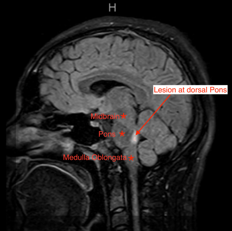
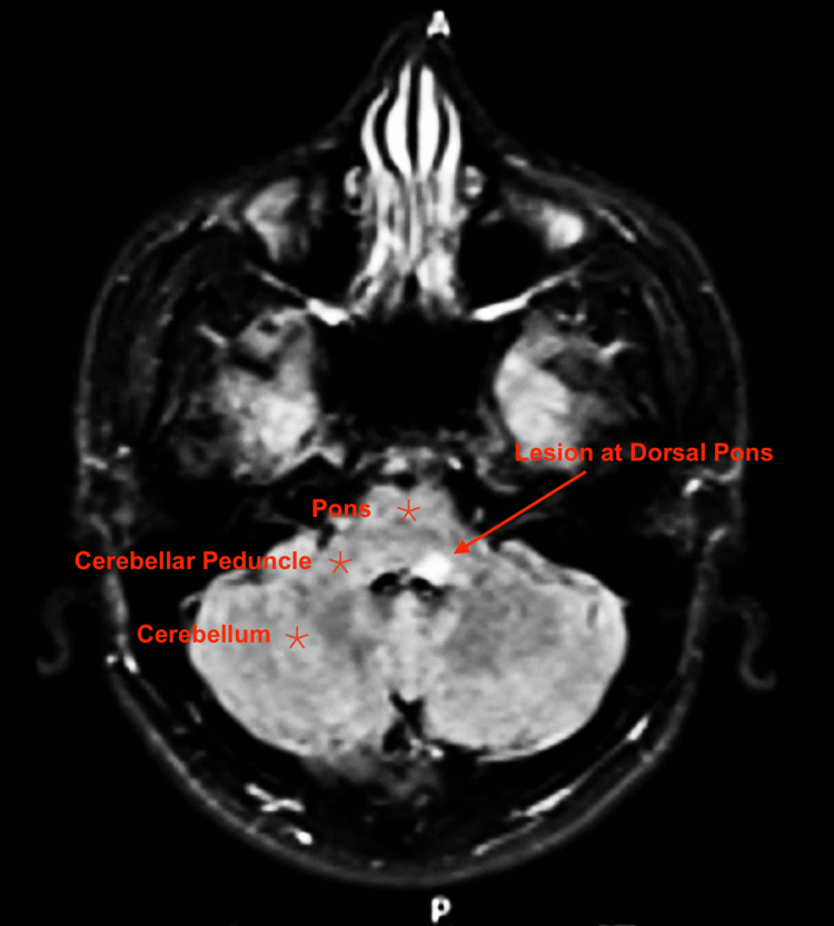
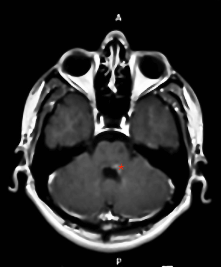
While multiple sclerosis (MS) commonly manifests with optic nerve involvement, it can also masquerade as diverse cranial nerve (CN) palsies. We present the case of a young male initially diagnosed with Bell's palsy based on unilateral facial nerve paralysis. Despite the presence of typical clinical features, the patient's evaluation took an unexpected turn. Subsequent brain MRI revealed demyelinating lesions, ultimately confirming the diagnosis of MS. This case underscores the importance of maintaining vigilance in diagnosing atypical presentations of MS, illustrating how meticulous evaluation and neuroimaging play pivotal roles in uncovering underlying pathologies when conventional diagnoses such as Bell's palsy raise uncertainties.
Keywords: bell`s palsy; facial palsy; neuro deficits; neuro-imaging; s: multiple sclerosis.
Copyright © 2024, Abdelrehim et al.
Conflict of interest statement
The authors have declared that no competing interests exist.
Figures
References
-
- Clinical practice. Bell's palsy. Gilden DH. N Engl J Med. 2004;351:1323–1331. - PubMed
-
- Bell's palsy: the spontaneous course of 2,500 peripheral facial nerve palsies of different etiologies. Peitersen E. Acta Oto-Laryngologica. 2002;549:4–30. - PubMed
-
- Multiple sclerosis. Noseworthy JH, Lucchinetti C, Rodriguez M, Weinshenker BG. N Engl J Med. 2000;343:938–952. - PubMed
-
- Clinical practice guideline: Bell's palsy. Baugh RF, Basura GJ, Ishii LE, et al. Otolaryngol Head Neck Surg. 2013;149:0–27. - PubMed
Publication types
LinkOut - more resources
Full Text Sources