

Addition of a Second Calcaneal Pin for Spanning Ankle External Fixation
- PMID: 38559505
- PMCID: PMC10981796
- DOI: 10.7759/cureus.55312
Addition of a Second Calcaneal Pin for Spanning Ankle External Fixation
Abstract
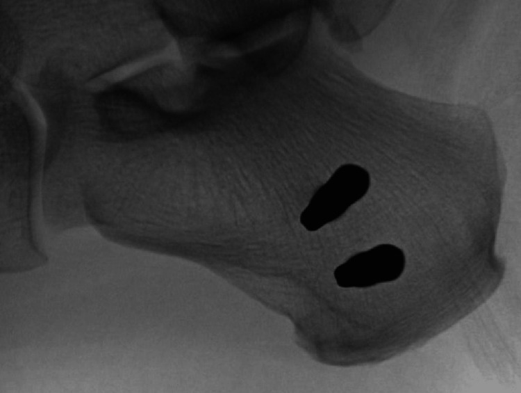
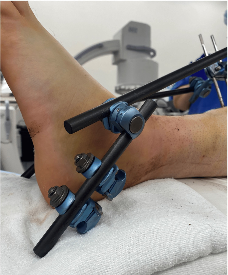
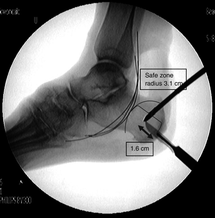
Spanning ankle external fixation is a commonly used technique for the treatment of fractures of the lower extremity. Traditionally, a single pin is placed in the safe zone of the calcaneus to provide a point of traction for fracture reduction and stabilization. Complications include infection and pin loosening with subsequent loss of fracture reduction. We aim to highlight the benefits and techniques of adding a second calcaneal pin to reduce the likelihood of infection, pin loosening, and possible loss of fracture reduction. Using the standard medial-to-lateral placement technique, two centrally threaded Schanz pins were placed within the safe zone of the calcaneus approximately 2 cm apart and were connected by clamps and a short carbon fiber rod. The remainder of the external fixation apparatus is assembled using a standard technique after obtaining fracture reduction. There is an increased incidence of infection and pin loosening with decreased bone quality and a longer duration within an external fixator. The addition of a second calcaneal pin can be used to reduce the incidence of pin loosening and associated sequela, especially in patients with decreased bone quality, thus improving outcomes for patients undergoing spanning ankle external fixation.
Keywords: ankle; calcaneus; external fixation; fracture; technique.
Copyright © 2024, Deluca et al.
Conflict of interest statement
The authors have declared that no competing interests exist.
Figures
References
-
- Tibial shaft fractures - management and treatment options: a review of the current literature. Bode G, Strohm PC, Südkamp NP, Hammer TO. http://www.achot.cz/dwnld/achot_2012_6_499_505.pdf. Acta Chirurgiae Orthopaedicae et Traumatologiae Cechoslovaca. 2012;79:499–505. - PubMed
-
- The role of external fixation in acute foot trauma. Klaue K. Foot Ankle Clin. 2004;9:583-94, x. - PubMed
-
- Operative treatment of femoral shaft fractures in children and adolescents. Beaty JH. Clin Orthop Relat Res. 2005:114–122. - PubMed
-
- Calcaneal tuberosity fractures through prior Schantz pin sites in patients with diabetic neuropathy. Ramsey DC, Laursen RK, Meeker J, Yoo B. Foot (Edinb) 2019;39:96–99. - PubMed
LinkOut - more resources
Full Text Sources