. 2024 Mar 6;9(3):322-338.
doi: 10.1016/j.jacbts.2024.01.006. eCollection 2024 Mar.

Myocardial Matrix Hydrogels Mitigate Negative Remodeling and Improve Function in Right Heart Failure Model

Affiliations

Myocardial Matrix Hydrogels Mitigate Negative Remodeling and Improve Function in Right Heart Failure Model

Jervaughn D Hunter et al. JACC Basic Transl Sci. .

Abstract

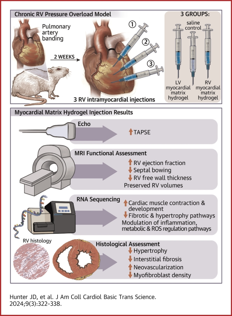
This study evaluates the effectiveness of myocardial matrix (MM) hydrogels in mitigating negative right ventricular (RV) remodeling in a rat model of RV heart failure. The goal was to assess whether a hydrogel derived from either the right or left ventricle could promote cardiac repair. Injured rat right ventricles were injected with either RV-or left ventricular-derived MM hydrogels. Both hydrogels improved RV function and morphology and reduced negative remodeling. This study supports the potential of injectable biomaterial therapies for treating RV heart failure.

Keywords: biomaterial; extracellular matrix; hydrogel; negative right ventricular remodeling; right ventricle.


Conflict of interest statement

This research was funded in part by the NIH National Heart, Lung, and Blood Institute (NHLBI) grant R01HL146147 (Drs Davis and Christman). Dr Hunter was supported by an NIH NHLBI Training Grant (T32HL105373) and an NIH NHLBI Predoctoral Fellowship (1F31HL158212). Dr Christman is co-founder, consultant, board member, and holds an equity interest in Ventrix Bio, Inc. All other authors have reported that they have no relationships relevant to the contents of this paper to disclose.

Figures

None
Graphical abstract
Figure 1
Figure 1
LV MM and RV MM Hydrogels Improve Systolic Function Over Time Echocardiographic imaging shows that both myocardial matrix (MM) hydrogels improve tricuspid annular plane systolic excursion (TAPSE) over saline control and over time. At the time of banding (day –14), all animal groups exhibited healthy TAPSE. At the time of injection (day 0), all banded animals had significantly reduced TAPSE. At day 14 and day 28, animals treated with either the left ventricular (LV)-derived MM hydrogels or the right ventricular (RV)-derived MM hydrogel showed significantly improved TAPSE compared with the saline control at the respective time point. Data are presented as mean ± SEM. N = 9-10. ∗∗∗P < 0.001 compared with saline-treated animals at the respective time point, #P < 0.05 compared with saline-treated animals at the injection time point, and $P < 0.05 compared with the same treatment at the prior time point with 2-way repeated measures analysis of variance with Tukey’s post hoc test.
Figure 2
Figure 2
LV MM and RV MM Hydrogels Mitigate Negative RV Remodeling and Improve RV Function (A) Short axis: representative images of study animals show the right ventricle (upper left of each image) of each treatment group at end-diastole. RV volumes, ejection fraction, and morphology were quantified by assessing changes in RV end-diastolic volume (RVEDV) (B), RV end-systolic volume (RVESV) (C), RV ejection fraction (RVEF) (D), LV end-diastolic eccentricity index (LVEDEI) (E), LV end-systolic eccentricity index (LVESEI) (F), and RV free wall thickness (G). Scale bar = 2 mm. Data are presented as mean ± SEM. N = 8-9. ∗P < 0.05, ∗∗P < 0.01, and ∗∗∗P < 0.001 compared with saline-treated animals with 1-way analysis of variance with Tukey’s post hoc test. Abbreviations as in Figure 1.
Figure 3
Figure 3
Functional Improvements Correlate Between Imaging Modalities Data from echocardiography and magnetic resonance imaging were compared to show the conserved functional benefit of each hydrogel. (A and B) Animals treated with LV MM or RV MM hydrogels showed clear separation from saline control group by exhibiting increased TAPSEs and reduced RVEDV (r = –0.71; P ≤ 0.001) (A) and RVESV (r = –0.87, P ≤ 0.001) (B). (C) Animals treated with the LV MM or RV MM hydrogel also showed clustering improvements in the 2 main measures of RV function (RVEF and TAPSE) (r = 0.85; P ≤ 0.001) being clearly separated from the saline-treated group. (D) Correlation plot reveals that animal treated with LV MM or RV MM hydrogels clustered with thinner right ventricles and higher TAPSEs than saline-treated animals (r = –0.91; P ≤ 0.001). N = 8 to 9. Abbreviations as in Figures 1 and 2.
Figure 4
Figure 4
Differential Gene Expression at 1 Week Postinjection (A) Enhanced volcano plot reveals 98 differentially expressed genes comparing LV MM and saline-treated animals. (B) Enhanced volcano plot shows that animals treated with the RV MM hydrogel exhibited 275 differentially expressed genes compared with saline. Gene ontology (GO) analysis reveals pathways affected by LV MM (C) and RV MM (D) treatments compared with saline-treated animals. N = 5-6. Abbreviations as in Figure 1.
Figure 5
Figure 5
LV MM and RV MM Hydrogels Affect Vascularization and Not Myofibroblast and Macrophage Density at 1 Week Postinjection (A) Representative image of CD68+ (cyan) and CD163+ (red) macrophages in the right ventricle. Scale bar = 20 μm. There were no differences in CD68 density (B), CD163 density (C), or CD163/CD68 ratio (D) between the groups. Representative images of endothelial cell staining (green; isolectin), smooth muscle cells (red; α-smooth muscle actin [SMA]), and nuclei (blue; 4′,6-diamidino-2-phenylindole [DAPI]) of saline-treated (E), LV MM–treated (F), and RV MM–treated (G) hearts. (H) LV MM– and RV MM–treated animals showed greater arteriole growth than saline-treated animals. There was no difference in capillary density (I) or myofibroblast density (J) between the groups. Scale bar = 50 μm. Data are presented as mean ± SEM. ∗P < 0.05 compared with saline-treated animals with 1-way analysis of variance with Tukey’s post hoc test. Abbreviations as in Figure 1.
Figure 6
Figure 6
LV MM and RV MM Hydrogels Reduce Hypertrophy and Interstitial Fibrosis in the RV Free Wall at 4.5 Weeks’ Postinjection Representative images of cardiomyocyte cell boundary staining (red; wheat germ agglutinin) and nuclei (blue; DAPI) of healthy (A), saline-treated (B), LV MM–treated (C), and RV MM–treated (D) hearts. (E) Animals with either MM hydrogel exhibited significantly smaller cardiomyocyte cross-sectional areas than animals treated with saline. Scale bar = 50 μm. Representative images of Masson’s trichrome staining of healthy (F), saline-treated (G), LV MM–treated (H), and RV MM–treated (I) hearts. (J) Animals treated with either MM hydrogel displayed significantly less RV free wall fibrosis. Scale bar = 2 mm. Data are presented as mean ± SEM. ∗P < 0.05 compared with saline-treated animals with 1-way analysis of variance with Tukey’s post hoc test. Abbreviations as in Figures 1 and 5.
Figure 7
Figure 7
LV MM and RV MM Hydrogels Increase Arteriole Density and Reduce Myofibroblast Density in the RV Free Wall at 4.5 Weeks’ Postinjection Representative images of endothelial staining (green; isolectin), smooth muscle cells (red; α-SMA), and nuclei (blue; DAPI) of healthy (A), saline-treated (B), LV MM–treated (C), and RV MM–treated (D) hearts. (E) Animal treated with either the LV MM or the RV MM hydrogel exhibited greater arteriole growth compared with saline. (F) There was no difference in capillary density between the groups. (G) LV MM– and RV MM–treated animals had significantly less myofibroblast density than saline-treated animals. Scale bar = 50 μm. Data are presented as mean ± SEM. ∗∗∗P < 0.001, compared with saline- treated animals with 1-way analysis of variance with Tukey’s post hoc test. Abbreviations as in Figures 1 and 5.

Similar articles

Cited by

References

    1. Ryan J.J., Archer S.L. The right ventricle in pulmonary arterial hypertension: disorders of metabolism, angiogenesis and adrenergic signaling in right ventricular failure. Circ Res. 2014;115:176–188. - PMC - PubMed
    1. Garcia A.M., Beatty J.-T., Nakano S.J. Heart failure in single right ventricle congenital heart disease: physiological and molecular considerations. Am J Physiol Heart Circ Physiol. 2020;318:H947–H965. - PMC - PubMed
    1. Voelkel N.F. In: Right Ventricular Physiology, Adaptation and Failure in Congenital and Acquired Heart Disease. Friedberg M.K., Redington A.N., editors. Springer Science + Business Media; 2018. How does the pressure-overloaded right ventricle adapt and why does it fail? Macro-and micro-molecular perspectives; pp. 19–27.
    1. Reddy S., Bernstein D. Molecular mechanisms of right ventricular failure. Circulation. 2015;132:1734–1742. - PMC - PubMed
    1. Dayer N., Ltaief Z., Liaudet L., Lechartier B., Aubert J.-D., Yerly P. Pressure overload and right ventricular failure: from pathophysiology to treatment. J Clin Med. 2023;12:4722. - PMC - PubMed

LinkOut - more resources