. 2024 Mar 12;2024(2):hoae014.
doi: 10.1093/hropen/hoae014. eCollection 2024.

Extracellular vesicles secreted by human aneuploid embryos present a distinct transcriptomic profile and upregulate MUC1 transcription in decidualised endometrial stromal cells

Affiliations

Extracellular vesicles secreted by human aneuploid embryos present a distinct transcriptomic profile and upregulate MUC1 transcription in decidualised endometrial stromal cells

Sofia Makieva et al. Hum Reprod Open. .

Abstract

Study question: Do extracellular vesicles (EVs) secreted by aneuploid human embryos possess a unique transcriptomic profile that elicits a relevant transcriptomic response in decidualized primary endometrial stromal cells (dESCs)?

Summary answer: Aneuploid embryo-derived EVs contain transcripts of PPM1J, LINC00561, ANKRD34C, and TMED10 with differential abundance from euploid embryo-derived EVs and induce upregulation of MUC1 transcript in dESCs.

What is known already: We have previously reported that IVF embryos secrete EVs that can be internalized by ESCs, conceptualizing that successful implantation to the endometrium is facilitated by EVs. Whether these EVs may additionally serve as biomarkers of ploidy status is unknown.

Study design size duration: Embryos destined for biopsy for preimplantation genetic testing for aneuploidy (PGT-A) were grown under standard conditions. Spent media (30 μl) were collected from euploid (n = 175) and aneuploid (n = 140) embryos at cleavage (Days 1-3) stage and from euploid (n = 187) and aneuploid (n = 142) embryos at blastocyst (Days 3-5) stage. Media samples from n = 35 cleavage-stage embryos were pooled in order to obtain five euploid and four aneuploid pools. Similarly, media samples from blastocysts were pooled to create one euploid and one aneuploid pool. ESCs were obtained from five women undergoing diagnostic laparoscopy.

Participants/materials setting methods: EVs were isolated from pools of media by differential centrifugation and EV-RNA sequencing was performed following a single-cell approach that circumvents RNA extraction. ESCs were decidualized (estradiol: 10 nM, progesterone: 1 µM, cAMP: 0.5 mM twice every 48 h) and incubated for 24 h with EVs (50 ng/ml). RNA sequencing was performed on ESCs.

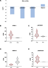
Main results and the role of chance: Aneuploid cleavage stage embryos secreted EVs that were less abundant in RNA fragments originating from the genes PPM1J (log2fc = -5.13, P = 0.011), LINC00561 (log2fc = -7.87, P = 0.010), and ANKRD34C (log2fc = -7.30, P = 0.017) and more abundant in TMED10 (log2fc = 1.63, P = 0.025) compared to EVs of euploid embryos. Decidualization per se induced downregulation of MUC1 (log2fc = -0.54, P = 0.0028) in ESCs as a prerequisite for the establishment of receptive endometrium. The expression of MUC1 transcript in decidualized ESCs was significantly increased following treatment with aneuploid compared to euploid embryo-secreted EVs (log2fc = 0.85, P = 0.0201).

Large scale data: Raw data have been uploaded to GEO (accession number GSE234338).

Limitations reasons for caution: The findings of the study will require validation utilizing a second cohort of EV samples.

Wider implications of the findings: The discovery that the transcriptomic profile of EVs secreted from aneuploid cleavage stage embryos differs from that of euploid embryos supports the possibility to develop a non-invasive methodology for PGT-A. The upregulation of MUC1 in dESCs following aneuploid embryo EV treatment proposes a new mechanism underlying implantation failure.

Study funding/competing interests: The study was supported by a Marie Skłodowska-Curie Actions fellowship awarded to SM by the European Commission (CERVINO grant agreement ID: 79620) and by a BIRTH research grant from Theramex HQ UK Ltd. The authors have no conflicts of interest to declare.

Keywords: aneuploidy; embryo; endometrium; extracellular vesicles; implantation; non-invasive preimplantation genetic testing for aneuploidy.


Conflict of interest statement

None to declare.

Figures

Figure 1.
Figure 1.
Experimental design using human embryo-secreted extracellular vesicles isolated from embryo culture medium. Human embryos created with IVF were cultured in sequential medium between Days 1 and 7 of development. According to this culture system, the embryos were transferred to new medium on Days 3 and 5 of development. The spent medium was collected on these days and EVs were isolated to define two versions of EVs: Day 3-EVs (D3 EVs) denoting the EVs isolated from medium where embryos were cultured between Days 1 and 3 of embryo development and Day 5-EVs (D5 EVs) denoting the EVs isolated from medium where embryos were cultured between Days 3 and 5 of development. We define three groups of EV samples depending on the purpose of experiments. In Group 1, D3 EVs were used for the experiments intended for the characterization of embryo-secreted EVs. Group 2 was used for the exploration of the diagnostic capacities of the RNA cargo in D3 EVs. In this group, all embryos were from PGT-A cycles and were diagnosed as euploid (D3 EVe) or aneuploid (D3 EVa). In Group 3, D5 EVs of diagnosed embryos (D5 EVe or D5 EVa) were used to treat dESCs. Graphic created with BioRender software. EV, extracellular vesicles; PGT-A, preimplantation genetic testing for aneuploidy; EVe, euploid EVs; EVa, aneuploid EVs; dESCs, decidualized endometrial stromal cells.
Figure 2.
Figure 2.
Exploration of human embryo-secreted extracellular vesicle RNA cargo. (A) Embryo-derived EVs were recovered from spent medium where single embryos were cultured for 48 h between Days 1 and 3 of development. Spent media samples from 35 single embryo cultures (i) were pooled to create a single pool (ii), which was used to isolate EVs (Group 1). (B) Representative NTA (nanoparticle tracking analysis) showing concentration (particles/ml) and size (nm) of embryo-derived EVs found in one pool sample (Aii). (C, D) TEM (transmission electron microscopy) showing EVs found in single embryo culture media (Ai). Black arrow indicates a larger EV and white arrow indicates a smaller EV. Scale bar is 200 nm (C) and 100 nm (D). (E) A representative Agilent 4200 TapeStation reading from a Total RNA library prepared using a pool of embryo EVs (Aii). (F) Biotype distribution across the RNAs present in EVs (n = 3 pools). (G) The 60 most abundant RNAs conserved across n = 3 pools of D3 EV and that code for proteins were used as an input in MCL (Markov cluster algorithm) analysis on STRING (protein–protein interaction networks functional enrichment analysis). An inflation parameter of 2 was used to identify four major clusters. The i (yellow) cluster consists of RNAs coding for mitochondrial proteins, the ii (red) RNA ribosomal processing and biogenesis, the iii (green) focal adhesion and intracellular trafficking, and the iv (blue) micromolecule biosynthesis. (H) The chromosomes most represented by the most abundant RNAs were 14 (20%) and mitochondria (MT; 12.7%). EV, extracellular vesicle; L, ladder; lincRNA, long intergenic non-coding RNAs.
Figure 3.
Figure 3.
Correlation of expression matrix between human embryo extracellular vesicle RNA and embryo cell-RNA. Single-cell RNA sequencing raw data on embryos was available in literature from Petropoulos et al. (2016) (A) and Yan et al. (2013) (B) and was used to examine correlation with RNA sequencing data from embryo-derived D3 EVs in our study to validate the hypothesis that EVs are proxy of the tissue of origin. A: Petropoulos et al. (2016) performed RNA sequencing on 13 single cells from embryos desegregated on Day 3 of development. The correlation of the expression matrix shows strong correlation between RNA expression in Day 3 (D3) embryo cells and RNA fragments identified in EVs of Day 3 embryos (n = 3). B: Yan et al. (2013) performed RNA sequencing on zygotes (n = 3) and single cells from different stages of embryo development: 2-cell (n = 3), 4-cell (n = 3) and 8-cell (n = 3). The correlation of the expression matrix shows strong correlation of RNA expression in 4- and 8-cell embryos with RNA fragments in D3 EVs. EV, extracellular vesicle.
Figure 4.
Figure 4.
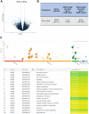
Differential abundance in extracellular vesicle RNA cargo between euploid and aneuploid Day 3 embryos. (A, B) Differential gene expression analysis with SEQC cut-off (sequencing quality control raw P-value < 0.01, fold change: 1) showing the genes differentially abundant between D3 EVs collected from aneuploid embryos (D3 EVa; n = 4 biological replicates) and the ones collected from euploid embryos (D3 EVe; n = 5 biological replicates). A: Volcano plot. B: The comparison was defined as ‘EVa vs. EVe’, using the euploid embryos-derived EVs as reference. The number of genes differentially abundant when implementing two different cut-offs is indicated. (C) Enrichment overrepresentation analysis results of D3 EVa and D3 EVe transcriptomes: GOSt multiquery plot of Manhattan. All differentially abundant genes were selected and tested against BP, CC, Reactome, and Wikipathway collections. The most significant results (FDR < 0.05) are highlighted. EV, extracellular vesicle; BP, biological process; CC, cellular component; FDR,false discovery rate; GOSt, gene ontology statistics.
Figure 5.
Figure 5.
Differentially abundant RNAs between aneuploid and euploid extracellular vesicles. According to differential gene expression analysis, five RNA species showed differential abundance (FDR: P adjusted < 0.05) in EVa (A). In lower abundance: PPM1J (B; FDR = 0.010), LINC00561 (C; FDR = 0.010), and ANKRD34C (D; FDR = 0.016) while in higher abundance: TMED10 (E; FDR = 0.025). CMP, counts per million; FDR, false discovery rate; PPM1J, protein phosphatase, Mg2+/Mn2+-dependent 1J; LINC00561, long intergenic non-protein coding RNA 561; ANKRD34C, ankyrin repeat domain 34C; TMED10, transmembrane p24 trafficking protein 10; EVa, aneuploid extracellular vesicles; EVe, euploid extracellular vesicles.
Figure 6.
Figure 6.
The response of decidualized endometrial cells to the uptake of extracellular vesicles secreted from Day 5 euploid and aneuploid embryos. (A, B) Individual EVe and EVa samples (not pools) were analyzed with ΝTA. A: NTA showing concentration (particles/ml) and size (nm) of D5 EVe (n = 5, red line) and D5 EVa (n = 5, grey line) embryos. B: Quantification of the NTA results; EVe mean concentration: 48868407976 ± 5443394193 particles/ml and EVa mean concentration: 33595295619 ± 3743527687 particles/ml (p = ns, fc = 1.45), ± values denote SEM. (C, D) Successful decidualization of ESCs is confirmed by DGE analysis between dESCs and ESCs. C: Volcano plot showing that decidualization of ESCs caused a remarkable transcriptomic response characterized by the upregulation of 1023 and downregulation of 1111 genes (n = 5 patients, FDR <0.05). D: Known markers of decidualization were upregulated in cells treated with the decidualization protocol: PRL (prolactin; FDR < 0.0001), IGFBP1 (insulin-like growth factor-binding protein 1; FDR < 0.0001), LIFR (leukemia inhibitory factor receptor; FDR = 0.02), IL11 (FDR < 0.0001), FOXO1 (forkhead box protein O1; FDR = 0.001). (E, F) DEG comparing dESCs treated with D5 EVe and D5 EVa (50 μg/ml; 24 h; n = 5 patients, cut-off set FDR < 0.05). E: Volcano plot showing MUC1(mucin 1) as the only gene upregulated in dESCs treated with D5 EVa (dot shown with arrow). F: MUC1 gene differential expression in the following comparisons: ESCs versus dESCs (log2FC = −0.54, FDR = 0.0028) and dESCs treated with D5 EVe versus dESCs treated with D5 EVa (log2FC = 0.85, FDR = 0.0201). MUC1 was downregulated in dESCs (when compared to ESCs) and upregulated in dESCs treated with EVa (when compared to EVe). EV, extracellular vesicle; FDR, false discovery rate; EVe, euploid EVs; EVa, aneuploid EVs; NTA, nanoparticle tracking analysis; DGE, differential gene expression; ESCs, endometrial stromal cells not decidualized; dESCs, decidualized endometrial stromal cells.
Figure 7.
Figure 7.
Gene set enrichment analysis of gene expression comparison of decidualized endometrial stromal cells treated with Day 5 extracellular vesicles secreted from aneuploid or euploid embryos. All the expressed genes were sorted based on their log2FC value for comparison between dESCs + EVa and dESCs + EVe, resulting in a pre-ranked gene list that was further used in a GSEA gene ontology (GO), cell component (CC), or pathway analysis. Only categories with an FDR < 0.05 and a minimum of 20 genes are presented. The NES (normalized enrichment score) of top enriched (blue bars) and top depleted (orange bars) terms in dESCs + EVa compared to dESCs + EVe are listed, categorized by GO, CC, or pathway annotations. WEB-based GEne SeT AnaLysis Toolkit (WebGestalt, http://www.webgestalt.org/) was utilized to perform the GSEA. dESCs, decidualized endometrial stromal cells; EVa, euploid EVs; EVa, aneuploid EVs; GSEA, gene set enrichment analysis; FDR, false discovery rate.

References

    1. Aleksejeva E, Zarovni N, Dissanayake K, Godakumara K, Vigano P, Fazeli A, Jaakma Ü, Salumets A.. Extracellular vesicle research in reproductive science: Paving the way for clinical achievements†. Biol Reprod 2022;106:408–424. - PubMed
    1. Belandres D, Shamonki M, Arrach N.. Current status of spent embryo media research for preimplantation genetic testing. J Assist Reprod Genet 2019;36:819–826. - PMC - PubMed
    1. Capra E, Lange-Consiglio A.. The biological function of extracellular vesicles during fertilization, early embryo-maternal crosstalk and their involvement in reproduction: review and overview. Biomolecules 2020;10:1510. - PMC - PubMed
    1. Cimadomo D, Rienzi L, Conforti A, Forman E, Canosa S, Innocenti F, Poli M, Hynes J, Gemmell L, Vaiarelli A. et al. Opening the black box: why do euploid blastocysts fail to implant? A systematic review and meta-analysis. Hum Reprod Update 2023;29:570–633. - PubMed
    1. Chauhan M, Balakrishnan M, Chan R, Yallampalli C.. Adrenomedullin 2 (ADM2) regulates mucin 1 at the maternal-fetal interface in human pregnancy. Biol Reprod 2015;93:136. - PMC - PubMed