. 2021 Dec 28:13:100081.
doi: 10.1016/j.ahjo.2021.100081. eCollection 2022 Jan.

Intracardiac masses: Single center experience within 12 years: I-MASS Study

Affiliations

Intracardiac masses: Single center experience within 12 years: I-MASS Study

Zehra Bugra et al. Am Heart J Plus. .

Abstract

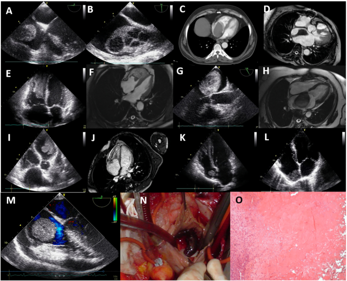
Objective: The aim of this cross-sectional, retrospective, descriptive study was to review and classify cardiac masses systematically and to determine their frequencies.

Methods: The medical records of 64,862 consecutive patients were investigated within 12 years. Every patient with a cardiac mass imaged by transthoracic echocardiography (TTE) and confirmed with an advanced imaging modality such as transesophageal echocardiography (TEE), computed tomography (CT) and/or cardiac magnetic resonance imaging (CMR) was included. Acute coronary syndromes triggering thrombus formation, vegetations, intracardiac device and catheter related thrombi were excluded.

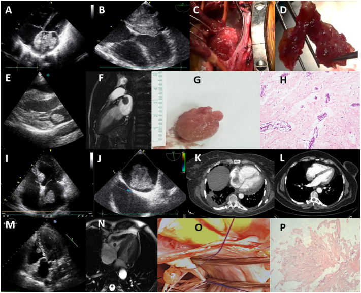
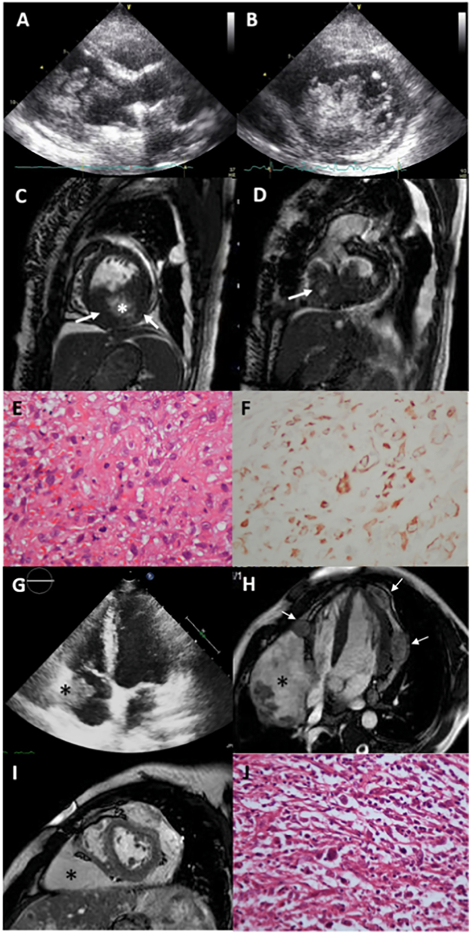
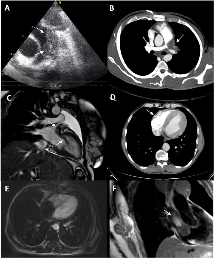
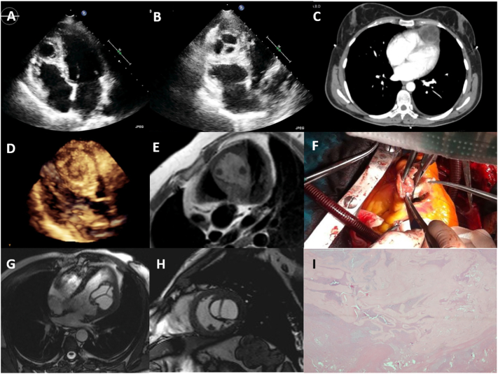
Results: Data demonstrated 127 (0.195%) intracardiac masses consisting of 33 (0.050%) primary benign, 3 (0.004%) primary malignant, 20 (0.030%) secondary tumors, 3 (0.004%) hydatid cysts and 68 (0.104%) thrombi respectively. The majority of primary cardiac tumors were benign (91.67%), predominantly myxomas (78.79%), and the less malignant (8.33%). Secondary cardiac tumors were common than the primary malignant tumors (20:3), with male dominancy (55%), lymphoma and lung cancers were the most frequent. Intracardiac thrombi was the majority of the cardiac masses, thrombi accompanying malignancies were in the first range (n = 17, 25%), followed by autoimmune diseases (n = 13, 19.12%) and ischemic heart disease with low ejection fraction (n = 12, 17.65%).

Conclusions: This retrospective analysis identified 127 patients with cardiac masses. The majority of benign tumors were myxoma, the most common tumors that metastasized to the heart were lymphoma and lung cancers, and the thrombi associated with malignancies and autoimmune diseases were the most frequent.

Keywords: Cardiac cysts; Cardiac thrombus; I-MASS Study; Primary cardiac tumors; Secondary cardiac tumors.


Conflict of interest statement

The authors declare that they have no known competing financial interests or personal relationships that could have appeared to influence the work reported in this paper.

Figures

Fig. 1
Fig. 1
Primary benign tumors of the heart. (A) 2D TTE giant left atrial myxoma; (B) 2D TEE polylobulated myxoma, attached with a pedicle to the fossa ovalis (C) intraoperative view; (D) gross examination of the tumor, a dark red, fragile, polylobulated mass; (E) 2D TTE left atrial mobile, well circumscribed, ball shaped myxoma; (F) CMR SSFP cine, a hypointense mass with a short pedicle originating from the superior wall of the left atrium; (G) gross view of the surgically resected mass, smooth surface, bright pink colour (H) histology shows intense perivascular benign neoplastic cells and rare same type cells in myxoid stroma, compatible with myxoma, hematoxylin-eosin × 100; (I) 2D TTE pediculated left atrial myxoma originating from the interatrial septum; (J) 2D TEE, pediculated left atrial myxoma adjacent to the superior vena cava and patent foramen ovale; (K) CT, right atrial lobulated myxoma originating from fossa ovalis; (L) CT, myxoma originating from the posterior right atrial wall; (M) 2D TTE, a well-defined, nodular, mobile papillary fibroelastoma, originating from the anterior tricuspid leaflet chorda in the right ventricle; (N) CMR SSFP cine of the papillary fibroelastoma; (O) gross examination of the surgically resected papillary fibroelastoma, a translucent and gelatinous mass; (P) histology shows a benign tumor with multiple papillary fronds of different size that consisted of an acellular matrix and surrounded by single layer of endothelial cells, hematoxylin-eosin × 100. 2D TTE: two-dimensional transthoracic echocardiography; 2D TEE: two-dimensional transesophageal echocardiography; CMR: cardiac magnetic resonance imaging; CT: computed tomography; SSFP: steady state free processing.
Fig. 2
Fig. 2
Primary malignant tumors of the heart. Left ventricular endocavitary rhabdomyosarcoma; (A) 2D TTE, a polylobulated, mobile tumor filling the left ventricle; (B) 2D TTE short-axis view, tumor mass and extensions; (C) CMR, tumor originating from the inferior wall of the left ventricle (arrow) with cystic and necrotic areas at the centrum (*); (D) tumor also infiltrates the right ventricle (arrow); (E) histology: pleomorphic malign tumor cells of rhabdomyosarcoma, mitotic figures and atypical cells, hematoxylin-eosin × 200; (F) positive desmin immunochemistry in tumoral cells × 200. High grade angiosarcoma; (G) 2D TTE tumor mass (*) in the right atrial cavity, infiltrating right atrial wall, (H) contrast-enhanced SSFP cine CMR, nodular lesions of the tumor (arrows) infiltrating left and right ventricular myocardium and epicardium, with a large mass of tumor in the atrioventricular groove and right ventricular myocardium (*) narrowing the right heart chambers; (I) CMR, short axis SSFP, large tumor mass (*); (J) High grade angiosarcoma, irregular malignant spindle cells haphazardly arranged, hematoxylin-eosin × 40. 2D TTE: two-dimensional transthoracic echocardiography; CMR: cardiac magnetic resonance imaging, SSFP: steady state free processing.
Fig. 3
Fig. 3
Secondary malignant tumors of the heart. (A) 2D TTE short axis, main PA infiltration of the B-cell lymphoma with granular appearance; (B) contrast CT, lymphoma infiltrating the main PA; (C) CMR, malignant mesenchymal intrabronchial tumor, cardiac invasion of the tumor via left pulmonary vein (18, with permission); (D) CT, right ventricular wall invasion of the renal cell carcinoma; (E) CMR, hepatocellular carcinoma, almost filling the whole right atrial cavity; (F) CMR, right atrial invasion of the papillary thyroid cancer. 2D TTE: two-dimensional transthoracic echocardiography; PA: pulmonary artery; CMR: cardiac magnetic resonance imaging; CT: computed tomography.
Fig. 4
Fig. 4
Cardiac hydatid cysts with various myocardial locations. (A and B) 2D TTE, (C) contrast-enhanced thoracic CT scan, a multiseptated anechoic cyst located at the apex; (D) 3D TTE, a granulated spheric mass, with patchy hypoechoic regions located within the interventricular septum, (19, with permission); (E) CMR, hydatid cyst located within the interventricular septum with water content and a few daughter cysts; (F) gross examination of the surgically resected hydatid cyst; (G) SSFP cine (20, with permission) (H) CMR, showing left ventricular hydatid cyst with septations originating from the lateral wall into the left ventricular cavity (I) histological examination of the echinococcal lesion with cuticular membranes in cystic content, hematoxylin-eosin × 40. 2D TTE: two-dimensional transthoracic echocardiography; CT: computed tomography; 3D TTE: three-dimensional transthoracic echocardiography; CMR: cardiac magnetic resonance imaging; SSFP: steady state free processing.
Fig. 5
Fig. 5
Intracardiac thrombi with noncardiac disorders. Non small cell lung cancer: (A) 2D TEE, a large thrombus in the right atrium with lytic areas; (B) 2D TEE, thrombus extending from vena cava superior through the right atrial cavity; renal cell carcinoma: (C) CT, thrombus almost completely filling the left atrium; endometrial cancer: (D) CMR, a thrombus extending from vena cava inferior through the right atrium; esophageal cancer: (E) 2D TTE, and (F) CMR, a thrombus filling the right ventricular cavity and extending through the mid ventricular level; ovarian cancer: (G) 2D TEE, and (F) CMR, left atrial thrombus with hypoechoic regions; Behçet's disease: (I) 2D TTE, a massive, lobulated, mobile left ventricular thrombus; (J) CMR, an immobile thrombus infiltrating the right atrial side of the tricuspid valve and endocardial fibroelastosis at the right ventricular wall; rheumatoid arthritis: (K) 2D TTE, right atrial immobile thrombus; systemic lupus erythematosus with antiphospholipid syndrome: (L) 2D TTE, right atrial immobile thrombus; chronic kidney disease, hemodialysis: (M) 2D TEE, right atrial giant, ball shaped thrombus, moderate pericardial effusion; (N) surgical image (O) thrombus histology with organized fibrin mass, hematoxylin-eosin × 200. 2D TTE: two-dimensional transthoracic echocardiography; 2D TEE: two-dimensional transesophageal echocardiography; CMR: cardiac magnetic resonance imaging.

References

    1. Reynen K. Frequency of primary tumors of the heart. Am. J. Cardiol. 1996;77:107. - PubMed
    1. Gowda R.M., Khan I.A., Nair C.K., Mehta N.J., Vasavada B.C., Sacchi T.J. Cardiac papillary fibroelastoma: a comprehensive analysis of 725 cases. Am. Heart J. 2003;146:404–410. - PubMed
    1. Strecker T., Rösch J., Weyand M., Agaimy A. Primary and metastatic cardiac tumors: imaging characteristics, surgical treatment, a histopathological spectrum: a 10 year-experience at a german heart center. Cardiovasc. Pathol. 2012;21:436–443. - PubMed
    1. Hudzik B., Miszalski-Jamka K., Glowacki J., Glowacki J., Lekston A., Gierlotka M., Zembala M., Polonski L., Gasior M. Malignant tumors of the heart. Cancer Epidemiol. 2015;39:665–672. - PubMed
    1. Yu L., Gu T., Shi E. Echocardiographic findings and clinical correlation with cardiac myxoma. J. Am. Coll. Cardiol. Imaging. 2016;9:618–620. - PubMed

LinkOut - more resources