

Pediatric Synovial Osteochondromatosis of the Knee with Leg Length Discrepancy: A Case Report
- PMID: 38560319
- PMCID: PMC10976523
- DOI: 10.13107/jocr.2024.v14.i03.4280
Pediatric Synovial Osteochondromatosis of the Knee with Leg Length Discrepancy: A Case Report
Abstract
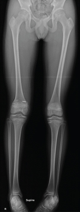
Introduction: Synovial chondromatosis (SC) is very rare among children. We are aware of no reports of patients with SC accompanied by leg length discrepancy (LLD).
Case report: We describe a case of synovial osteochondromatosis of a 7-year-old boy complicated by LLD. We performed epiphysiodesis of the distal femur and arthroscopic resection of loose bodies and total synovectomy. Three years after surgery, LLD had been corrected and there was no sign of recurrence.
Conclusion: Physicians should be aware of synovial osteochondromatosis complicated by LLD in childhood and take radiographs of the whole length of lower legs when this condition is suspected.
Keywords: Synovial osteochondromatosis; child; knee joint; leg length discrepancy.
Copyright: © Indian Orthopaedic Research Group.
Conflict of interest statement
Conflict of Interest: Nil
Figures


Similar articles
-
The use of posteromedial portal for arthroscopic treatment of synovial chondromatosis of the knee: a case report.J Med Case Rep. 2022 Dec 10;16(1):457. doi: 10.1186/s13256-022-03667-2. J Med Case Rep. 2022. PMID: 36494697 Free PMC article.
-
Arthroscopic Management of Synovial Osteochondromatosis of the Hip.Orthopedics. 2015 Jun;38(6):e536-8. doi: 10.3928/01477447-20150603-91. Orthopedics. 2015. PMID: 26091229
-
A Missed Case of Synovial Osteochondromatosis.Cureus. 2023 Nov 8;15(11):e48498. doi: 10.7759/cureus.48498. eCollection 2023 Nov. Cureus. 2023. PMID: 38073924 Free PMC article.
-
Arthroscopic Retrieval of over 100 Loose Bodies in Shoulder Synovial Chondromatosis: A Case Report and Review of Literature.Orthop Surg. 2016 Nov;8(4):511-515. doi: 10.1111/os.12294. Orthop Surg. 2016. PMID: 28032713 Free PMC article. Review.
-
Synovial osteochondromatosis mimicking juvenile idiopathic arthritis in an adolescent: a case-based review.Clin Rheumatol. 2022 Aug;41(8):2571-2580. doi: 10.1007/s10067-022-06224-w. Epub 2022 May 31. Clin Rheumatol. 2022. PMID: 35641775 Free PMC article. Review.
Cited by
-
Inflammation-induced leg length discrepancy in children: from molecular mechanisms to clinical implications.Front Med (Lausanne). 2025 May 20;12:1542822. doi: 10.3389/fmed.2025.1542822. eCollection 2025. Front Med (Lausanne). 2025. PMID: 40463981 Free PMC article. Review.
-
Neglected knee swelling: A case report of massive synovial chondromatosis.Int J Surg Case Rep. 2024 Dec;125:110636. doi: 10.1016/j.ijscr.2024.110636. Epub 2024 Nov 21. Int J Surg Case Rep. 2024. PMID: 39577318 Free PMC article.
-
Bilateral Knee Synovial Chondromatosis in a 2-Year-Old Girl: First Reported Case and Literature Review.Am J Case Rep. 2025 Mar 19;26:e945921. doi: 10.12659/AJCR.945921. Am J Case Rep. 2025. PMID: 40106396 Free PMC article. Review.
References
-
- Milgram JW. Synovial osteochondromatosis:A histopathological study of thirty cases. J Bone Joint Surg Am. 1977;59:792–801. - PubMed
-
- Maurice H, Crone M, Watt I. Synovial chondromatosis. J Bone Joint Surg Br. 1988;70:807–11. - PubMed
-
- Davis RI, Hamilton A, Biggart JD. Primary synovial chondromatosis:A clinicopathologic review and assessment of malignant potential. Hum Pathol. 1998;29:683–8. - PubMed
-
- Carey RP. Synovial chondromatosis of the knee in childhood. A report of two cases. J Bone Joint Surg Br. 1983;65:444–7. - PubMed
-
- Kistler W. Synovial chondromatosis of the knee joint:A rarity during childhood. Eur J Pediatr Surg. 1991;1:237–9. - PubMed
Publication types
LinkOut - more resources
Full Text Sources