. 2024 Apr 1;14(1):7632.
doi: 10.1038/s41598-024-58004-y.

CircGPC3 promotes hepatocellular carcinoma progression and metastasis by sponging miR-578 and regulating RAB7A/PSME3 expression

Affiliations

CircGPC3 promotes hepatocellular carcinoma progression and metastasis by sponging miR-578 and regulating RAB7A/PSME3 expression

Linling Ju et al. Sci Rep. .

Abstract

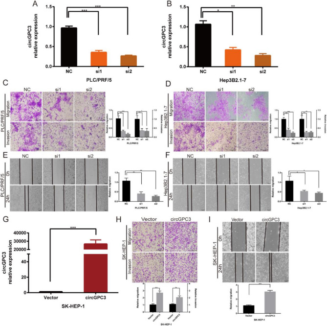
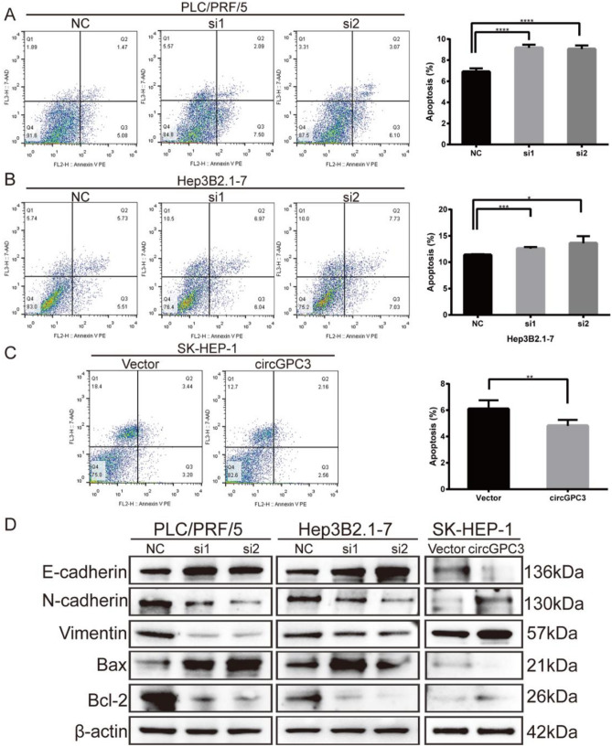
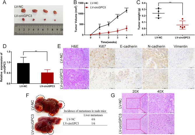
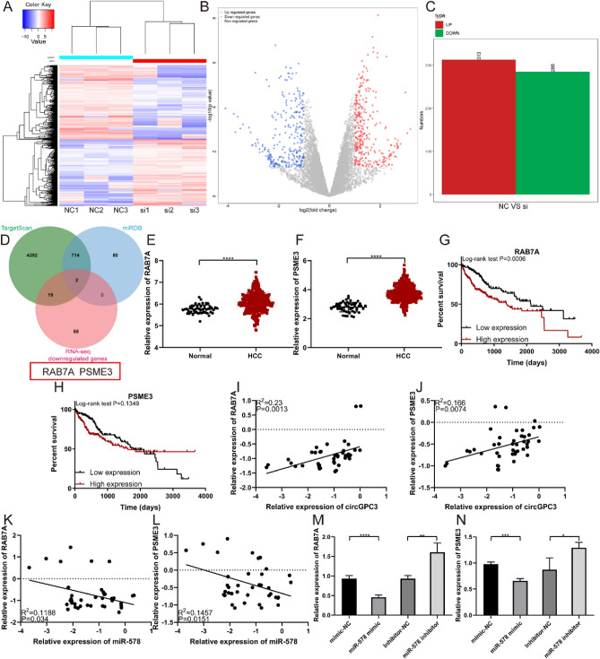
CircRNAs are a class of highly stable noncoding RNAs that play an important role in the progression of many diseases, especially cancer. In this study, high-throughput sequencing was used to screen for abnormally expressed circRNAs, and we found that circGPC3 was overexpressed in HCC tissues. However, the underlying mechanism of circGPC3 in the development and metastasis of hepatocellular carcinoma (HCC) remains unknown. In our study, we found that circGPC3 was significantly upregulated in HCC tissues and cells and that its overexpression was positively correlated with overall survival, TNM stage and lymph node metastasis. In vivo and in vitro experiments showed that circGPC3 knockdown repressed HCC cell migration, invasion and proliferation and promoted apoptosis. Mechanistically, circGPC3 promoted HCC proliferation and metastasis through the miR-578/RAB7A/PSME3 axis. Our results demonstrate that circGPC3 contributes to the progression of HCC and provides an intervention target for HCC.


Conflict of interest statement

The authors declare no competing interests.

Figures

Figure 1
Figure 1
Expression and clinical significance of circGPC3 in HCC. (A) A circRNA microarray was used to identify the differentially expressed circRNAs between HCC and matched adjacent normal tissues. (B) qRT‒PCR was used to evaluate the expression of circGPC3 in paired HCC and matched adjacent normal tissues. (C) FISH images of circGPC3 expression in paired HCC and matched adjacent normal tissues. The nuclei were stained with DAPI (blue), and cytoplasmic circGPC3 was stained red. Right panel, scale bar 50 μm. (D) Kaplan–Meier analysis of the OS rate in HCC patients with high or low expression of circGPC3. (E) Representative FISH images of circGPC3 expression in HCC tissues with different TNM stages. **p < 0.01.
Figure 2
Figure 2
Identification of circGPC3 expression and analysis of its characteristics in HCC cells. (A) qRT‒PCR was used to evaluate the expression of circGPC3 in HCC cell lines and a normal liver cell line. (B) Convergent and divergent primers were used to amplify the back-spliced and linear products to verify the circular form of circGPC3 using PLC/PRF/5 and Hep3B2.1-7 cells. (C) The back-splice junction of circGPC3 was identified by Sanger sequence analysis. (D) FISH assay was performed to observe the subcellular localization of circGPC3. (E) qRT‒PCR was used to measure the expression of circGPC3 in the nucleus and cytoplasm of PLC/PRF/5 cells. (F, G) The expression of circGPC3 and GPC3 mRNA was measured by qRT‒PCR in PLC/PRF/5 and Hep3B2.1-7 cells treated with or without RNase R. (H) Verification of the circular form of circGPC3 after treatment with RNase R by agarose gel electrophoresis. *p < 0.05, **p < 0.01, ****p < 0.0001, ns, not significant.
Figure 3
Figure 3
circGPC3 knockdown inhibits HCC cell migration and invasion. (A, B) qRT‒PCR was used to determine the expression of circGPC3 in PLC/PRF/5 and Hep3B2.1-7 cells transfected with two independent siRNAs targeting circGPC3. (C, D) Transwell assays were used to evaluate the migratory and invasive capabilities of PLC/PRF/5 and Hep3B2.1-7 cells when circGPC3 was downregulated. (E, F) Wound healing assays at 0 h and 24 h were performed to assess cell migratory capability after knocking down circGPC3 in PLC/PRF/5 and Hep3B2.1-7 cells. (G) qRT‒PCR was used to determine the expression of circGPC3 in SK-HEP-1 cells transfected with the circGPC3 overexpression plasmid. (H) Transwell assays were used to evaluate the migratory and invasive capabilities of SK-HEP-1 cells when circGPC3 was upregulated. (I) Cell migratory capability was assessed by the wound healing assay after upregulating the expression of circGPC3 in SK-HEP-1 cells. *p < 0.05, **p < 0.01, ***p < 0.001.
Figure 4
Figure 4
circGPC3 knockdown inhibits HCC cell proliferation. (A, B) EdU assays were used to evaluate the proliferative capacities of PLC/PRF/5 and Hep3B2.1-7 cells when circGPC3 was downregulated. (C, D) Colony formation assays were used to evaluate the proliferative capacities of PLC/PRF/5 and Hep3B2.1-7 cells. (E) EdU assays were used to evaluate the proliferative capacities of SK-HEP-1 cells when circGPC3 was upregulated. (F) Colony formation assays were used to evaluate the proliferative capacities of SK-HEP-1 cells. *p < 0.05, **p < 0.01, ***p < 0.001.
Figure 5
Figure 5
circGPC3 knockdown promotes HCC cell apoptosis. (A, B) Cell apoptosis was measured by flow cytometry in PLC/PRF/5 and Hep3B2.1-7 cells transfected with siRNAs. (C) Apoptosis in SK-HEP-1 cells was analyzed by flow cytometry when circGPC3 was upregulated. (D) Western blot analysis was performed to detect the protein levels of E-cadherin, N-cadherin, vimentin, Bax and Bcl-2 in PLC/PRF/5, Hep3B2.1-7 and SK-HEP-1 cells after circGPC3 knockdown or overexpression. *p < 0.05, **p < 0.01, ***p < 0.001, ****p < 0.0001.
Figure 6
Figure 6
circGPC3 knockdown suppresses HCC growth and metastasis in vivo. (A) Image of subcutaneous tumors in nude mice after PLC/PRF/5 cell injection. (B) The volume of tumors in nude mice was calculated weekly for 4 weeks. (C) The weight of tumors was recorded after dissection of the tumors. (D) qRT‒PCR was used to detect the expression of circGPC3 in both the LV-circGPC3 and LV-NC groups. (E) H&E stained images of transplanted tumor tissues and IHC stained images of tumor tissues stained with Ki-67, E-cadherin, N-cadherin and vimentin. Scale bar, 100 μm. (F) Images of liver metastasis in nude mice after injection of PLC/PRF/5 cells stably transfected with LV-circGPC3 and LV-NC vectors into the spleen. (G) H&E stained images of micrometastases. Scale bar: left, 200 μm; right, 100 μm. **p < 0.01, ***p < 0.001, ****p < 0.0001.
Figure 7
Figure 7
CircGPC3 regulates HCC growth and metastasis by sponging miR-578. (A) Venn diagrams showing overlapping miRNAs that bind to circGPC3 identified by circBank and circInteractome. (B) CircGPC3 biotin-labeled probes could capture more circGPC3. (C, D) In the RNA pull-down assay, qRT‒PCR analysis was performed on PLC/PRF/5 and Hep3B2.1-7 cells to predict circGPC3-target miRNAs. (D) Schematic illustration of the binding sites of circGPC3 and miR-578. (E–G) Relative luciferase activities were calculated in PLC/PRF/5 and Hep3B2.1-7 cells cotransfected with miR-578 mimics or mimic-NC and circGPC3-WT or circGPC3-MUT vectors. (H) FISH assay was used to observe the subcellular localization of circGPC3 (red) and miR-578 (green). (I) qRT‒PCR was performed to evaluate the expression of miR-578 in HCC and adjacent normal tissues. (J, K) Transwell assays were used to evaluate the migratory and invasive capabilities of PLC/PRF/5 and Hep3B2.1-7 cells by transfecting these cells with miR-578 mimics or inhibitors. (L, M) Relative migration and invasion rates of PLC/PRF/5 and Hep3B2.1-7 cells were calculated for cells transfected with miR-578 mimics or inhibitors. (N) EdU assays were used to evaluate the proliferative capacities of PLC/PRF/5 cells after transfection with miR-578 mimics or inhibitors. **p < 0.01, ***p < 0.001, ****p < 0.0001, ns, not significant.
Figure 8
Figure 8
miR-578 rescues the tumor-inhibition effects caused by circGPC3 knockdown in HCC cells. (A, B) Transwell assays were used to evaluate the migratory and invasive capabilities of PLC/PRF/5 and Hep3B2.1-7 cells cotransfected with si-circGPC3 or si-NC and miR-578 inhibitor or inhibitor-NC. (C, D) EdU assays were used to evaluate the proliferative capacities of PLC/PRF/5 and Hep3B2.1-7 cells cotransfected with si-circGPC3 or si-NC and miR-578 inhibitor or inhibitor-NC. *p < 0.05, **p < 0.01, ***p < 0.001.
Figure 9
Figure 9
CircGPC3 regulates RAB7A/PSME3 expression through miR-578. (A) Heatmap of differentially expressed genes in PLC/PRF/5 cells transfected with si-NC and si-circGPC3. The analysis was performed using the R package DESeq (version 1.28.0). (B) Volcano plots of differentially expressed genes. (C) Differentially expressed upregulated and downregulated genes identified by RNA sequencing. (D) A Venn diagram was used to screen the potential target genes by TargetScan, miRDB and RNA-seq. (E, F) Expression levels of RAB7A and PSME3 in HCC based on TCGA samples. (G, H) Kaplan–Meier analysis of the OS rate in HCC patients with high or low expression of RAB7A and PSME3. (I, J) Correlation analysis of RAB7A and PSME3 expression with circGPC3 expression. (K, L) Correlation analysis of RAB7A and PSME3 expression with miR-578 expression. (M, N) qRT‒PCR was used to detect the expression levels of RAB7A and PSME3 in HCC cells transfected with miR-578 mimic or inhibitor. **p < 0.01, ***p < 0.001, ****p < 0.0001.

Similar articles

Cited by

References

    1. Yang JD, et al. A global view of hepatocellular carcinoma: Trends, risk, prevention and management. Nat. Rev. Gastroenterol. Hepatol. 2019;16:589–604. doi: 10.1038/s41575-019-0186-y. - DOI - PMC - PubMed
    1. Wang W, Wei C. Advances in the early diagnosis of hepatocellular carcinoma. Genes Dis. 2020;7:308–319. doi: 10.1016/j.gendis.2020.01.014. - DOI - PMC - PubMed
    1. Chalasani NP, et al. A Novel blood-based panel of methylated DNA and protein markers for detection of early-stage hepatocellular carcinoma. Clin. Gastroenterol. Hepatol. Off. Clin. Pract. J. Am. Gastroenterol. Assoc. 2021;19:2597–2605.e2594. doi: 10.1016/j.cgh.2020.08.065. - DOI - PubMed
    1. Singal AG, et al. International liver cancer association (ILCA) white paper on biomarker development for hepatocellular carcinoma. Gastroenterology. 2021;160:2572–2584. doi: 10.1053/j.gastro.2021.01.233. - DOI - PMC - PubMed
    1. Chen L, et al. The bioinformatics toolbox for circRNA discovery and analysis. Brief. Bioinform. 2021;22:1706–1728. doi: 10.1093/bib/bbaa001. - DOI - PMC - PubMed