. 2024 Apr 1;31(1):34.
doi: 10.1186/s12929-024-01022-9.

Significance of hepatitis B virus capsid dephosphorylation via polymerase

Affiliations

Significance of hepatitis B virus capsid dephosphorylation via polymerase

Chih-Hsu Chang et al. J Biomed Sci. .

Abstract

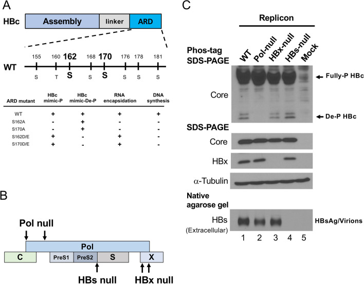
Background: It is generally believed that hepatitis B virus (HBV) core protein (HBc) dephosphorylation (de-P) is important for viral DNA synthesis and virion secretion. HBV polymerase contains four domains for terminal protein, spacer, reverse transcriptase, and RNase H activities.

Methods: HBV Polymerase mutants were transfected into HuH-7 cells and assayed for replication and HBc de-P by the Phos-tag gel analysis. Infection assay was performed by using a HepG2-NTCP-AS2 cell line.

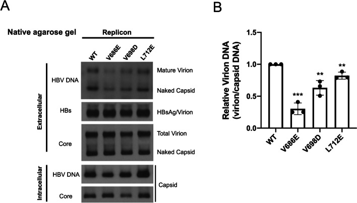
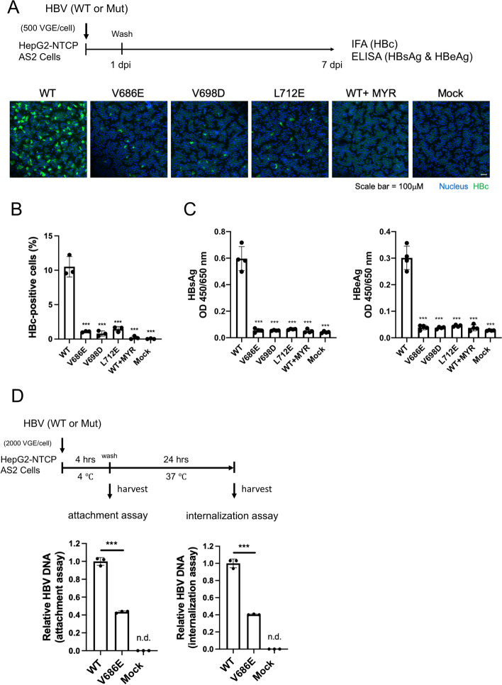
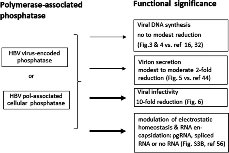
Results: Here, we show that a novel phosphatase activity responsible for HBc de-P can be mapped to the C-terminal domain of the polymerase overlapping with the RNase H domain. Surprisingly, while HBc de-P is crucial for viral infectivity, it is essential for neither viral DNA synthesis nor virion secretion. The potential origin, significance, and mechanism of this polymerase-associated phosphatase activity are discussed in the context of an electrostatic homeostasis model. The Phos-tag gel analysis revealed an intriguing pattern of "bipolar distribution" of phosphorylated HBc and a de-P HBc doublet.

Conclusions: It remains unknown if such a polymerase-associated phosphatase activity can be found in other related biosystems. This polymerase-associated phosphatase activity could be a druggable target in clinical therapy for hepatitis B.

Keywords: Capsids dephosphorylation (de-P); HBV core protein (HBc); Hepatitis B virus (HBV); Phosphatase; Polymerase (pol); RNase H domain.


Conflict of interest statement

We declare no competing financial or non-financial interests.

Figures

Fig. 1
Fig. 1
Dephosphorylation of HBV core protein (HBc) is correlated only with the presence of HBV polymerase, but not with the HBs envelope or the HBx protein. A The C-terminus of HBc contains three major and four minor phosphorylation sites. A summary of previous phosphomimicking mutagenesis studies on the major phosphoacceptor sites of HBc serine-162 and serine-170. Mutant S162D/E and mutant S170D/E enabled HBV pregenomic RNA encapsidation, but not viral DNA synthesis. B Cartoon illustration of three HBV mutant plasmids used in Fig. 1C. A mutant pol null is ablated at the first ATG initiation codon and the second ATG codon is engineered into a stop codon TAG. Mutant HBx null contains two engineered TAA stop codons in the HBx gene. Mutant HBs null lost the initiation codon of the small envelope protein. C Top panel: Phos-tag gel electrophoresis separated the fully phosphorylated HBc from HBc with partial or no phosphorylation. Completely dephosphorylated (De-P) HBc migrated faster than phosphorylated HBc. Different mutants in Fig. 1B produced similar amounts of HBc by Western blot analysis. Middle panel: Lack of HBx protein from mutant plasmid HBx null by Western blot using an anti-HBx antibody. Bottom panel: Mutant HBs null produced no HBs envelope protein in the medium by NAGE and Western blot using an anti-HBs antibody. Pol is known to be expressed at a very low level and no good antibody for pol is available
Fig. 2
Fig. 2
Dephosphorylation of HBc core protein is dependent on the encapsidation signal of the pregenomic RNA. A Encapsidation of pregenomic RNA (pgRNA) requires a highly ordered structure of the RNA packaging signal (epsilon) and polymerase. Epsilon mutant R and mutant L each contain a disrupted stem-loop structure of epsilon [68]. Double mutant R + L restored the wild type-like stem-loop structure of epsilon. Mutant pkex-1 contains a disrupted base pairing in the lower portion of the upper stem loop structure of the epsilon RNA [14]. B Upper panel: Single mutant R and single mutant L are defective in pgRNA encapsidation by Northern blot analysis, while the double mutant R + L restored pgRNA encapsidation. Mutant pkex-1 exhibited stronger signal intensity of encapsidated pgRNA than WT-HBV. Middle panel: Total cytoplasmic viral RNAs by Northern blot analysis. Lower panels: Viral DNA synthesis was detected only in WT-HBV and mutant R + L. RC: relaxed circle; DL: double-strand linear; SS: single-strand DNA
Fig. 3
Fig. 3
Dephosphorylation of HBc core protein is not required for viral DNA synthesis. A Different pol domain mutants were characterized for their viral DNA synthesis and HBc dephosphorylation. B HBV DNA synthesis can be detected in WT and mutant V686E (red asterisk) by Southern blot analysis. Due to the leaky polymerase expression in the CMV-driven replicon plasmid pol-null (Fig. 1; see Methods), trace amount of residual SS DNA can be detected after longer exposure of the X-ray film in the negative controls (lane 1 and 3). C A replication competent RNase H domain mutant V686E (red asterisk) exhibited no detectable dephosphorylated HBc by the Phos-tag gel. D No dephosphorylated HBc was detected in the double mutant Y63D/V686E (red asterisk)
Fig. 4
Fig. 4
HBc dephosphorylation is neither necessary nor sufficient for viral DNA synthesis. A Eleven RNase H domain mutants were designed to contain substitutions from a hydrophobic to an acidic residue. B Viral DNA synthesis of these RNase H mutants was analyzed by Southern blot analysis. Red asterisks highlight mutants with dissociated activities of viral DNA synthesis in B) and HBc dephosphorylation in C). C Phos-tag gel analysis detected the presence or absence of dephosphorylated HBc protein. D A summary table of five RNase H domain mutants (red asterisk) which exhibited dissociation between HBc dephosphorylation and viral DNA synthesis
Fig. 5
Fig. 5
A moderate effect of HBc core dephosphorylation on genome-containing virion secretion. A Upper panel: Three putative phosphatase-deficient RNase H domain mutants secreted significantly less amount of virion-associated viral DNA than wild type HBV, while HBsAg/virions and naked capsids were similar between wild type and mutants. Middle panel: Extracellular viral and subviral particles were collected from the media of HuH-7 cells on day 5 post-transfection with WT-HBV and mutants. PEG precipitated particles were resolved by native agarose gel, followed by Western blot analyses using anti-core and anti-HBs antibodies. Lower panel: Intracellular capsid particles were analyzed for viral DNA and core by Southern and Western blot analyses. B A bar graph comparison of virion-associated viral DNAs between wild type and three RNase H domain mutants. Signal intensities of HBV DNA associated with mature virions in the top panel of A) were normalized to HBV DNAs associated with intracellular capsids at the bottom panel of A). Signal intensities were quantified by densitometry and an Image J software, *** p < 0.001, ** p < 0.01. p value refers to the Student’s t-test statistical analysis
Fig. 6
Fig. 6
Core dephosphorylation-deficient RNase H mutants exhibited a 10-fold reduction in viral infectivity. A & (B) A 10-fold reduction in HBc core protein signal was detected by confocal IFA in RNase H  domain mutants deficient in putative phosphatase activity at 7 dpi. None of the pol mutation sites overlaps with the S envelope ORF. HepG2-NTCP-AS2 cells were in vitro infected with equal amounts of virions from WT-HBV and mutants defective in core dephosphorylation. MYR: a preS1 inhibitory control peptide. C The ELISA assays for HBsAg (left) and HBeAg (right) in the media detected a 10-fold reduction in cells infected with RNase H domain mutants. D RNase H domain mutant V686E exhibited a 2.5-fold reduction in attachment (left) and subsequent internalization (right). Naked capsids do not bind to NTCP and are present in near equal proportion in WT and mutant virions. HBV DNA was measured by qPCR. *** p < 0.001, p value refers to the Student’s t-test statistical analysis
Fig. 7
Fig. 7
A summary of the functional significance of HBc de-phosphorylation  A novel putative phosphatase with an HBc de-P activity was detected at the C-terminal domain of HBV polymerase by site-directed mutagenesis and the Phos-tag gel assay. Depending on the specific position of the missense point mutation, the phosphatase-defective pol mutants exhibited various degrees of reduction in viral replication, virion secretion and viral infectivity. The origin of this putative phosphatase could come from an unknown intrinsic phosphatase encoded by the HBV pol ORF. Alternatively, it could come from a cellular phosphatase physically associated with HBV polymerase. In one of our previous reports [56], hyperphosphorylated capsids are biased to preferentially encapsidate shorter spliced RNAs, instead of the full-length 3.5 kb pgRNA. This putative phosphatase could play a regulatory role in pgRNA packaging and capsid assembly through modulating HBc dephosphorylation and thus maintaining electrostatic homeostasis in the capsid interior. Relative to the parental single mutant Y63D, core-particle-associated viral RNA is significantly reduced in the double mutant Y63D/V686E (Fig. S3B). This result suggests that the loss of HBc de-P by V686E can result in a reduced amount of viral RNA encapsidation. The thickness of arrows reflect their respective degree of functional effects upon the loss of HBc de-P

Similar articles

Cited by

References

    1. Baltimore D. RNA-dependent DNA polymerase in virions of RNA tumour viruses. Nature. 1970;226(5252):1209–11. doi: 10.1038/2261209a0. - DOI - PubMed
    1. Baltimore D, Smoler DF. Association of an endoribonuclease with the avian myeloblastosis virus deoxyribonucleic acid polymerase. J Biol Chem. 1972;247(22):7282–7. doi: 10.1016/S0021-9258(19)44626-7. - DOI - PubMed
    1. Bartenschlager R, Schaller H. Hepadnaviral assembly is initiated by polymerase binding to the encapsidation signal in the viral RNA genome. EMBO J. 1992;11(9):3413–20. doi: 10.1002/j.1460-2075.1992.tb05420.x. - DOI - PMC - PubMed
    1. Basagoudanavar SH, Perlman DH, Hu J. Regulation of hepadnavirus reverse transcription by dynamic nucleocapsid phosphorylation. J Virol. 2007;81(4):1641–9. doi: 10.1128/JVI.01671-06. - DOI - PMC - PubMed
    1. Blumberg BS, Hepatitis B. Virus, the vaccine, and the control of primary cancer of the liver. Proc Natl Acad Sci U S A. 1997;94(14):7121–5. doi: 10.1073/pnas.94.14.7121. - DOI - PMC - PubMed

MeSH terms