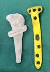
Changes in Patellar Height and Tibial Posterior Slope after Biplanar High Tibial Osteotomy with Computer-Designed Personalized Surgical Guides: A Retrospective Study
- PMID: 38561920
- PMCID: PMC11062865
- DOI: 10.1111/os.14049
Changes in Patellar Height and Tibial Posterior Slope after Biplanar High Tibial Osteotomy with Computer-Designed Personalized Surgical Guides: A Retrospective Study
Abstract
Objective: Medial opening-wedge high tibial osteotomy (MOWHTO) is a surgical procedure to treat medial compartment osteoarthritis in the knee with varus deformity. However, factors such as patellar height (PH) and the sagittal plane's posterior tibial slope angle (PTSA) are potentially overlooked. This study investigated the impact of alignment correction angle guided by computer-designed personalized surgical guide plate (PSGP) in MOWHTO on PH and PTSA, offering insights for enhancing surgical techniques.
Methods: This retrospective study included patients who underwent 3D-printed PSGP-assisted MOWHTO at our institution from March to September 2022. The paired t-tests assessed differences in all preoperative and postoperative measurement parameters. Multivariate linear regression analysis examined correlations between PTSA, CDI (Caton-Deschamps Index), and the alignment correction magnitude. Receiver operating characteristic (ROC) curve analysis determined the threshold of the correction angle, calculating sensitivity, specificity, and area under the curve.
Results: A total of 107 patients were included in our study. The CDI changed from a preoperative mean of 0.97 ± 0.13 (range 0.70-1.34) to a postoperative mean of 0.82 ± 0.13 (range 0.55-1.20). PTSA changed from a preoperative mean of 8.54 ± 2.67 (range 2.19-17.55) to a postoperative mean of 10.54 ± 3.05 (range 4.48-18.05). The t-test revealed statistically significant changes in both values (p < 0.05). A significant alteration in patellar height occurred when the correction angle exceeded 9.39°. Moreover, this paper illustrates a negative correlation between CDI change and the correction angle and preoperative PTSA. Holding other factors constant, each 1-degree increase in the correction angle led to a 0.017 decrease in postoperative CDI, and each 1-degree increase in preoperative PTSA resulted in a 0.008 decrease in postoperative CDI. PTSA change was positively correlated only with the correction angle; for each 1-degree increase in the opening angle, postoperative PTS increased by 0.188, with other factors constant.
Conclusion: This study highlights the effectiveness and precision of PSGP-assisted MOWHTO, focusing on the impact of alignment correction on PH and PTSA. These findings support the optimization of PSGP technology, which offers simpler, faster, and safer surgeries with less radiation and bleeding than traditional methods. However, PSGP's one-time use design and the learning curve required for its application are limitations, suggesting areas for further research.
Keywords: Knee osteoarthritis; Medial opening‐wedge high tibial osteotomy; Patellar height; Personalized surgical guide plate; Posterior tibial slope angle.
© 2024 The Authors. Orthopaedic Surgery published by Tianjin Hospital and John Wiley & Sons Australia, Ltd.
Conflict of interest statement
We declare that we have no financial and personal relationships with other people or organizations that can inappropriately influence our work, there is no professional or other personal interest of any nature or kind in any product, service and/or company that could be construed as influencing the position presented in, or the review of, the manuscript entitled.
Figures


Similar articles
-
Osteotomy Correction Angle Cut-off Points Can Guide the Operation to Prevent a Significant Decrease in Patella Height.Orthop Surg. 2024 Mar;16(3):628-636. doi: 10.1111/os.14000. Epub 2024 Feb 7. Orthop Surg. 2024. PMID: 38326241 Free PMC article.
-
Patella height is not altered by descending medial open-wedge high tibial osteotomy (HTO) compared to ascending HTO.Knee Surg Sports Traumatol Arthrosc. 2018 Jun;26(6):1859-1866. doi: 10.1007/s00167-017-4548-0. Epub 2017 Apr 17. Knee Surg Sports Traumatol Arthrosc. 2018. PMID: 28417183
-
Effect of the amount of correction on posterior tibial slope and patellar height in open-wedge high tibial osteotomy.J Orthop Surg (Hong Kong). 2021 Sep-Dec;29(3):23094990211049571. doi: 10.1177/23094990211049571. J Orthop Surg (Hong Kong). 2021. PMID: 34670434
-
Distal tibial tubercle osteotomy can lessen change in patellar height post medial opening wedge high tibial osteotomy? A systematic review and meta-analysis.J Orthop Surg Res. 2022 Jul 6;17(1):341. doi: 10.1186/s13018-022-03231-0. J Orthop Surg Res. 2022. PMID: 35794572 Free PMC article.
-
The effects of open wedge high tibial osteotomy for knee osteoarthritis on the patellofemoral joint. A systematic review.Knee. 2023 Jan;40:201-219. doi: 10.1016/j.knee.2022.11.023. Epub 2022 Dec 10. Knee. 2023. PMID: 36512892
Cited by
-
Combined Distal Femoral Osteotomy and Medial Patellofemoral Ligament Reconstruction for Patellar Instability and Genu Valgus: A Case Report and Literature Review.Orthop Surg. 2025 Jul;17(7):2201-2208. doi: 10.1111/os.70057. Epub 2025 May 31. Orthop Surg. 2025. PMID: 40448506 Free PMC article. Review.
-
A systematic review and meta-analysis examining alterations in medial meniscus extrusion and clinical outcomes following high tibial osteotomy.J Orthop. 2025 Feb 12;68:121-130. doi: 10.1016/j.jor.2025.01.036. eCollection 2025 Oct. J Orthop. 2025. PMID: 40070518 Review.
References
MeSH terms
Grants and funding
- TJWJ2022MS025/Tianjin Municipal Health Science and Technology Project, top-level project: research on personalized knee preservation for knee osteoarthritis based on osteotomy orthosis
- 22ZYJDSY00110/Central Guided Local Science and Technology Development Funds, Science and Technology Innovation Base Project, Research on Key Technology and Clinical Application of Precise Osteotomy Correction around Knee Joint
LinkOut - more resources
Full Text Sources

