[Preprint]. 2024 Mar 21:2024.03.21.586181.
doi: 10.1101/2024.03.21.586181.

Asynchronous microexon splicing of LSD1 and PHF21A during neurodevelopment

Affiliations

Asynchronous microexon splicing of LSD1 and PHF21A during neurodevelopment

Masayoshi Nagai et al. bioRxiv. .

Update in

Abstract

LSD1 histone H3K4 demethylase and its binding partner PHF21A, a reader protein for unmethylated H3K4, both undergo neuron-specific microexon splicing. The LSD1 neuronal microexon weakens H3K4 demethylation activity and can alter the substrate specificity to H3K9 or H4K20. Meanwhile, the PHF21A neuronal microexon interferes with nucleosome binding. However, the temporal expression patterns of LSD1 and PHF21A splicing isoforms during brain development remain unknown. In this work, we report that neuronal PHF21A isoform expression precedes neuronal LSD1 isoform expression during human neuron differentiation and mouse brain development. The asynchronous splicing events resulted in stepwise deactivation of the LSD1-PHF21A complex in reversing H3K4 methylation. We further show that the enzymatically inactive LSD1-PHF21A complex interacts with neuron-specific binding partners, including MYT1-family transcription factors and post-transcriptional mRNA processing proteins such as VIRMA. The interaction with the neuron-specific components, however, did not require the PHF21A microexon, indicating that the neuronal proteomic milieu, rather than the microexon-encoded PHF21A segment, is responsible for neuron-specific complex formation. These results indicate that the PHF21A microexon is dispensable for neuron-specific protein-protein interactions, yet the enzymatically inactive LSD1-PHF21A complex might have unique gene-regulatory roles in neurons.

Keywords: LSD1; PHF21A; histone demethylase; histone methylation; microexon splicing.


Conflict of interest statement

Conflict of interest The authors declare that they have no conflicts of interest with the contents of this article.

Figures
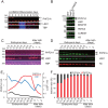
Figure 1:
Figure 1:. The expression of PHF21A-n increases faster than that of LSD1-n.
(A) mRNA levels of LSD1 and PHF21A isoforms in LUHMES cells. Cells were differentiated into neurons as indicated and harvested from day 3 to day 12, analyzed by RT-PCR (n=2). (B) Expression of PHF21A and associated proteins in LUHMES cells and 293T cells examined by Western blot analysis using antibodies as indicated. (C) mRNA levels of LSD1 and PHF21A isoforms in the developing mouse brain. Whole-cell lysates prepared from mouse whole brains (E12.5 and E13.5) and cortices (from E14.5 to P2) at indicated periods were analyzed by RT-PCR. (D) Expression of PHF21A and LSD1 proteins in the developing mouse brain. Whole-cell lysates were subjected to Western blot analysis. (E) Quantification of Western signals for PHF21A-c, PHF21A-n, and LSD1 normalized by histone H3. (F) The ratio of PHF21A protein isoforms in the developing mouse brain based on the Western signals.
Figure 2:
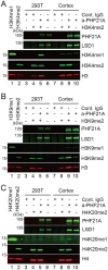
Figure 2:. A weaker H3K4 demethylation activity of the intermediate complex with PHF21A-n and LSD1-c.
(A) Schematic of the demethylation assay. The PHF21A-containing complex was immunoprecipitated from LHUMES cell nuclear extracts and incubated with designer nucleosomes with specific lysine di-methylations. (B-D) Western blot using the antibodies for indicated histone methylations to detect demethylase activity. The reactions were carried out using H3K4me2- (B), H3K9me2- (C), and H4K20me2- (D) nucleosomes. The appearance of mono-methylated lysine indicates the demethylation activity. The abundance of LSD1, PHF21A, and total H3 (c-term) were examined with specific antibodies. H3K4me1/me2 designer nucleosomes and 293T nuclear extract serve as specificity and positive control for the histone antibodies, respectively.
Figure 3:
Figure 3:. No detectable demethylation activity of the mature neuronal complex with PHF21A-n and LSD1-n.
The demethylation assay of immunoprecipitated PHF21A complexes from the P0 mouse cortices using the designer nucleosomes carrying H3K4me2 (A), H3K9me2 (B), and H4K20me2 (C). Neither a reduction of di-methylation nor the appearance of mono-methylation was observed with any demethylation reactions.
Figure 4:
Figure 4:. Proteomics analysis of PHF21A-containing complex in MEFs and neurons.
(A) Silver staining of proteins co-precipitated by an anti-PHF21A antibody. Nuclear extracts from MEFs and cortical neurons (DIV7) were used. Asterisks indicate proteins specifically found in PHF21A-immunoprecipitates, (B-C) Co-IP-MS analysis of PHF21A-associated proteins in MEF (B) and cortical neurons (C). Volcano plots of identified proteins (n=3). Green dots: the bait protein, PHF21A. Black and red dots: the interactors (Cutoff: FC > 1.5, P-value < 0.1, and peptide ≥ 6). Red dots: neuron-specific interactors with the same cut-off. Gray dots: proteins that did not pass the cutoff. The x-axis shows the log2 FC (a-PHF21A antibody/control IgG), and the y-axis denotes −log10 P-values. (D) Venn diagram of PHF21A interactor in MEFs (orange) versus neurons (blue). (E) Domain organization of neuron-specific PHF21A interactors. (F) Co-IP-Western assays to test the interaction between PHF21A, BRAF35, or iBRAF. Asterisks indicate nonspecific bands. (G) Expression of neuron-specific PHF21A interactors in the developing mouse brains. Whole-cell lysates prepared from mouse whole brains (E12.5 and E13.5) and cortices (from E14.5 to after birth 2) at indicated periods were subjected to Western blot analysis using antibodies as indicated.
Figure 5:
Figure 5:. Binding partners of PHF21-n and ectopic PHF21A-c in neurons are highly similar.
(A) Generation of Phf21a-neuronal exon knockout allele (Δn). Black arrows: PCR primers. Red triangles: CRISPR cut sites. (B) Genotyping PCR analysis of Phf21a+/+, Phf21a+/Δn, and Phf21aΔn/Δn mice using the primer sets shown in Fig. 5B. (C) Representative picture of P15 Phf21a+/+ (left) and Phf21aΔn/Δn (right) mice. (D) Western blot analysis of lysates prepared from cortical neurons (DIV7) isolated from Phf21a+/+, Phf21a+/Δn, and Phf21aΔn/Δn mice using antibodies as indicated. PHF21A-c instead of PHF21A-n is expressed in Phf21a-nΔn/Δn neuron. (E) Silver staining of proteins co-precipitated by anti-PHF21A antibody using the nuclear extracts from P0 cortices of the indicated genotypes. (F) Functional enrichment analysis of PHF21A-interacting proteins with Metascape (41). The statistically significant PHF21A interactors in Phf21a+/+ cortices were used. Log10 P-value < −10. The molecular networks that contain proteins validated by reciprocal IP experiment are presented. (G) Reciprocal Co-IP-Western assays to validate the interaction between PHF21A and newly-identified interaction partners. (H and I) Volcano plots of Co-IP-MS analysis of PHF21A-interacting proteins (n=3-4) using Phf21a+/+ (H) and Phf21aΔn/Δn (I) cortices. Green dots: the bait protein, PHF21A. Black and red dots: statistically significant interactors (Cutoff: FC > 2, Padj-value < 0.01, and peptide ≥ 6). Red dots: neuron-specific proteins identified in Fig. 4C that pass the cutoff. Gray dots: proteins that do not pass the threshold. The x-axis shows the log2 FC (a-PHF21A antibody/control IgG), and the y-axis denotes −log10 P-values. (J) Venn diagram of PHF21A interactor in Phf21a+/+ cortex (Blue) versus Phf21aΔn/Δn cortex (orange). (K) Scatter plot comparing the log2FC (a-PHF21A antibody/control IgG) of all PHF21A-associated proteins between WT and Phf21aΔn/Δn cortices. The R2 value and the fit of the linear regression are indicated. Green dots: the bait protein, PHF21A. Black and red dots: statistically significant interactors in Phf21a+/+ a-PHF21A and Phf21aΔn/Δn (Cutoff: Padj-value < 0.01, and peptide ≥ 6). Red dots: neuron-specific proteins identified in Fig. 4C that pass the cutoff. Blue dots: statistically significant interactors in Phf21a+/+ a-PHF21A. Orange dots: statistically significant interactors in Phf21aΔn/Δn. The x-axis shows the log2 FC of Phf21a+/+ (a-PHF21A antibody/control IgG). The y-axis shows the log2 FC of Phf21aΔn/Δn (a-PHF21A antibody/control IgG).

References

    1. Najmabadi H., Hu H., Garshasbi M., Zemojtel T., Abedini S. S., Chen W., Hosseini M., Behjati F., Haas S., Jamali P., Zecha A., Mohseni M., Püttmann L., Vahid L. N., Jensen C., Moheb L. A., Bienek M., Larti F., Mueller I., Weissmann R., Darvish H., Wrogemann K., Hadavi V., Lipkowitz B., Esmaeeli-Nieh S., Wieczorek D., Kariminejad R., Firouzabadi S. G., Cohen M., Fattahi Z., Rost I., Mojahedi F., Hertzberg C., Dehghan A., Rajab A., Banavandi M. J. S., Hoffer J., Falah M., Musante L., Kalscheuer V., Ullmann R., Kuss A. W., Tzschach A., Kahrizi K., and Ropers H. H. (2011) Deep sequencing reveals 50 novel genes for recessive cognitive disorders. Nature. 478, 57–63 - PubMed
    1. McCarthy S. E., Gillis J., Kramer M., Lihm J., Yoon S., Berstein Y., Mistry M., Pavlidis P., Solomon R., Ghiban E., Antoniou E., Kelleher E., O’Brien C., Donohoe G., Gill M., Morris D. W., McCombie W. R., and Corvin A. (2014) De novo mutations in schizophrenia implicate chromatin remodeling and support a genetic overlap with autism and intellectual disability. Mol Psychiatry. 19, 652–658 - PMC - PubMed
    1. De Rubeis S., He X., Goldberg A. P., Poultney C. S., Samocha K., Ercument Cicek A., Kou Y., Liu L., Fromer M., Walker S., Singh T., Klei L., Kosmicki J., Fu S.-C., Aleksic B., Biscaldi M., Bolton P. F., Brownfeld J. M., Cai J., Campbell N. G., Carracedo A., Chahrour M. H., Chiocchetti A. G., Coon H., Crawford E. L., Crooks L., Curran S. R., Dawson G., Duketis E., Fernandez B. A., Gallagher L., Geller E., Guter S. J., Sean Hill R., Ionita-Laza I., Jimenez Gonzalez P., Kilpinen H., Klauck S. M., Kolevzon A., Lee I., Lei J., Lehtimäki T., Lin C.-F., Ma’ayan A., Marshall C. R., McInnes A. L., Neale B., Owen M. J., Ozaki N., Parellada M., Parr J. R., Purcell S., Puura K., Rajagopalan D., Rehnström K., Reichenberg A., Sabo A., Sachse M., Sanders S. J., Schafer C., Schulte-Rüther M., Skuse D., Stevens C., Szatmari P., Tammimies K., Valladares O., Voran A., Wang L.-S., Weiss L. A., Jeremy Willsey A., Yu T. W., Yuen R. K. C., Cook E. H., Freitag C. M., Gill M., Hultman C. M., Lehner T., Palotie A., Schellenberg G. D., Sklar P., State M. W., Sutcliffe J. S., Walsh C. A., Scherer S. W., Zwick M. E., Barrett J. C., Cutler D. J., Roeder K., Devlin B., Daly M. J., and Buxbaum J. D. (2014) Synaptic, transcriptional and chromatin genes disrupted in autism. Nature. 515, 209–215 - PMC - PubMed
    1. Lister R., Mukamel E. A., Nery J. R., Urich M., Puddifoot C. A., Johnson N. D., Lucero J., Huang Y., Dwork A. J., Schultz M. D., Yu M., Tonti-Filippini J., Heyn H., Hu S., Wu J. C., Rao A., Esteller M., He C., Haghighi F. G., Sejnowski T. J., Behrens M. M., and Ecker J. R. (2013) Global Epigenomic Reconfiguration During Mammalian Brain Development. Science. 341, 1237905. - PMC - PubMed
    1. Stroud H., Su S. C., Hrvatin S., Greben A. W., Renthal W., Boxer L. D., Nagy M. A., Hochbaum D. R., Kinde B., Gabel H. W., and Greenberg M. E. (2017) Early-Life Gene Expression in Neurons Modulates Lasting Epigenetic States. Cell. 171, 1151–1164.e16 - PMC - PubMed

Publication types