[Preprint]. 2024 Mar 22:rs.3.rs-4077218.
doi: 10.21203/rs.3.rs-4077218/v1.

Functional Redundancy in Candida auris Cell Surface Adhesins Crucial for Cell-Cell Interaction and Aggregation

Affiliations

Functional Redundancy in Candida auris Cell Surface Adhesins Crucial for Cell-Cell Interaction and Aggregation

Tristan W Wang et al. Res Sq. .

Update in

Abstract

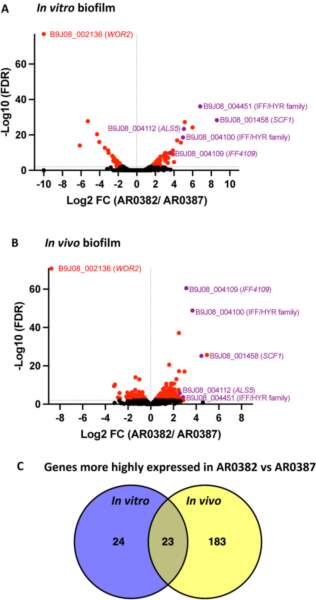
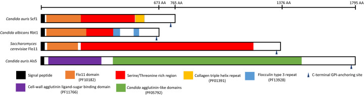
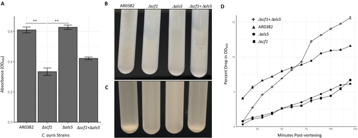
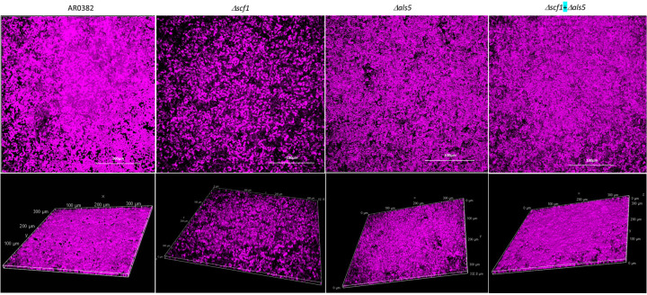
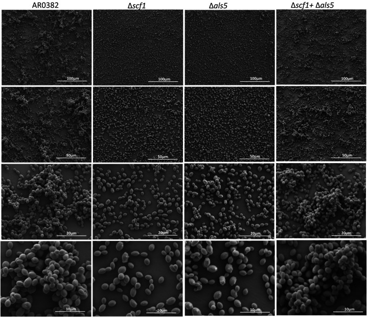
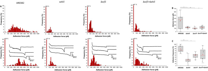
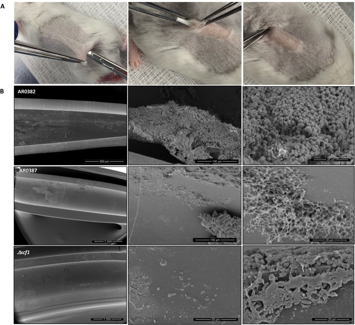
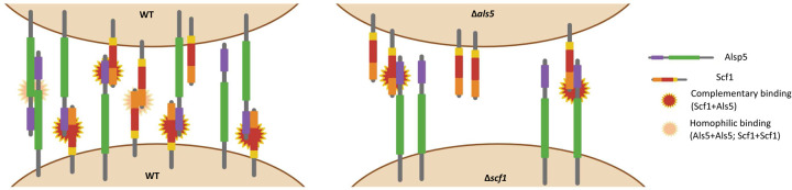
Candida auris is an emerging nosocomial fungal pathogen associated with life-threatening invasive disease due to its persistent colonization, high level of transmissibility and multi-drug resistance. Aggregative and non-aggregative growth phenotypes for C. auris strains with different biofilm forming abilities, drug susceptibilities and virulence characteristics have been described. Using comprehensive transcriptional analysis we identified key cell surface adhesins that were highly upregulated in the aggregative phenotype during in vitro and in vivo grown biofilms using a mouse model of catheter infection. Phenotypic and functional evaluations of generated null mutants demonstrated crucial roles for the adhesins Als5 and Scf1 in mediating cell-cell adherence, coaggregation and biofilm formation. While individual mutants were largely non-aggregative, in combination cells were able to co-adhere and aggregate, as directly demonstrated by measuring cell adhesion forces using single-cell atomic force spectroscopy. This co-adherence indicates their role as complementary adhesins, which despite their limited similarity, may function redundantly to promote cell-cell interaction and biofilm formation. Functional diversity of cell wall proteins may be a form of regulation that provides the aggregative phenotype of C. auris with flexibility and rapid adaptation to the environment, potentially impacting persistence and virulence.


Conflict of interest statement

Additional Declarations: There is NO Competing Interest.

Figures

Fig. 1.
Fig. 1.. RNA-seq analysis of in vitro and in vivo grown biofilms depicting genes differentially regulated in the aggregative C. auris strain AR0382 compared to the non-aggregative strain AR0387.
Volcano plots of comparative differential gene expressions during (A) in vitro and (B) in vivo biofilm growth. LFC, log (base 2) fold change. FDR, false-discovery rate. Black: not statistically significant (FDR > 0.01); Red: Statistically significant (FDR < 0.01); Purple: Statistically significant and an adhesin. (C) Venn diagrams representing the overlap in the numbers of genes that are more highly expressed in strain AR0382 in vitro and in vivo.
Figure 2.
Figure 2.. Scf1 adhesin domain organization.
Diagram comparing the C. auris Scf1 domain structure to that of the C. albicans Rbt1 adhesin and the Saccharomyces cerevisiae Flo11 depicting a common Flo11 domain and a serine-threonine rich region (>50%) recognized by Als5. Pfam database code is in parentheses; signal peptides and GPI-anchors were predicted using the prediction softwares SignalP 6.0 and NetGPI-1.1, respectively. Functional domains of adhesin proteins were identified via InterProtScan (https://www.ebi.ac.uk/interpro/search/sequence/) (accessed February 12, 2024). Uniprot entries: A0A2H1A319 (Scf1); A0A8H6F4R1 (Rbt1); P08640 (Flo11); A0A2H0ZHZ9 (Als5).
Figure 3.
Figure 3.. Comparative evaluation of biofilm formation, aggregation and sedimentation rate of Δscf1 and Δals5 mutants individually and in combination compared to the wild-type (AR0382).
(A) Measurement of the metabolic activity of 24 h biofilms based on values of OD490 comparing wild-type AR0382 to Δscf1 and Δals5 mutants and Δscf1als5 combination. Statistical analysis was performed by one-way ANOVA and post-hoc Tukey test with p-values representing significant differences. Bar-plots show mean and SEM of n = 3 biological replicates, each as an average of 4 technical replicates. P = 2.61×10⁻³, 1.75×10⁻³. (B) Cell aggregation 2 min after vigorous vortexing and (C) 10 min post-vortexing. (D) Measurement of rate of cell sedimentation over 2 hr. Values represented are mean OD plus SEM of three technical replicates. **0.001 < P ≤ 0.01.
Figure. 4.
Figure. 4.. Representative images from confocal laser scanning microscopy analysis of biofilms formed by the C. auris AR0382 wild-type (WT) strain and the Δscf1 and Δals5 mutants grown individually and in combination (Δscf1+Δals5).
Z-stack reconstructions of biofilms stained with polysaccharide stain concanavalin A (fuchsia).
Figure. 5.
Figure. 5.. Representative images from scanning electron microscopy analysis.
24 h biofilms formed by the C. auris AR0382 wild-type (WT) strain and the Δscf1 and Δals5 mutants grown individually and in combination (Δscf1als5).
Figure 6.
Figure 6.. Single-cell force spectroscopy of C. auris cell-cell adhesion.
(A) Adhesion force histograms with representative retraction profiles (inset) obtained for the interaction between AR0382 wild-type cells, cells of Δals5, cells of Δscf1 and between cells of Δals5 and Δscf1 (Δals5scf1); 2 representative cell pairs are shown for each strain. (B) Adhesion force boxplots show data on n = 6 pairs of AR0382 cells, Δals5 cells, and Δscf1 cells and n = 8 cell pairs combining Δals5 and Δscf1. Statistical analysis was performed by one-way ANOVA and post-hoc Tukey test with p-values representing significant differences. P = 1.42×10⁻², 7.69×10⁻³, 1.01×10⁻². (C) As in (B), adhesion frequency boxplots show data on n = 7 pairs of AR0382 cells, n = 5 pairs of Δals5 cells, n = 6 pairs of Δscf1 cells and n = 8 pairs between Δals5scf1 cells. P=1.71×10⁻³, 3.77×10⁻². Red stars represent the mean values, red lines are the medians, boxes are the 25−75% quartiles and whiskers the standard deviation from mean. *0.01 < P ≤ 0.05, **0.001 < P ≤ 0.01.
Figure. 7.
Figure. 7.. Infection and biofilm formation in catheters implanted in mice.
(A) A small incision is made in a shaved area in the dorsum of anesthetized mice and catheter fragments (0.5 cm) are inserted within a formed subcutaneous tunnel. (B) Scanning electron microscopy of explanted catheters. Representative low- and high-magnification SEM images demonstrating mature biofilm formed within lumen of catheters infected with AR0382 wild-type strain consisting of aggregates of yeast cells.
Figure. 8.
Figure. 8.. Hypothetical mechanistic model depicting complementary Scf1/Als5 binding.
(left) Adherence between wild-type (WT) cells involving Scf1+Als5 complementary binding and homophilic interactions between Als5+Als5 and Scf1+Scf1; (right) Complementary binding between Scf1 and Als5 on the Δals5 and Δscf1 mutant cells, respectively. Domain designations and colors are consistent with those in Fig. 2.

Similar articles

References

    1. Vila T., Sultan A.S., Montelongo-Jauregui D. & Jabra-Rizk M.A. Candida auris: A fungus with identity crisis Pathog Dis 78, ftaa034 (2020). - PMC - PubMed
    1. Lockhart S.R. et al. Simultaneous emergence of multidrug-resistant Candida auris on 3 continents confirmed by whole-genome sequencing and epidemiological analyses. Clin Infect Dis 64, 134–140 (2017). - PMC - PubMed
    1. Cortegiani A., Misseri G., Giarratano A., Bassetti M. & Eyre D. The global challenge of Candida auris in the intensive care unit. Crit Care Med. 23, 150 (2019). - PMC - PubMed
    1. Lyman M. et al. Worsening Spread of Candida auris in the United States, 2019 to 2021.Ann Intern Med 176, 489–495 (2023). - PMC - PubMed
    1. de Jong A.W. & Hagen F. Attack, defend and persist: How the fungal pathogen Candida auris was able to emerge globally in healthcare environments. Mycopathologia. 148, 353–365. (2019). - PubMed

Publication types

LinkOut - more resources